. 2006 Jan-Mar;5(1):31-40.

Imaging molecular expression on vascular endothelial cells by in vivo immunofluorescence microscopy

Affiliations

Imaging molecular expression on vascular endothelial cells by in vivo immunofluorescence microscopy

Judith M Runnels et al. Mol Imaging. 2006 Jan-Mar.

Abstract

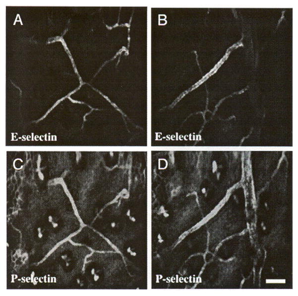
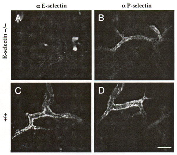
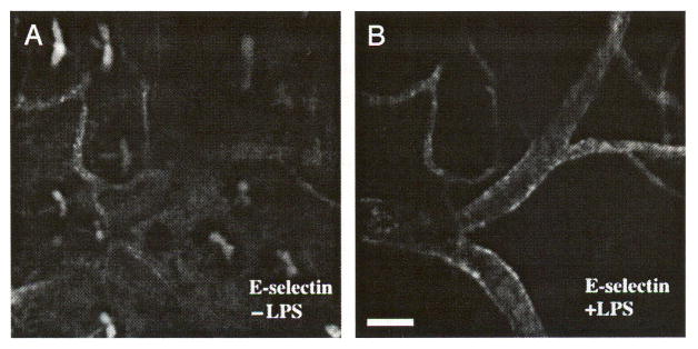
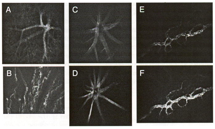
Molecular expression on the vascular endothelium is critical in regulating the interaction of circulating cells with the blood vessel wall. Leukocytes as well as circulating cancer cells gain entry into tissue by interacting with adhesion molecules on the endothelial cells (EC). Molecular targets on the EC are increasingly being explored for tissue-specific delivery of therapeutic and imaging agents. Here we use in vivo immunofluorescence microscopy to visualize the endothelial molecular expression in the vasculature of live animals. High-resolution images are obtained by optical sectioning through the intact skin using in vivo confocal and multiphoton microscopy after in situ labeling of EC surface markers with fluorescent antibodies. Other vascular beds such as the bone marrow and ocular blood vessels can be imaged with little or no tissue manipulation. Live imaging is particularly useful for following the dynamic expression of inducible molecules such as E-selectin during an inflammatory response.


Figures

Figure 1
Figure 1
Chemical structure of IRDye 38.
Figure 2
Figure 2
Optical sectioning by in vivo confocal microscopy. (A) Autofluorescent surface hair, (B) autofluorescent epidermal keratinocytes, and (C) an artery and vein pair in a plane deeper than the keratinocyte layer. The vessels are stained with Cy5.5-conjugated anti-PECAM-1 antibody that had been intravenously injected 16 hr before the images were taken. Bars indicate 100 μm.
Figure 3
Figure 3
Skin autofluorescence decreases at longer near-IR wavelengths. The tissue autofluorescence is most obvious in the FITC channel (A) compared to the Cy5.5 channel (C, D), and least obvious in the IR38 channel (F). (B, E, and G) Absorption and emission spectra for FITC, Cy5.5, and IR38fluorophores, respectively (A) FITC-conjugated anti-PECAM-1, (C) Cy5.5-conjugated anti-E-selectin, (D) Cy5.5-conjugated anti-E-selectin, and (F) IR38-conjugated anti-E-selectin with LPS treatment. Panels A and C are of the same site, as are panels D and F. The bowtie-like structures in A, C, and D are hair follicles. The honeycombed structures in A, C, and D are keratinocytes.
Figure 4
Figure 4
Coexpression of constitutive E-selectin with PECAM-1. (A–E) Cy5.5-conjugated anti-E-selectin was injected into the tail vein followed by FITC-conjugated anti-PECAM-1 injection 2 days later. On the third day, the ear skin was imaged. (B, E) E-selectin is constitutively expressed in a subset of vessels expressing (A, D) PECAM-1. (C) Overlay of A and B. Images A–C from one mouse, D, E from a second mouse. Bars indicate 100 μm.
Figure 5
Figure 5
Coexpression of E- and P-selectin in quiescent mouse skin. IR38-conjugated anti-E-selectin and Cy5.5-conjugaled anti-P-selectin were intravenously coinjected. Two days later, the vasculature in the mouse ear was imaged. (A, B) Expression of E-selectin is restricted to small venules, whereas (C, D) P-selectin is expressed in those venules as well as larger venules. Images A and C, and B and D are of the same sites, respectively. Bar indicates 100 μm.
Figure 6
Figure 6
Specificity of Cy5.5 conjugated E- and P-selectin antibodies in vivo is demonstrated in the skin of Balb/c E-selectin knockout mice. (B) A characteristic quiescent P-selectin expression pattern is present in E-selectin knockout mouse skin, but no E-selectin (A) expression is detected. Both (C) E-selectin and (D) P-selectin are expressed on skin vessel walls in the control mice in the quiescent state. Each panel represents a separate animal. Bar indicates 50 μm.
Figure 7
Figure 7
LPS induces E-selectin expression in venules larger than 50 μm. Intravenous injection of Cy5.5-conjugated anti-E-selectin antibody was followed by intravenous injection of IR38-conjugated anti-E-selectin and intraperitoneal injection of LPS 9 days later. On Day 12. the mouse was imaged. E-selectin is constitutively expressed in postcapillary venules in (A) skin, but is expressed on larger venules after LPS induction (B). Images A and B are of the same site in the mouse ear skin. Bar indicates 100 μm.
Figure 8
Figure 8
In vivo fluorescence confocal microscopy can be performed on ocular tissues. PECAM-1 expression in the retina (A) and choroid (B) of a mouse 24 hr after intravenous injection of Cy5.5-conjugated anti-PECAM antibody. Cy5.5-conjugated anti-VCAM in a normal C57BL/6 mouse (C) and in a LPS-treated mouse retina (D). Cy5.5-conjugated anti-E-selectin in the conjunctiva of a normal mouse (E) and IR38-conjugated anti-E-selectin 24 hr after systemic administration of LPS (F).

References

    1. Hynes RO. Integrins, versatility, modulation, and signalling in cell adhesion. Cell. 1992;69:11–25. - PubMed
    1. Rouslahti E. Extracellular matrix/growth factor interactions. Cold Spring Harb Symp Quant Biol. 1992;57:309–315. - PubMed
    1. Bullard DC, Kunkel EJ, Kubo H, Hicks MJ, Lorenzo I, Doyle NA, Doerschuk CM, Ley K, Beaudet AL. Infectious susceptibility and severe deficiency of leukocyte rolling and recruitment in E-selectin and P-selectin double mutant mice. J Exp Med. 1996;183:2329–2396. - PMC - PubMed
    1. Schweitzer KM, Drager AM, van der Valk P, Thijsen SFT, Zevenbergen A, Theijsmeijer AP, van der Schoot CE, Langenhuisen MMAC. Constitutive expression of E-selectin and vascular cell adhesion molecule-1 on endothelial cells of hematopoietic tissues. Am J Pathol. 1996;148:165–175. - PMC - PubMed
    1. Rice GE, Bevilacqua MP. An inducible endothelial cell surface glycoprotein mediates melanoma adhesion. Science. 1989;24:1303–1306. - PubMed

Publication types

MeSH terms

LinkOut - more resources