. 2006 Oct 1;399(1):59-67.
doi: 10.1042/BJ20060464.

Direct binding of Cbl to Tyr568 and Tyr936 of the stem cell factor receptor/c-Kit is required for ligand-induced ubiquitination, internalization and degradation

Affiliations

Direct binding of Cbl to Tyr568 and Tyr936 of the stem cell factor receptor/c-Kit is required for ligand-induced ubiquitination, internalization and degradation

Kristina Masson et al. Biochem J. .

Abstract

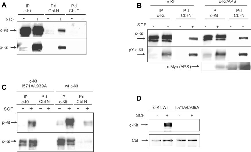
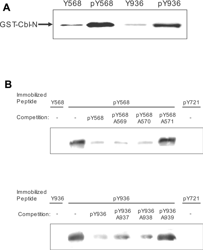
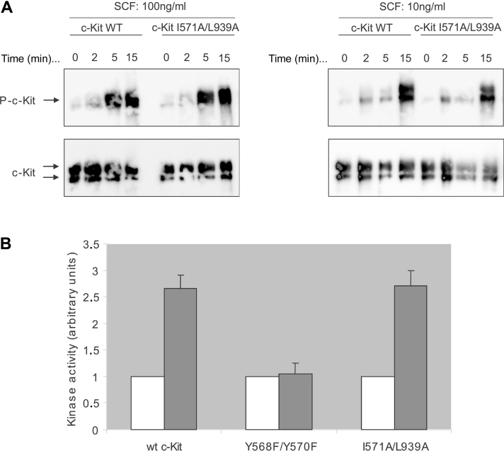
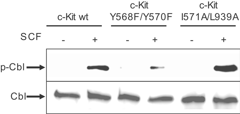
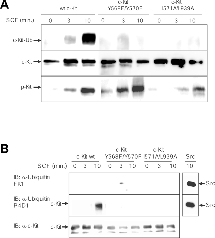
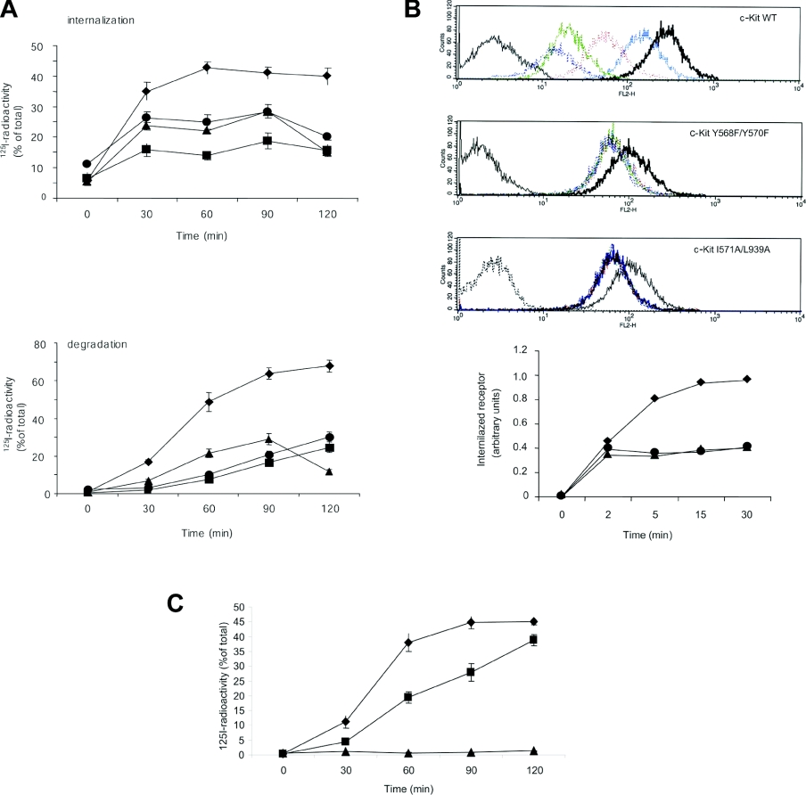
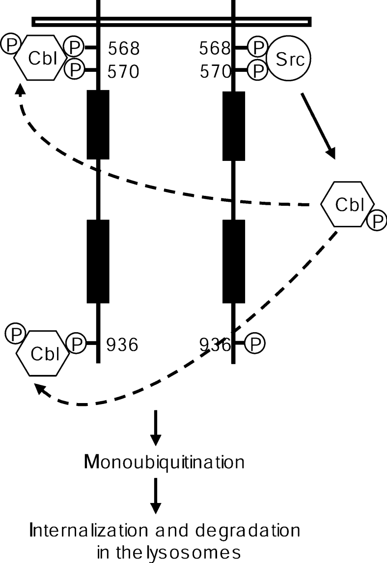
The ubiquitin E3 ligase Cbl has been shown to negatively regulate tyrosine kinase receptors, including the stem cell factor receptor/c-Kit. Impaired recruitment of Cbl to c-Kit results in a deregulated positive signalling that eventually can contribute to carcinogenesis. Here, we present results showing that Cbl is activated by the SFKs (Src family kinases) and recruited to c-Kit in order to trigger receptor ubiquitination. We demonstrate that phosphorylated Tyr568 and Tyr936 in c-Kit are involved in direct binding and activation of Cbl and that binding of the TKB domain (tyrosine kinase binding domain) of Cbl to c-Kit is specified by the presence of an isoleucine or leucine residue in position +3 to the phosphorylated tyrosine residue on c-Kit. Apart from the direct association between Cbl and c-Kit, we show that phosphorylation of Cbl by SFK members is required for activation of Cbl to occur. Moreover, we demonstrate that Cbl mediates monoubiquitination of c-Kit and that the receptor is subsequently targeted for lysosomal degradation. Taken together, our findings reveal novel insights into the mechanisms by which Cbl negatively regulates c-Kit-mediated signalling.


Figures

Figure 1
Figure 1. Interaction of the N-terminal half of Cbl with c-Kit at position Tyr568 and Tyr936
(A) COS-1 cells were transiently transfected with wild-type c-Kit expression plasmids and stimulated with SCF as indicated. After lysis, cell lysates were subjected to either an immunoprecipitation (IP) with c-Kit antibody or a pulldown (Pd) with GST fusion proteins of either the N-terminal or C-terminal half of Cbl. Immunocomplexes were separated on an SDS/8% polyacrylamide gel, blotted and sequentially probed for c-Kit (upper panel) or anti-phosphotyrosine (lower panel). (B) COS-1 cells were co-transfected with expression plasmid for c-Kit and Myc-tagged APS and stimulated with SCF as indicated. After lysis, cell lysates were subjected to immunoprecipitation with anti-c-Kit as well to a pulldown with GST–Cbl (N-term) fusion protein. Complexes were analysed by Western blotting and the membranes were sequentially probed for c-Kit, phosphorylated c-Kit and APS. (C) COS-1 cells were transfected with the c-Kit mutant I571A/L939A and samples were treated as described above. (D) Cbl was immunoprecipitated from COS-1 cells transfected with either wild-type c-Kit or the I571A/L939A mutant and samples were analysed by Western blotting.
Figure 2
Figure 2. Cbl binds directly to phosphorylated Tyr568 and Tyr936 and the specificity is determined by the amino acids at the +3 positions to the tyrosine residues respectively
(A) Phosphopeptides pY568 and pY936 (sequences in the Experimental section) immobilized to Affigel 10 beads were incubated (3 h at 4 °C) with the indicated purified GST fusion proteins in the presence of either unphosphorylated or phosphorylated peptides in solution as indicated. Samples were analysed by SDS/PAGE and Western blotting, and membranes were probed with an anti-GST antibody. (B) Pulldown experiment performed as described above with competition by soluble peptides (100 μM) as indicated.
Figure 3
Figure 3. Intrinsic kinase activity and ability to phosphorylate Src of the c-Kit double mutant I571A/L939A
(A) COS-1 cells were transfected with wild-type c-Kit (lanes 1 and 2) as well as the I571A/L939A (lanes 3 and 4) double mutant and stimulated with SCF for the indicated periods of time (min; values above lanes) and concentrations. Lysates were immunoprecipitated with a c-Kit antibody and analysed by Western blotting. The membrane was probed for tyrosine-phosphorylated residues (4G10) and reprobed for c-Kit as loading control. (B) Densitometric analysis of an Src in vitro kinase assay. Src was immunoprecipitated from an aliquot of cell lysate prepared as described above, followed by incubation (3 h at 4 °C) with 50 μM [γ-32P]ATP in kinase buffer (described in detail in the subsection ‘In vitro kinase assay’ in the Experimental section). Samples were separated on an SDS/8% polyacrylamide gel and blotted on to Immobilon. The filter was incubated with 1 M KOH for 1 h at 55 °C to hydrolyse non-specific phosphoserine present in the proteins, exposed to the PhosphoImager and densitometrically analysed. Src activity upon stimulation in the respective mutant (grey bars) is expressed as fold activity in unstimulated cells (white bars). Mean values and standard deviations for three independent experiments are shown.
Figure 4
Figure 4. The effect of the wild-type c-Kit versus the two c-Kit double mutants Y568F/Y570F and I571A/L939A on Cbl phosphorylation
Transiently transfected COS-1 cells expressing wild-type c-Kit or one of the two double mutants Y568F/Y570F or I571A/L939A respectively were stimulated with SCF for the indicated periods of time, immunoprecipitated with a Cbl antibody and subjected to Western blotting. The membrane was probed for tyrosine phosphorylation and Cbl respectively.
Figure 5
Figure 5. Monoubiquitination of c-Kit upon phosphorylation of Tyr568 and Tyr936 and recruitment of Cbl
(A) PAE cells expressing wild-type, Y568F/Y570F or I571A/L939A c-Kit were stimulated with SCF for the indicated time points, followed by lysis and immunoprecipitation with c-Kit antibody. Samples were subjected to a Western-blot analysis for ubiquitin (P4D1) and c-Kit. (B) An identical experiment was performed, with the exception that the membrane was probed with a selective polyubiquitin antibody (FK1) as well as the P4D1 ubiquitin antibody recognizing both poly- and mono-ubiquitin. Immunoprecipitated Src was included as a positive control for the polyubiquitin antibody. IB, immunoblot.
Figure 6
Figure 6. Analysis of degradation of internalized SCF in cells expressing wild-type c-Kit versus I571A/L939A mutant c-Kit and their intracellular processing
(A) PAE cells expressing wild-type (◆), Y568F/Y570F (▲) or I571A/L939A (●) c-Kit were incubated with 125I-SCF for 60 min on ice, followed by incubation at 37 °C for the indicated periods of time. Cells were washed with an acidic buffer (see the subsection ‘Internalization and degradation experiments’ in the Experimental section) and surface-released radioactivity was determined. Loss of surface-bound radioactivity was taken as a measure of internalization. The medium was collected and subjected to precipitation with 10% (w/v) trichloroacetic acid. Trichloroacetic acidsoluble radioactivity was taken as a measure of degradation of 125I-SCF. As a negative control, wild-type c-Kit-expressing PAE cells were treated with 2 μM SU6656 (■) to inhibit Src activity before examining internalization and SCF degradation. (B) Flow cytometry analysis of internalization. Ba/F3 cells transfected with either wild-type (◆), Y568F/Y570F (▲) or I571A/L939A (●) mutant c-Kit were incubated with 100 ng/ml SCF for the indicated periods of time, followed by flow cytometry analysis using an antibody against the extracellular part of c-Kit. The following colour coding of the histograms was used: grey: negative control; black; 0 min; blue: 2 min; red: 5 min; green: 15 min; purple: 30 min. (C) PAE cells expressing wild-type c-Kit were pre-incubated in the absence of inhibitor (◆) or in the presence of either 100 μM lactacystin (■) or 20 μM chloroquine (▲) for 4 h, followed by the assessment of binding, internalization and degradation of 125I-SCF as described above. Mean values and standard deviations for three independent experiments are shown.
Figure 7
Figure 7. Proposed model for the sequential Src- and Cbl-dependent steps during the ubiquitination of c-Kit
Activation of SFKs, initiated by Src binding to pY568 of ligand-stimulated c-Kit, leads to phosphorylation of Cbl. Phosphorylated Cbl is then recruited to pY568 and pY936 of c-Kit, which in turn results in monoubiquitination of the receptor and its subsequent internalization and degradation in the lysosomes.

References

    1. Lennartsson J., Jelacic T., Linnekin D., Shivakrupa R. Normal and oncogenic forms of the receptor tyrosine kinase kit. Stem Cells. 2005;23:16–43. - PubMed
    1. Rönnstrand L. Signal transduction via the stem cell factor receptor/c-Kit. Cell. Mol. Life Sci. 2004;61:2535–2548. - PMC - PubMed
    1. Lennartsson J., Voytyuk O., Heiss E., Sundberg C., Sun J., Rönnstrand L. c-Kit signal transduction and involvement in cancer. Cancer Ther. 2005;3:5–28.
    1. Thien C. B., Langdon W. Y. Cbl: many adaptations to regulate protein tyrosine kinases. Nat. Rev. Mol. Cell Biol. 2001;2:294–307. - PubMed
    1. Lill N. L., Douillard P., Awwad R. A., Ota S., Lupher M. L., Jr, Miyake S., Meissner-Lula N., Hsu V. W., Band H. The evolutionarily conserved N-terminal region of Cbl is sufficient to enhance down-regulation of the epidermal growth factor receptor. J. Biol. Chem. 2000;275:367–377. - PubMed

Publication types