. 2006 Aug;120(2):171-8.
doi: 10.1016/j.clim.2006.05.002. Epub 2006 Jun 16.

Long-lived effector/central memory T-cell responses to severe acute respiratory syndrome coronavirus (SARS-CoV) S antigen in recovered SARS patients

Affiliations

Long-lived effector/central memory T-cell responses to severe acute respiratory syndrome coronavirus (SARS-CoV) S antigen in recovered SARS patients

Li-Tao Yang et al. Clin Immunol. 2006 Aug.

Abstract

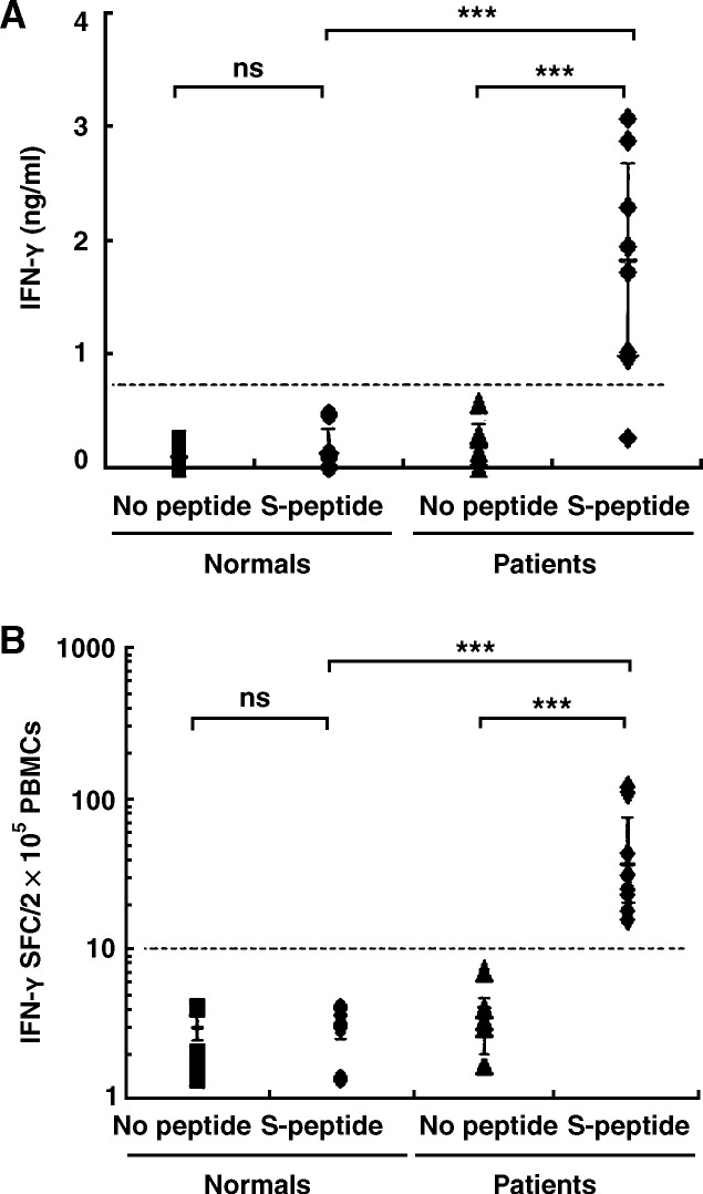
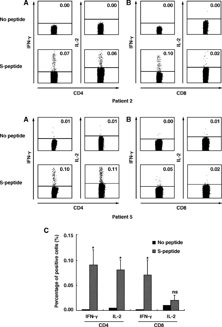
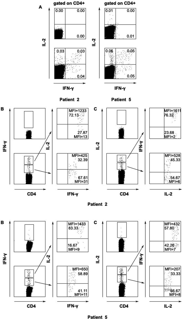
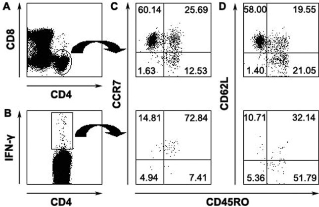
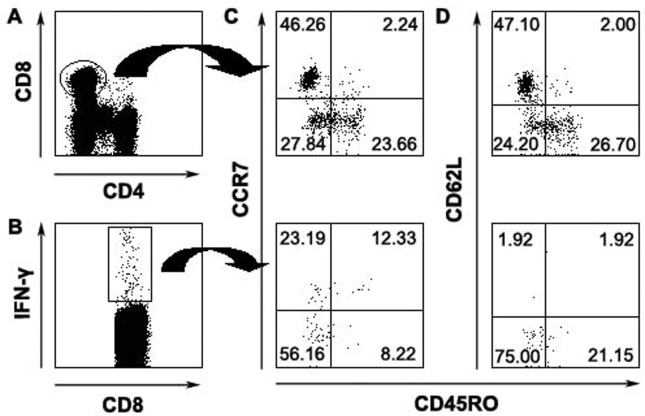
The role of cell-mediated immunity in human SARS-CoV infection is still not well understood. In this study, we found that memory T-cell responses against the spike (S) protein were persistent for more than 1 year after SARS-CoV infection by detecting the production of IFN-gamma using ELISA and ELISpot assays. Flow cytometric analysis showed that both CD4(+) and CD8(+) T cells were involved in cellular responses against SARS-CoV infection. Interestingly, most of SARS-CoV S-specific memory CD4(+) T cells were central memory cells expressing CD45RO(+) CCR7(+) CD62L(-). However, the majority of memory CD8(+) T cells revealed effector memory phenotype expressing CD45RO(-) CCR7(-) CD62L(-). Thus, our study provides the evidence that SARS-CoV infection in humans can induce cellular immune response that is persistent for a long period of time. These data may have an important implication in the possibility of designing effective vaccine against SARS-CoV infection, specifically in defining T-cell populations that are implicated in protective immunity.


Figures

Figure 1
Figure 1
SARS-CoV S-peptide-specific IFN-γ production by PBMCs. Fresh PBMCs were isolated from fully recovered SARS patients and normal individuals and cultured in vitro with or without a pool of 169 SARS-CoV S peptides in the presence of anti-CD28 (1 μg/ml) and anti-CD49d mAbs (1 μg/ml). After incubation for 72 h, the level of IFN-γ in culture supernatants was assayed by ELISA (A). The frequency of IFN-γ-secreting cells was detected by ELISpot assay at the single cell level after stimulation for 20 h (B). Student's t test was used for statistical analysis. The significance was set at P < 0.01 (***). ns, not significant.
Figure 2
Figure 2
Expression of intracellular IFN-γ and IL-2 in PBMCs of recovered SARS patients after stimulation with SARS-CoV S peptides in vitro. Fresh PBMCs from recovered SARS patients were cultured for 5 h in vitro with a mixture of 169 SARS-CoV S peptides and stained with anti-CD4, CD8, IFN-γ and IL-2 mAbs. The expression of IFN-γ and IL-2 on CD4+ (A) and CD8+ (B) T cells was analyzed. The numbers in each quadrant were indicated as percentage. Data were representative of two of five recovered SARS individuals with similar results. The percentage (mean ± SE) of IFN-γ or IL-2-secreting cells from 5 independent experiments was shown within CD4 or CD8 T cells (C). Student's t test was used for statistical analysis. Statistical significance was set at P < 0.05 (*), ns, not significant.
Figure 3
Figure 3
The correlation of intensity of IFN-γ and IL-2 within SARS-CoV S-specific CD4 T cells. The subsets of SARS-CoV S-specific CD4+ T cells based on the expression of IFN-γ and IL-2 (A), the expression of IL-2 in both IFN-γhigh or IFN-γlow subpopulations (B) and frequency as well as intensity of IFN-γ-producing cells in IL-2high and IL-2low subsets (C) (A, top panels, unstimulated cells; bottom panels, S-peptide-stimulated cells; B–C, left panels top quadrant, unstimulated cells; left panels bottom quadrant, S-peptide-stimulated cells). Percentage of indicated cells and the mean fluorescent intensity (MFI) were shown. Data were representative of two of five recovered SARS individuals.
Figure 4
Figure 4
Phenotypic analyses of memory CD4 T cells specific for SARS-CoV S peptides. Total CD4+ (A) or CD4+ IFN-γ+ cells (B) were analyzed for the expression of CD45RO and CCR7 (C) or CD45RO and CD62L (D). The numbers in each quadrant were indicated as percentage. Data were representative for 3 separate experiments with similar results.
Figure 5
Figure 5
Characteristics of SARS-CoV S peptides-specific memory CD8 T cells. Total CD8+ (A) or CD8+ IFN-γ+ (B) were analyzed for the expression of CD45RO and CCR7 expression (C) or CD45RO and CD62L (D). Data were representative of 1 of 3 independent experiments with similar results.

References

    1. Peiris J.S.M., Lai S.T., Poon L.L.M., Guan Y., Yam L.Y.C., Lim W., Nicholls J., Yee W.K.S., Yan W.W., Cheung M.T., Chan K.H., Tsang D.N.C., Yung R.W.H., Ng T.K., Yuen K.Y. Coronavirus as a possible cause of severe acute respiratory syndrome. Lancet. 2003;361:1319–1325. - PMC - PubMed
    1. Drosten C., Günther S., Preiser W., Werf S., Brodt H.R., Becker S., Rabenau H., Panning M., Kolesnikova L., Fouchier R.A.M., Berger A., Burguiere A.M., Cinatl J., Eickmann M., Escriou N., Grywna K., Kramme S., Manuguerra J.C., Müller S., Rickerts V., Stürmer M., Vieth S., Klenk H.D., Osterhaus A.D.M.E., Schmitz H., Doerr H.W. Identification of a novel coronavirus in patients with severe acute respiratory syndrome. N. Engl. J. Med. 2003;348(20):1967–1976. - PubMed
    1. Peiris J.S.M., Yuen K.Y., Osterhaus A.D.M.E., Stöhr K. The severe acute respiratory syndrome. N. Engl. J. Med. 2003;349(25):2431–2441. - PubMed
    1. Yang Z.Y., Kong W.P., Huang Y., Roberts A., Murphy B.R., Subbarao K., Nabel G.J. A DNA vaccine induces SARS coronavirus neutralization and protective immunity in mice. Nature. 2004;428:561–564. - PMC - PubMed
    1. Bisht H., Roberts A., Vogel L., Bukreyev A., Collins P.L., Murphy B.R., Subbarao K., Moss B. Severe acute respiratory syndrome coronavirus spike protein expressed by attenuated vaccinia virus protectively immunizes mice. Proc. Natl. Acad. Sci. 2004;101(17):6641–6646. - PMC - PubMed

Publication types

MeSH terms