. 2006 Jun 20;16(12):1194-200.
doi: 10.1016/j.cub.2006.04.043.

Mitotic checkpoint slippage in humans occurs via cyclin B destruction in the presence of an active checkpoint

Affiliations

Mitotic checkpoint slippage in humans occurs via cyclin B destruction in the presence of an active checkpoint

Daniela A Brito et al. Curr Biol. .

Abstract

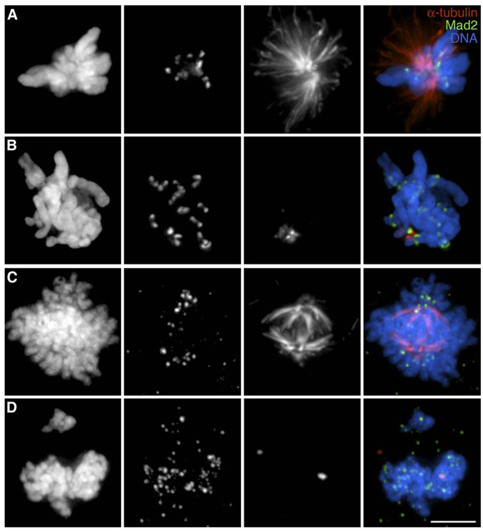
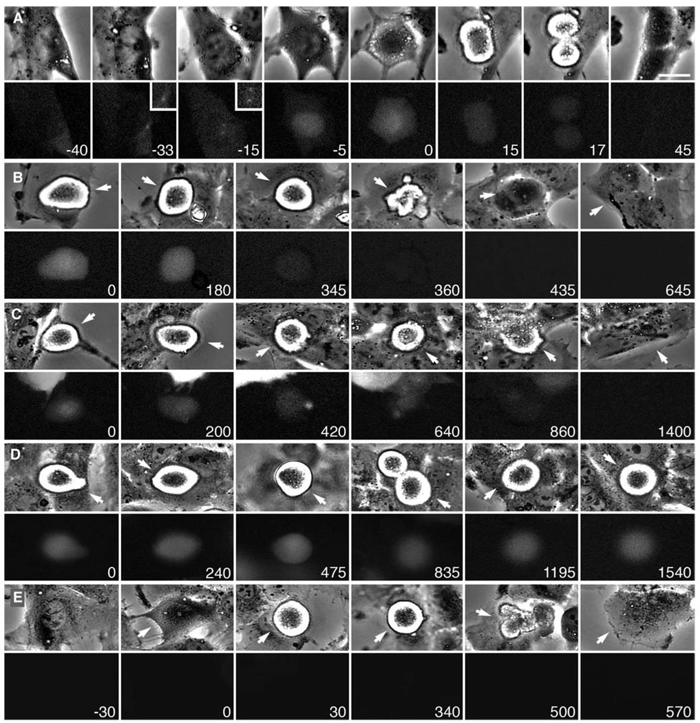
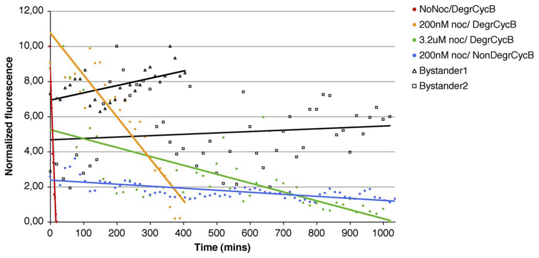
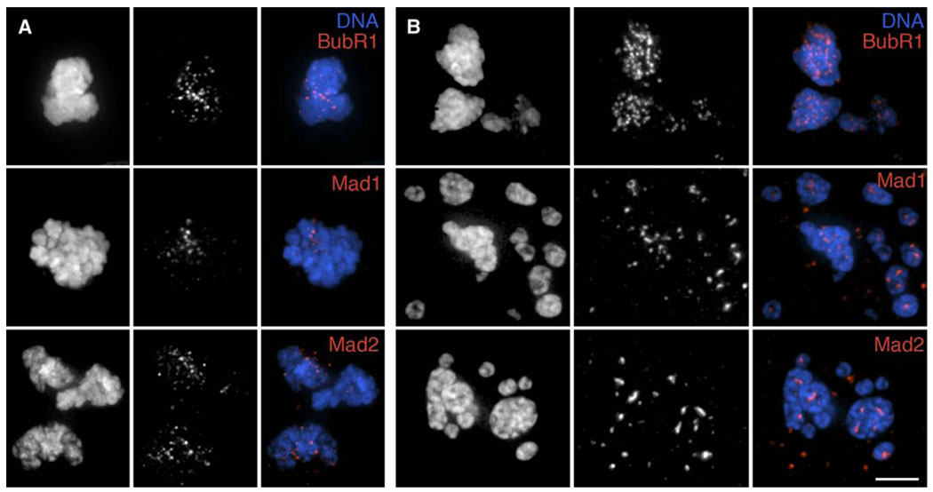
In the presence of unattached/weakly attached kinetochores, the spindle assembly checkpoint (SAC) delays exit from mitosis by preventing the anaphase-promoting complex (APC)-mediated proteolysis of cyclin B, a regulatory subunit of cyclin-dependent kinase 1 (Cdk1). Like all checkpoints, the SAC does not arrest cells permanently, and escape from mitosis in the presence of an unsatisfied SAC requires that cyclin B/Cdk1 activity be inhibited. In yeast , and likely Drosophila, this occurs through an "adaptation" process involving an inhibitory phosphorylation on Cdk1 and/or activation of a cyclin-dependent kinase inhibitor (Cdki). The mechanism that allows vertebrate cells to escape mitosis when the SAC cannot be satisfied is unknown. To explore this issue, we conducted fluorescence microscopy studies on rat kangaroo (PtK) and human (RPE1) cells dividing in the presence of nocodazole. We find that in the absence of microtubules (MTs), escape from mitosis occurs in the presence of an active SAC and requires cyclin B destruction. We also find that cyclin B is progressively destroyed during the block by a proteasome-dependent mechanism. Thus, vertebrate cells do not adapt to the SAC. Rather, our data suggest that in normal cells, the SAC cannot prevent a slow but continuous degradation of cyclin B that ultimately drives the cell out of mitosis.


Figures

Figure 1
Figure 1. Abnormal Spindles with Mad2-Positive Kinetochores Form in the Presence of Nocodazole
PtK1 (A, B) and RPE1 (C, D) cultures were incubated with 400 nM (A), 10 µM (B), 200 nM (C), or 3.2 µM nocodazole for 6 hr (A and B), 9 hr (C), or 15 hr (D), respectively, prior to fixation and immunostaining for Mad2 and α-tubulin. First column, DNA; second column, Mad2; third column, α-tubulin; fourth column, merged images. Note that a rudimentary spindle forms in both cell types at the lower but not higher nocodazole concentrations. See text for details. Scale bar equals 5 µm.
Figure 2
Figure 2. Cyclin B Degradation Is Required for RPE1 Cells to Exit Mitosis in the Presence of an Unsatisfied SAC
Selected phase-contrast (top) and fluorescence (bottom) frames from time-lapse video recordings. (A–C) RPE1 cells were transfected with a cyclin B-GFP plasmid and GFP-expressing cells were followed. (A) Non-drug-treated control cell in which cyclin B-GFP accumulates in the nucleus during prophase (−5 min) and is then destroyed after the SAC is satisfied and the cell enters anaphase (15–17 min). Insets show centrosomes at a higher magnification. Time, relative to nuclear envelope breakdown, is shown in minutes. Scale bar equals 5 µm. (B and C) GFP-expressing mitotic cells in the presence of 200 nM (B) or 3.2 µM (C) nocodazole. The arrow in each frame marks the cell of interest. Time is shown in minutes. Note that the cyclin B-GFP fluorescence begins to visibly decline throughout the block and is barely detectable near the time the cells finally begin to exit mitosis (360 min in [B] and 860 min in [C]). (D and E) RPE1 cultures were transfected with a GFP-cyclin B-Δ85 plasmid. 24 hr after transfection, the cultures were incubated with 200 nM nocodazole and areas were subsequently followed by dual-mode time-lapse microscopy. In the same field of view, we could find GFP-expressing and nonexpressing cells. (D) GFP-expressing cells (e.g., arrow) remained in mitosis until filming was terminated after more than 24 hr, and throughout this time the GFP fluorescence remained relatively level (see Figure 3). (E) In contrast, cells in the same culture that were not transfected with the plasmid (e.g., arrows) escaped mitosis after ~8 hr. See also Figure 3.
Figure 3
Figure 3. Cyclin B Proteolysis Occurs throughout a Nocodazole-Induced Block in Mitosis
RPE1 cultures were transfected with a plasmid carrying either a degradable or nondegradable (Δ85) GFP-cyclin B, and progress through mitosis was followed in GFP-expressing cells. Red line, GFP-cyclin B-expressing control cell not treated with nocodazole; orange line, the GFP-cyclin B-expressing cell shown in Figure 2B that was treated with 200 nM nocodazole; green line, the GFP-expressing cell shown in Figure 2C that was treated with 3.2 µM nocodazole. Areas containing mitotic cells were then followed by dual-mode time-lapse microscopy. Fluorescence was measured at each time point in a fixed size area of the cell corresponding to the mitotic spindle region in mitotic cells or the cytoplasm in interphase cells. Fluorescence levels of both mitotic and interphase cells were normalized to the background. Fluorescence of the mitotic cells was further normalized to a transfected interphase bystander cell (black lines) to allow comparison of intensities from independent recordings. The data shown here are representative of 11 control cells, 5 cells expressing GFP-cyclin B and incubated in 200 nM nocodazole, 3 cells expressing GFP-cyclin B and incubated in 3.2 µM nocodazole, and 3 cells expressing nondegradable GFP-cyclin B-Δ85 and incubated with 200 nM nocodazole (blue line). Note that the higher the nocodazole concentration, the slower cyclin B is degraded and the longer the cells take to exitmitosis (cf. orange and green lines). Also, degradation of the GFP fluorescence associated with the nondegradable form of cyclin B (blue line) was largely inhibited. Cells transfected with this construct were unable to escape mitosis during the recording interval (up to 1740 min).
Figure 4
Figure 4. BubR1, Mad1, and Mad2 Remain Associated with RPE1 Kinetochores as Cells Exit Mitosis in the Presence of an Unsatisfied SAC
RPE1 cultures were incubated with 200 nM (A) or 3.2 µM(B) nocodazole for 9 or 15 hr, respectively, before fixation and immunostaining for BubR1 (top), Mad1 (middle), or Mad2 (bottom). Cells that had just exitedmitosis (see text for details) were then located and photographed. Left columns in each panel, DNA; middle columns, BubR1, Mad1, or Mad2; right columns, merged images. Scale bar equals 5 µm.
Figure 5
Figure 5. Tpx2, an APC Substrate, Is Not Degraded in Cells that Exit Mitosis without Satisfying the SAC
(A) In untreated RPE1 cells, Tpx2 (bottom) is found in S/G2 but not G1 nuclei. During mitosis, this MT-associated protein localizes to spindle MTs, while during anaphase it is found on midbody MTs and centrosomes. Tpx2 is largely degraded by the time the nuclear envelope reforms around daughter nuclei during late telophase, although some remains associated with the centrosomes (far right row). (B) After treatment with 3.2 µM nocodazole, Tpx2 (bottom row) is found in the nuclei of G2 and prophase cells. In the absence of spindle MT formation, this protein then remains diffuse in the cytoplasm throughout the prolonged mitosis. However, once the cells exitmitosis and reflatten on the substrate (last three images in each row), it is found concentrated in the G1 micronuclei. Top row, merged images; middle row, DNA; bottom row, Tpx2. Scale bar equals 5 µm.

Similar articles

Cited by

References

    1. Rudner AD, Murray AW. The spindle assembly checkpoint. Curr. Opin. Cell Biol. 1996;8:773–780. - PubMed
    1. Minshull J, Straight A, Rudner AD, Dernburg AF, Belmont A, Murray AW. Protein phosphatase 2A regulates MPF activity and sister chromatid cohesion in budding yeast. Curr. Biol. 1996;6:1609–1620. - PubMed
    1. Rudner AD, Hardwick KG, Murray AW. Cdc28 activates exit from mitosis in budding yeast. J. Cell Biol. 2000;149:1361–1376. - PMC - PubMed
    1. Novak B, Toth A, Csikasz-Nagy A, Gyorffy B, Tyson JJ, Nasmyth K. Finishing the cell cycle. J. Theor. Biol. 1999;199:223–233. - PubMed
    1. Wolfe BA, Gould KL. Fission yeast Clp1p phosphatase affects G(2)/M transition and mitotic exit through Cdc25p inactivation. EMBO J. 2004;23:919–929. - PMC - PubMed

Publication types