. 2006 Aug 11;281(32):22786-93.
doi: 10.1074/jbc.M512486200. Epub 2006 Jun 16.

Carba analogs of cyclic phosphatidic acid are selective inhibitors of autotaxin and cancer cell invasion and metastasis

Affiliations

Carba analogs of cyclic phosphatidic acid are selective inhibitors of autotaxin and cancer cell invasion and metastasis

Daniel L Baker et al. J Biol Chem. .

Abstract

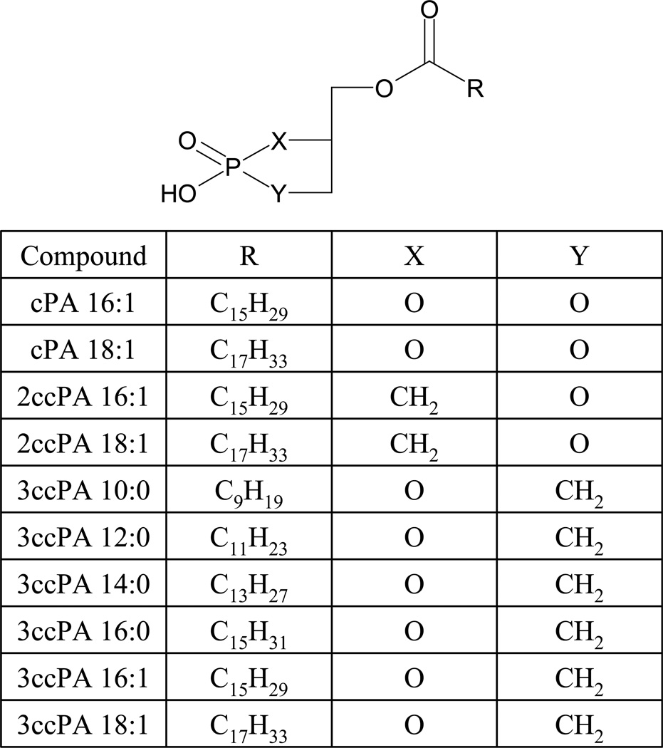
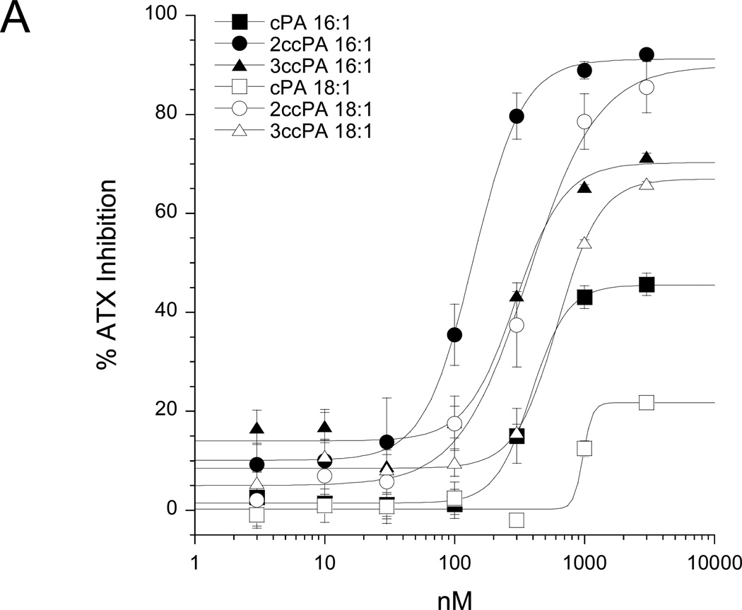
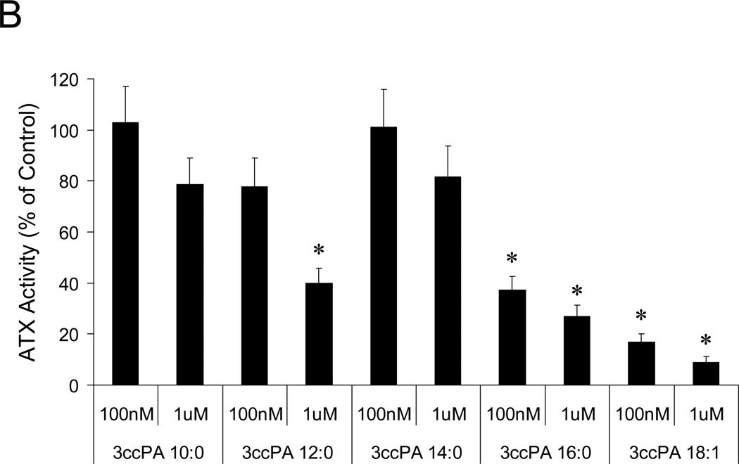
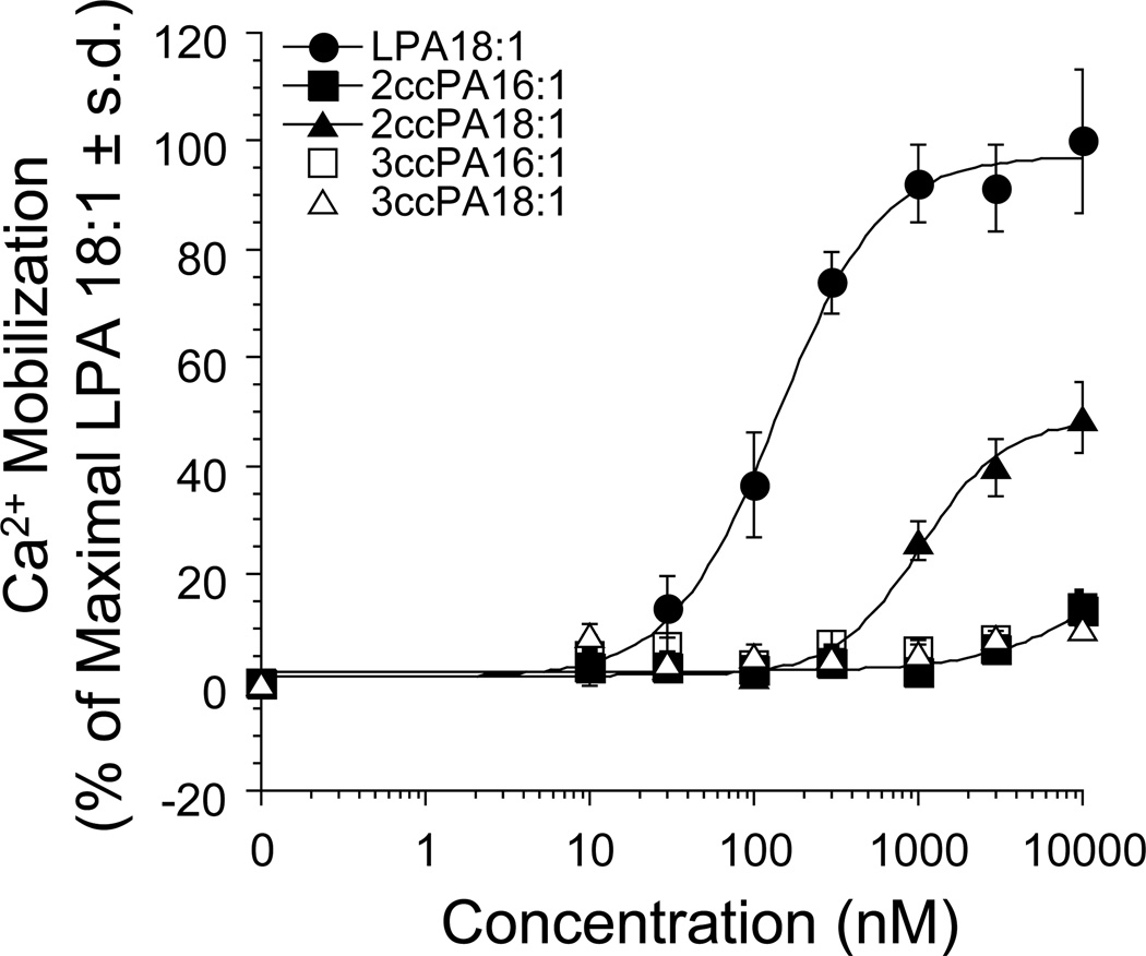
Autotaxin (ATX, nucleotide pyrophosphate/phosphodiesterase-2) is an autocrine motility factor initially characterized from A2058 melanoma cell-conditioned medium. ATX is known to contribute to cancer cell survival, growth, and invasion. Recently ATX was shown to be responsible for the lysophospholipase D activity that generates lysophosphatidic acid (LPA). Production of LPA is sufficient to explain the effects of ATX on tumor cells. Cyclic phosphatidic acid (cPA) is a naturally occurring analog of LPA in which the sn-2 hydroxy group forms a 5-membered ring with the sn-3 phosphate. Cellular responses to cPA generally oppose those of LPA despite activation of apparently overlapping receptor populations, suggesting that cPA also activates cellular targets distinct from LPA receptors. cPA has previously been shown to inhibit tumor cell invasion in vitro and cancer cell metastasis in vivo. However, the mechanism governing this effect remains unresolved. Here we show that 3-carba analogs of cPA lack significant agonist activity at LPA receptors yet are potent inhibitors of ATX activity, LPA production, and A2058 melanoma cell invasion in vitro and B16F10 melanoma cell metastasis in vivo.


Figures

Figure 1
Figure 1
Structures of the cPA analogs tested.
Figure 2
Figure 2
Inhibition of ATX by cPA analogs. (A) Conditioned media from HEK293 cells transiently transfected with ATX-HIS6 was incubated with 1 mM BPNPP in presence of increasing amounts of individual cPA analogs complexed 1:1 with BSA. Data were normalized to the appropriate BSA control and are reported as percent ATX inhibition (n=3 for each data point (mean ± standard deviation), results are representative of two independent experiments). (B) Recombinant ATX-HA was incubated with 1 µM FS-3 in the presence of 3ccPA analogs complexed 1:1 with BSA (n=3 for each bar (mean ± standard deviation), results are representative of two independent experiments, * p<0.05, T-test).
Figure 2
Figure 2
Inhibition of ATX by cPA analogs. (A) Conditioned media from HEK293 cells transiently transfected with ATX-HIS6 was incubated with 1 mM BPNPP in presence of increasing amounts of individual cPA analogs complexed 1:1 with BSA. Data were normalized to the appropriate BSA control and are reported as percent ATX inhibition (n=3 for each data point (mean ± standard deviation), results are representative of two independent experiments). (B) Recombinant ATX-HA was incubated with 1 µM FS-3 in the presence of 3ccPA analogs complexed 1:1 with BSA (n=3 for each bar (mean ± standard deviation), results are representative of two independent experiments, * p<0.05, T-test).
Figure 3
Figure 3
Inhibition of ATX-mediated LPA production by cPA analogs. Purified, recombinant ATX-HA was incubated with 2.5 µM ADMAN-LPC, a synthetic, fluorescent LPC analog in the absence or presence of 1 µM individual cPA analogs for 4 h. Lipids were extracted using a modified Bligh-Dyer protocol, separated by TLC (chloroform/ methanol/ ammonium hydroxide, 65:30:8 v/v/v), and visualized by UV illumination. (A) Representative TLC separation (colors are inverted). (B) Quantitative analysis of four independent experiments (* p<0.05, T-test).
Figure 4
Figure 4
The effect of increasing amounts of ccPA analogs on intracellular Ca2+ mobilization was determined in RH7777 cells stably expressing the LPA1 receptor. 100% represents maximal Ca2+ mobilization elicited by LPA 18:1. Each data point represents the mean ± standard deviation of triplicate wells and are representative of at least 3 independent experiments.
Figure 5
Figure 5
Panel A. Inhibition of cell invasion by cPA analogs. Optimized conditions were used to assess the inhibition of individual cPA analogs on A2058 melanoma cell invasion. The data are means ± standard deviations of triplicate wells and are representative of 3 separate experiments (* p<0.01, T-test). Panel B. Exogenously added LPA bypasses the inhibition by 3ccPA 16:1. 1 µM LPC in 0.1% BSA was present in the lower chamber of the invasion chamber and recombinant purified ATX, LPA 18:1, 3-ccPA 16:1 (3 µM), or their combination was included. LPA 18:1 dose dependently enhanced invasion. In contrast, 3ccPA 16:1 inhibited ATX-induced migration. Exogenously added LPA 18:1 (30–300 nM) bypassed the inhibitory effect of 3ccPA 16:1. Vehicle (0.1 % BSA without ATX) did not induce invasion when added with 1 µM LPC, and 3ccPA 16:1 (3 µM) in the absence of ATX had no effect on basal A2058 cell invasion. The data are means ± standard deviations of triplicate wells and are representative of 3 separate experiments (* p<0.01, T-test).
Figure 6
Figure 6
Inhibition of melanoma cell metastasis in vivo. B16F10 melanoma cells (5 × 10⁵) were injected in the tail vein of C57BL/6 mice, followed by i.p. injections of PBS (vehicle control) or 3ccPA 16:1 or 3ccPA 18:1 (250 µg/kg, 5 µg per dose) 15 min and 48 h after inoculation. Animals were sacrificed 3 weeks later and lungs were washed, fixed in formalin, and lung nodules were counted. The data are means ± standard deviations from groups of 6 (PBS) or 5 (3ccPA 16:1 and 3ccPA 18:1) animals (* p < 0.01, ANOVA).

References

    1. Stracke ML, Krutzsch HC, Unsworth EJ, Arestad A, Cioce V, Schiffmann E, Liotta LA. J Biol Chem. 1992;267:2524–2529. - PubMed
    1. Murata J, Lee HY, Clair T, Krutzsch HC, Arestad AA, Sobel ME, Liotta LA, Stracke ML. J Biol Chem. 1994;269:30479–30484. - PubMed
    1. Nam SW, Clair T, Campo CK, Lee HY, Liotta LA, Stracke ML. Oncogene. 2000;19:241–247. - PubMed
    1. Nam SW, Clair T, Kim YS, McMarlin A, Schiffmann E, Liotta LA, Stracke ML. Cancer Res. 2001;61:6938–6944. - PubMed
    1. Hama K, Aoki J, Fukaya M, Kishi Y, Sakai T, Suzuki R, Ohta H, Yamori T, Watanabe M, Chun J, Arai H. J Biol Chem. 2004;279:17634–17639. - PubMed

Publication types