. 2006 Oct;54(10):1095-103.
doi: 10.1369/jhc.5A6905.2006. Epub 2006 Jun 16.

Immunohistochemical characterization of elastic system fibers in rat molar periodontal ligament

Affiliations

Immunohistochemical characterization of elastic system fibers in rat molar periodontal ligament

Takashi Sawada et al. J Histochem Cytochem. 2006 Oct.

Abstract

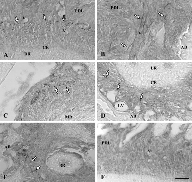
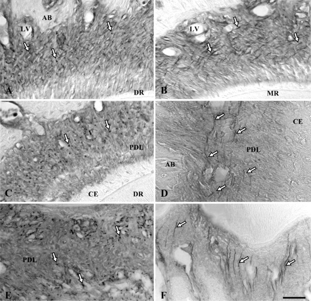
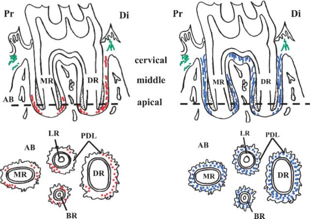
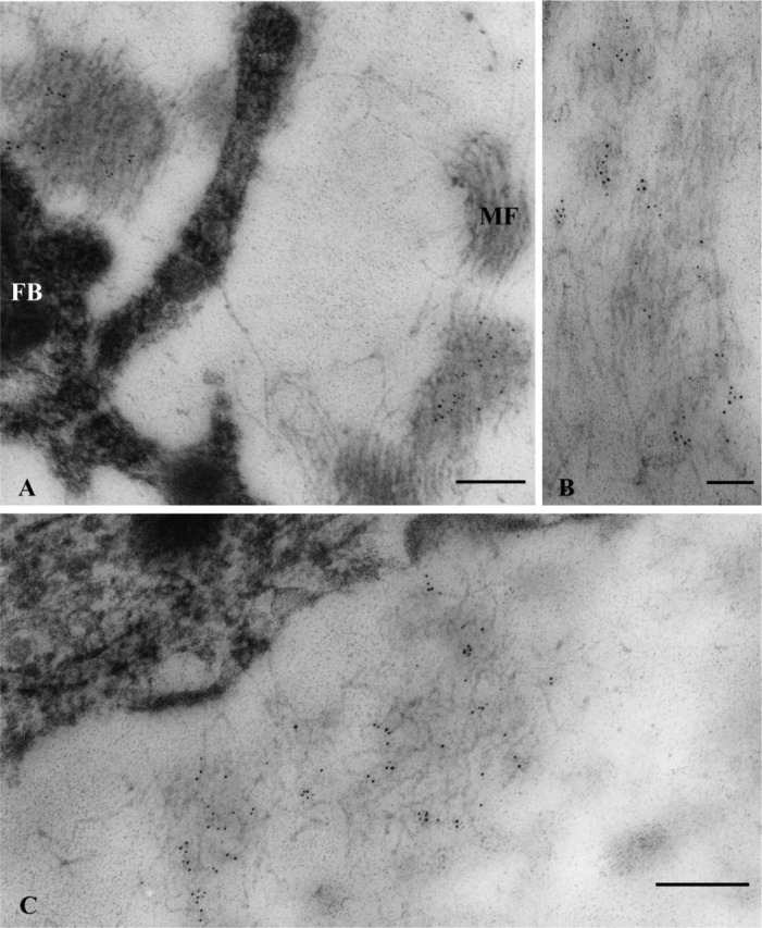
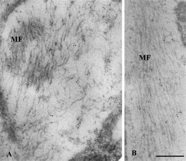
Among elastic system fibers, oxytalan fibers are known as a ubiquitous component of the periodontal ligament, but the localization and role of elastin-containing fibers, i.e., elastic and elaunin fibers, has yet to be clarified. In this study, we immunohistochemically investigated the localization of elastin and fibrillin, major proteins of elastin-containing fibers in the periodontal ligament of rat lower first molars. At the light microscope level, distribution of elastin-positive fibers was not uniform but often concentrated in the vicinity of blood vessels in the apical region of the ligament. In contrast, fibrillin-positive fibers were more widely distributed throughout the ligament, and the pattern of their distribution was comparable to the reported distribution of oxytalan fibers. At the ultrastructural level, assemblies or bundles of abundant fibrillin-containing microfibrils were intermingled with a small amount of elastin. This observation indicated that elastin-positive fibers observed under the light microscope were elaunin fibers. No mature elastic fibers, however, were found in the ligament. These results show that the major components of elastic system fibers in the periodontal ligament of the rat mandibular first molar were oxytalan and elaunin fibers, suggesting that the elastic system fibers play a role in the mechanical protection of the vascular system.


Figures

Figure 1
Figure 1
Immunoperoxidase staining of rat molar periodontal ligament for elastin (A-E) with control section (F). AB, alveolar bone; CE, cellular cementum; PDL, periodontal ligament; V, small blood vessels; LV, large blood vessels. (A) Distal side of periodontal ligament of distal root (DR) sectioned horizontally. Dot-like stains (white arrows) are concentrated mainly in the central zone of the ligament where small blood vessels are localized. (B) Elastin-positive fibers (white arrows) are oriented in the apico-occulusal direction along blood vessels. (C) Stained dotlike structures (white arrows) are concentrated on the distal side of the periodontal ligament of the mesial root (MR). (D) On the proximal side of the ligament of the lingual root (LR), dot-like stains (white arrows) are distributed in the central zone of the periodontal ligament but not around large blood vessels localized in areas close to alveolar bone. (E) Immunoreaction products (white arrows) are present in the periodontal ligament around the apex of the buccal root (BR). (F) Control section in which no immunoreaction product is present. Bar = 80 μm.
Figure 2
Figure 2
Immunoperoxidase staining of rat molar periodontal ligament for fibrillin. Dot-like or fibrous stained structures (white arrows) are evenly distributed throughout the central zone of the ligament mainly around blood vessels. AB, alveolar bone; CE, cellular cementum; PDL, periodontal ligament; V, small blood vessels; LV, large blood vessels. (A) Horizontal section of the cervical region of the distal root (DR). (B) Cervical region of the mesial root (MR) with obliquely sectioned stained fibrilous structures. (C) Apical region of the distal root (DR). (D) Longitudinal section of the apical region of the distal root. (E) Horizontal section of the periodontal ligament of interradicular bone. (F) Longitudinal section of the periodontal ligament of interradicular bone. Bar = 80 μm.
Figure 3
Figure 3
Schematic drawings of distribution of elastin and fibrillin in the periodontal ligament of rat mandibular first molars. Top: Sagittal section of roots of first molar. Bottom: Section along the horizontal plane at a level indicated by the broken line in the top drawing. Elastin (left, indicated in red) is mainly concentrated in the apical region of the periodontal ligament. Fibrillin (right, indicated in blue) is more widely distributed throughout the ligament. Elastin of gingival connective tissue is indicated in green. AB, alveolar bone; PDL, periodontal ligament; DR, distal root; MR, mesial root; LR, lingual root; BR, buccal root; Di, distal side; Pr, proximal side.
Figure 4
Figure 4
Immunogold localization of elastin in the distal region of the periodontal ligament of the distal root. (A) Five-nm gold particles are scattered within assemblies of microfibrils (MF) localized very close to a fibroblast (FB) in the vicinity of blood vessels. (B) Longitudinal section of assemblies of microfibrils onto which clusters of gold particles are localized. (C) Clusters of gold particles are more clearly seen within assemblies of oblique-transversely sectioned microfibrils. Bars: A,C = 200 nm; B = 100 nm.
Figure 5
Figure 5
Immunogold localization of fibrillin in the distal region of the ligament of the distal root. Gold particles are associated with microfibrils (MF) sectioned obliquely (A) and longitudinally (B). Bar = 200 nm.

References

    1. Beertsen W, Everts V, van den Hooff A. (1974) Fine structure of fibroblasts in the periodontal ligament of the rat incisor and their possible role in tooth eruption. Arch Oral Biol 19:1087–1098 - PubMed
    1. Bradamante Z, Svajger A. (1977) Pre-elastic (oxytalan) fibres in the developing elastic cartilage of the external ear of the rat. J Anat 123:735–743 - PMC - PubMed
    1. Carmichael GG, Fullmer HM. (1966) The fine structure of the oxytalan fiber. J Cell Biol 28:33–36 - PMC - PubMed
    1. Chantawiboonchai P, Warita H, Ohya K, Soma K. (1998) Confocal laser scanning-microscopic observations on the three-dimensional distribution of oxytalan fibres in mouse periodontal ligament. Arch Oral Biol 43:811–817 - PubMed
    1. Chavrier C. (1990) The elastic system fibres in healthy human gingiva. Arch Oral Biol 35:223–225s - PubMed