. 2006 Jul;26(13):4782-93.
doi: 10.1128/MCB.00069-06.

p53 downregulates its activating vaccinia-related kinase 1, forming a new autoregulatory loop

Affiliations

p53 downregulates its activating vaccinia-related kinase 1, forming a new autoregulatory loop

Alberto Valbuena et al. Mol Cell Biol. 2006 Jul.

Abstract

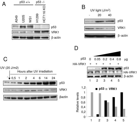
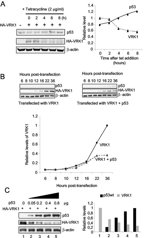
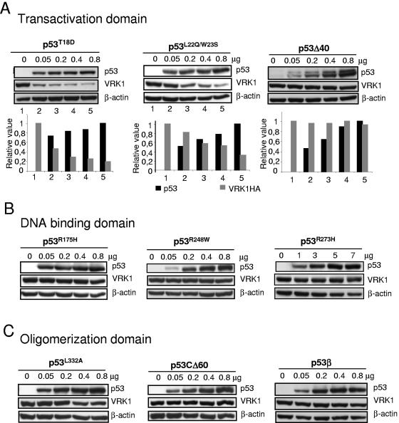
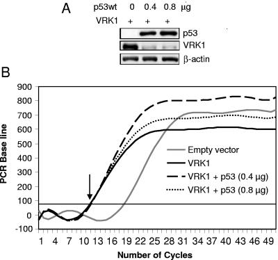
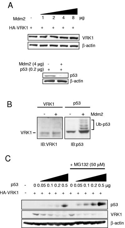
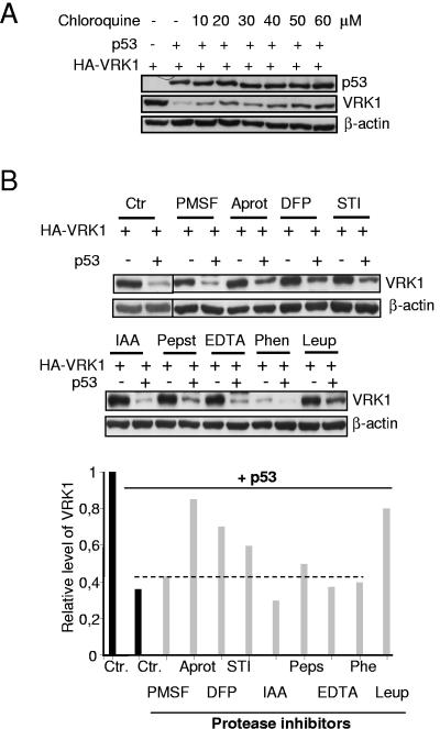
The stable accumulation of p53 is detrimental to the cell because it blocks cell growth and division. Therefore, increases in p53 levels are tightly regulated, mainly by its transcriptional target, mdm2, that downregulates p53. Elucidation of new signaling pathways requires the characterization of the members and the nature of their connection. Vaccinia-related kinase 1 (VRK1) contributes to p53 stabilization by partly interfering with its mdm2-mediated degradation, among other mechanisms; therefore, it is likely that some form of autoregulation between VRK1 and p53 must occur. We report here the identification of an autoregulatory loop between p53 and its stabilizing VRK1. There is an inverse correlation between VRK1 and p53 levels in cell lines, and induction of p53 by UV light downregulates VRK1 in fibroblasts. As the amount of p53 protein increases, there is a downregulation of the VRK1 protein level independent of its promoter. This effect is indirect but requires a transcriptionally active p53. The three most common transcriptionally inactive mutations detected in hereditary (Li-Fraumeni syndrome) and sporadic human cancer, p53(R175H), p53(R248W), and p53(R273H), as well as p53(R280K), are unable to induce downregulation of VRK1 protein. The p53 isoforms Delta40p53 and p53beta, lacking the transactivation and oligomerization domains, respectively, do not downregulate VRK1. VRK1 downregulation induced by p53 is independent of mdm2 activity and proteasome-mediated degradation since it occurs in the presence of proteasome inhibitors and in mdm2-deficient cells. The degradation of VRK1 is sensitive to chloroquine, an inhibitor of the late endosome-lysosome transport, and to serine protease inhibitors of the lysosomal pathway.


Figures

FIG. 1.
FIG. 1.
Expression of endogenous p53 and VRK1 in tumor cell lines and in UV treated fibroblasts. (A) Expression of VRK1 and p53 in cell lines that were p53 wild type (A549, U2OS, and WS1) or p53−/− (H1299 or HCT116-p53 knockout). VRK1 and p53 were determined by Western blotting, using actin as internal control. (B) Levels of p53 and VRK1 in human fibroblasts treated with the indicated dose of UV light; the extracts were prepared 8 h after treatment and analyzed by Western blotting. (C) The levels of endogenous p53 and VRK1 in WS1 fibroblasts were detected at different times after treatment with 20 J/m2 UV light. The proteins were identified by Western blotting with the corresponding antibodies indicated in the methods section. (D) Normal human fibroblasts, cell line WS1, were transfected with increasing amounts of pCB6+p53 as indicated and 5 μg of pCEFL-HA-VRK1. Cell extracts were prepared 36 h after transfection. The proteins were detected with the corresponding antibodies by Western blotting, and to the right is shown the quantification of the normalized values.
FIG. 2.
FIG. 2.
Downregulation of VRK1 by increasing levels of p53. (A) Inducible endogenous p53 downregulated VRK1. H1299 cells (p53Tet inducible) were transfected with 5 μg of pCEFL-HA-VRK1, and the p53 gene was induced with tetracycline. As the level of p53 increased, there was a noticeable decrease of VRK1 expression levels. Cell extracts were prepared at different time points after induction, and the proteins were detected by immunoblot analysis with the quantification shown in the graph to the right. (B) Time course of p53 and VRK1 protein levels. H1299 cells were transfected with pCEFL-HA-VRK1 (4 μg) with and without pCB6+p53 (200 ng). At different hours after transfection, cell extracts were prepared and analyzed by Western blotting for the amount of each protein. The quantification of the blots is shown in the graph to illustrate the trend in protein levels. The accumulation of p53 interferes with the accumulation of VRK1. The proteins were detected with the corresponding antibodies by Western blotting, and to the right is shown the quantification of the normalized values. (C) H1299 cells (p53−/−) were transfected with a fixed amount (5 μg) of pCEFL-HA-VRK1 and increasing amounts of p53 (wild type) to detect the effect of increasing p53 on VRK1 expressed from different promoters.
FIG. 3.
FIG. 3.
Implication of p53 protein domains in the induction of VRK1 downregulation. (A) Role of the transactivation domain. The effects of the phosphorylation mimicking mutant p53(T18D), the conformational double mutant p53(L22Q, W23S), and the p53 isoform lacking the transactivation domain (Δ40p53) were tested in combination with pCEFL-HA-VRK1 (5 μg). The Δ40p53 isoform was detected with the CM1 polyclonal antibody. At the bottom is shown the quantification of the blots to illustrate the changes in both proteins. (B) Contribution of the p53 DNA binding domain. Lack of effect of the most common p53 transcriptional mutants, p53(R175H), p53(R248W), and p53(R273H) on the level of VRK1 protein expressed from plasmid pCEFL-HA-VRK1 (5 μg). Cell extracts were prepared 36 h after transfection, and the levels of both proteins were determined by Western blotting. The transfected VRK1 was detected with an antibody specific for the HA epitope. (C) Contribution of the p53 oligomerization domain. The conformational mutant p53(L322A) and isoforms lacking the C-terminal region p53β and p53CΔ60 were studied.
FIG. 4.
FIG. 4.
The p53 protein does not affect VRK1 gene transcription levels. (A) Levels of p53 and VRK1 protein at the time point used for RNA determination. The levels of VRK1 protein in cells transfected with either 0.4 or 0.8 μg of plasmid pCB6+p53 are shown. The extracts were prepared 36 h after transfection. The proteins were detected by immunoblotting. (B) The levels of VRK1 mRNA, whether after p53 overexpression or not, were determined by RT-PCR as described in Materials and Methods. The results obtained in cells transfected with 0.4 (p53) and 0.8 (p53) μg of plasmid pCB6+p53 are shown.
FIG. 5.
FIG. 5.
p53 siRNA blocks the downregulation of VRK1 in H1299 cells. (A) H1299 cells were transfected with pCB6+p53 and an siRNA specific for p53 (pSUPER.retro.p53) as well as pCEFL-HA-VRK1 (5 μg) in different combinations to show that the level of the cotransfected p53 expressed from plasmid pCB6+p53 is downregulated. The cell extracts were prepared 36 h after transfection and analyzed by Western blotting with the corresponding antibodies. (B) H1299 cells were transfected with pCB6+p53 and 5 μg of pCEFL-HA-VRK1. The increase in p53 protein, as expected, downregulated the level of VRK1, but when cells were cotransfected with increasing amounts of siRNA specific for p53, expression plasmid pSUPER.retro.p53, the downregulation of VRK1 was reduced. The graph shows the quantification of results for both proteins.
FIG. 6.
FIG. 6.
Mdm2 is not implicated in p53 downregulation of VRK1. (A) Mdm2 protein overexpression does not produce VRK1 degradation by itself. H1299 cells were transfected with 5 μg of pCDNA-VRK1 expression plasmid alone or together with increasing amounts of plasmid encoding pCOC-Mdm2 where indicated. One microgram of plasmid (pUbiquitin-His) encoding ubiquitin was added in all cases. Also 0.2 μg of pCB6+p53 plasmid was cotransfected with Mdm2-encoding plasmid under similar conditions (lower panel). Whole-cell extracts were prepared 36 h after transfection and analyzed by Western blotting with the corresponding antibody. (B) Detection of ubiquitination in p53 but not VRK1 in H1299 cells. The cells were transfected with plasmid pCOC-Mdm2 (4 μg), pCEFL-HA-VRK1 (3 μg), or pCB6+p53 (1 μg) in the presence of 1 μg of pUbiquitin-His. The cell extracts were analyzed by immunoblotting with an anti-VRK1 antibody or a mix of antibodies for p53 to detect their potential change in migration in the gel if there was ubiquitination. (C) VRK1 downregulation by p53 is independent of Mdm2 protein and proteasome-mediated degradation. Mouse embryo fibroblasts derived from double knockout mice (p53−/− mdm2/) were cotransfected with the indicated plasmids pCEFL-HA-VRK1 (3 μg) or pCB6+p53. Where indicated, the proteasome inhibitor MG132 was added at 50 μM. Cells were processed as described in panel A, and levels of p53 and VRK1 proteins were detected.
FIG. 7.
FIG. 7.
VRK1 downregulation occurs in the lysosomal pathway. (A) Sensitivity of VRK1 downregulation to the inhibition of the late endosome to lysosome traffic by chloroquine. H1299 cells were transfected with pCB6+p53 and pCEFL-HA-VRK1. Chloroquine at the indicated concentrations was added 12 h after transfection, and cell extracts were prepared for immunoblot analysis 36 h after transfection. (B) Effect of protease inhibitors on the downregulation of VRK1 induced by p53. H1299 cells were transfected with pCB6+p53 and pCEFL-HA-VRK1, and different protease inhibitors were added to the culture 18 h after transfection. The inhibitors were used at the specified concentrations for 5 h. The serine protease inhibitors were the following: 1 mM PMSF, 10 μl/ml aprotinin (Aprot), 0.1 mM DFP, and 10 μl/ml STI. The following protease inhibitors were also used: for serine and cysteine, 50 μM leupeptin (Leup); for cysteine, 50 μM IAA; and for aspartyl, 1 μM pepstatin (Peps). As metalloprotease inhibitors, 5 mM EDTA and 5 mM 1,10-phenantroline (Phen) were used. Calpain inhibitors were also tested with no effect. The cells were collected 23 h after transfection and lysed for determination of VRK1 and p53 levels by Western blotting. Ctr, controls without inhibitors.

Similar articles

Cited by

References

    1. Agarwal, M. L., W. R. Taylor, M. V. Chernov, O. B. Chernova, and G. R. Stark. 1998. The p53 network. J. Biol. Chem. 273:1-4. - PubMed
    1. Ashcroft, M., M. H. Kubbutat, and K. H. Vousden. 1999. Regulation of p53 function and stability by phosphorylation. Mol. Cell. Biol. 19:1751-1758. - PMC - PubMed
    1. Balint, E. E., and K. H. Vousden. 2001. Activation and activities of the p53 tumour suppressor protein. Br. J. Cancer 85:1813-1823. - PMC - PubMed
    1. Barcia, R., S. Lopez-Borges, F. M. Vega, and P. A. Lazo. 2002. Kinetic properties of p53 phosphorylation by the human vaccinia-related kinase 1. Arch. Biochem. Biophys. 399:1-5. - PubMed
    1. Beinke, S., M. J. Robinson, M. Hugunin, and S. C. Ley. 2004. Lipopolysaccharide activation of the TPL-2/MEK/extracellular signal-regulated kinase mitogen-activated protein kinase cascade is regulated by IκB kinase-induced proteolysis of NF-κB1 p105. Mol. Cell. Biol. 24:9658-9667. - PMC - PubMed

Publication types

MeSH terms