. 2006 Jul;26(13):4882-94.
doi: 10.1128/MCB.02402-05.

Mechanisms directing the nuclear localization of the CtBP family proteins

Affiliations

Mechanisms directing the nuclear localization of the CtBP family proteins

Alexis Verger et al. Mol Cell Biol. 2006 Jul.

Abstract

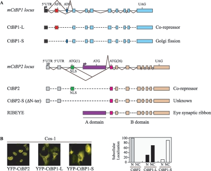
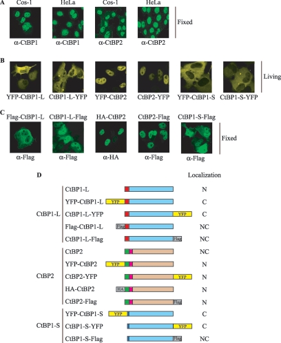
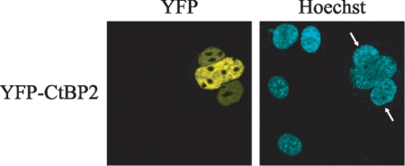
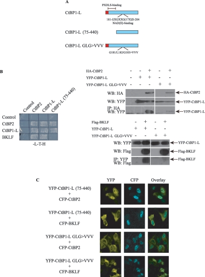
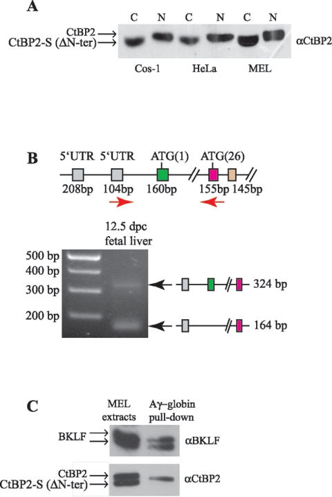
The C-terminal binding protein (CtBP) family includes four proteins (CtBP1 [CtBP1-L], CtBP3/BARS [CtBP1-S], CtBP2, and RIBEYE) which are implicated both in transcriptional repression and in intracellular trafficking. However, the precise mechanisms by which different CtBP proteins are targeted to different subcellular regions remains unknown. Here, we report that the nuclear import of the various CtBP proteins and splice isoforms is differentially regulated. We show that CtBP2 contains a unique nuclear localization signal (NLS) located within its N-terminal region, which contributes to its nuclear accumulation. Using heterokaryon assays, we show that CtBP2 is capable of shuttling between the nucleus and cytoplasm of the cell. Moreover, CtBP2 can heterodimerize with CtBP1-L and CtBP1-S and direct them to the nucleus. This effect strongly depends on the CtBP2 NLS. PXDLS motif-containing transcription factors, such as BKLF, that bind CtBP proteins can also direct them to the nucleus. We also report the identification of a splice isoform of CtBP2, CtBP2-S, that lacks the N-terminal NLS and localizes to the cytoplasm. Finally, we show that mutation of the CtBP NADH binding site impairs the ability of the proteins to dimerize and to associate with BKLF. This reduces the nuclear accumulation of CtBP1. Our results suggest a model in which the nuclear localization of CtBP proteins is influenced by the CtBP2 NLS, by binding to PXDLS motif partner proteins, and through the effect of NADH on CtBP dimerization.


Figures

FIG. 1.
FIG. 1.
Cellular localization of exogenously expressed CtBP1-L, CtBP2, and CtBP1-S. (A) Schematic representation (not to scale) of the exon structure of the genomic locus and splicing pattern, relative to mouse CtBP1 and mouse CtBP2, adapted from references , , , , and . Exons used exclusively for CtBP1-L (red box), CtBP2 (green box), CtBP2-S (CtBP2 ΔN-ter) (pink box), CtBP1-S (dark blue box), and RIBEYE (purple box) are color coded. The unique NLS of CtBP2 is indicated. (B) CtBP2 localizes into the nucleus of the cells, CtBP1-L is distributed throughout the nucleus and the cytoplasm, and CtBP1-S is predominantly cytoplasmic. Cos-1 cells were transfected with the indicated expression vectors, and the expressed proteins were visualized by confocal microscopy. The images shown in each panel are representative of all of the transfected cells. The graph depicts the quantitative data obtained from three independent experiments. In each experiment, more than 100 cells were counted in each group, with <10% standard deviation. N, exclusive nuclear staining; NC, cytoplasmic and/or nuclear staining.
FIG. 2.
FIG. 2.
Divergent effects of N-terminal and C-terminal Flag tag or YFP tag on CtBP cellular localization. (A to C) Endogenous CtBP1-L is mainly nuclear in Cos-1 and HeLa cells. Cos-1 and HeLa cells were either nontransfected (A) or transfected with the indicated expression vectors (B and C), and the expressed proteins were visualized by fluorescence microscopy either directly (B, yellow) or by using the indicated primary mouse antibodies and a fluorescein isothiocyanate-labeled secondary antibody (A and C, green). Cells were fixed with formaldehyde (A and C); living cells (B) were used directly for microscopic analysis. (D) Schematic representation of the fusion proteins used in this study. The constructs were generated as Flag-tagged (gray boxes), HA-tagged (gray boxes), and YFP fusion (yellow boxes) proteins as indicated. The unique N-terminal regions of CtBP1-L, CtBP2, and CtBP1-S are color coded as in Fig. 1A. Subcellular localization of each construct is summarized at right. C, predominantly cytoplasmic; N, predominantly nuclear; NC, nuclear and cytoplasmic.
FIG. 3.
FIG. 3.
Characterization of the CtBP2 NLS. (A) Alignment of the N-terminal region of CtBP family members with the box indicating the conserved NLS. Prefix r, rat; h, human; m, mouse; b, Bos taurus; d, Drosophila sp.; z, zebra fish; x, Xenopus sp. The start of the CtBP2-S (ΔN-ter CtBP2) isoform is indicated by an arrow. Shown at the lower part of the panel is the sequence comparison between NF-κB family members, highlighting the homology to the CtBP2 NLS, adapted from reference . (B to D) Cellular localization of YFP-CtBP fusion proteins. Cos-1 cells were transfected with the indicated expression vectors, and the expressed proteins were visualized by confocal microscopy. The images shown in each panel are representative of all of the transfected cells. Schematic illustrations of the constructs used are indicated below each microscopic image.
FIG. 4.
FIG. 4.
CtBP2 is capable of nuclear shuttling. Heterokaryon assays were carried out between untransfected mouse NIH 3T3 cells and monkey Cos-1 cells expressing YFP-CtBP2. Mouse cells (arrows) were identified by their speckled nuclei when stained with Hoechst 33258.
FIG. 5.
FIG. 5.
CtBP1-L and CtBP1-S become nuclear when cells are cotransfected with CtBP2 or BKLF. The NLS of CtBP2 is required for directing the nuclear localization of CtBP1-L and CtBP1-S. Cos-1 cells were cotransfected with the indicated CFP- and YFP-containing expression vectors, and localization of the expressed proteins was visualized by confocal microscopy. The images shown in each panel are representative of all of the transfected cells.
FIG. 6.
FIG. 6.
NADH regulates CtBP oligomerization. (A) Schematic illustration of CtBP1-L and mutants. The PXDLS-binding region and the NADH binding site are indicated. (B) CtBP1-L derivatives were tested for their ability to homodimerize, to heterodimerize with CtBP2, and to interact with a recognized PXDLS-motif-containing partner protein, BKLF. Left panel, yeast growth was monitored and the results reflecting association of the proteins are shown. Right panel, immunoprecipitation analysis. Cos-1 cells were transfected with YFP-CtBP1-L, YFP-CtBP1-L GLG>VVV, HA-CtBP2, and Flag-BKLF as indicated. Cell lysates were prepared and HA-CtBP2 or YFP-CtBP1-L protein complexes isolated using anti-HA and anti-YFP agarose affinity resin, respectively. Proteins in the cellular lysates and immunoprecipitated (IP) proteins were subsequently analyzed by Western blot (WB) analysis using the indicated antibodies. (C) Cos-1 cells were cotransfected with the indicated CFP and YFP expression vectors, and localization of the expressed proteins was visualized by confocal microscopy. The images shown in each panel are representative of all of the transfected cells.
FIG. 7.
FIG. 7.
Identification of a new CtBP2 splice form. (A) Cytoplasmic (C) and nuclear (N) protein extracts of Cos-1, HeLa, and MEL cells were subjected to Western blot analysis with anti-CtBP2 antibodies. A faster migrating band is consistently found only in the cytoplasmic fraction and likely represents a splice form of CtBP2 without the first coding exon containing the NLS. Full-length CtBP2 and CtBP2-S (ΔN-ter) are indicated (arrows). (B) Expression of mouse CtBP2. RT-PCR was performed with cDNA templates from mouse fetal liver at 12.5 dpc. PCR products were electrophoresed on agarose gels and visualized by ethidium bromide staining. The genomic organization of the mCtBP2 locus is indicated as in Fig. 1A, and the primers used for RT-PCR are indicated by red arrows. Two differentially expressed bands are detected, the upper one (324 bp) corresponding to CtBP2 containing both exon 1 (methionine M1, green) and exon 2 (methionine M26, pink) and the lower one (164 bp) corresponding to the CtBP2 isoform missing the first coding exon (methionine M1, 160 bp). The two splice forms are indicated at right. (C) A magnetic bead-DNA pull-down assay showing that full-length CtBP2, but not CtBP2-S (ΔN-ter), is able to localize to the proximal Aγ-globin promoter. MEL cell extract was incubated with DNA containing the BKLF binding sites (CACCC boxes) coupled to magnetic beads. Complexes were blotted with antibodies against BKLF and CtBP2.

Similar articles

Cited by

References

    1. Alpatov, R., G. C. Munguba, P. Caton, J. H. Joo, Y. Shi, Y. Shi, M. E. Hunt, and S. P. Sugrue. 2004. Nuclear speckle-associated protein Pnn/DRS binds to the transcriptional corepressor CtBP and relieves CtBP-mediated repression of the E-cadherin gene. Mol. Cell. Biol. 24:10223-10235. - PMC - PubMed
    1. Balasubramanian, P., L. J. Zhao, and G. Chinnadurai. 2003. Nicotinamide adenine dinucleotide stimulates oligomerization, interaction with adenovirus E1A and an intrinsic dehydrogenase activity of CtBP. FEBS Lett. 537:157-160. - PubMed
    1. Barnes, C. J., R. K. Vadlamudi, S. K. Mishra, R. H. Jacobson, F. Li, and R. Kumar. 2003. Functional inactivation of a transcriptional corepressor by a signaling kinase. Nat. Struct. Biol. 10:622-628. - PubMed
    1. Berthelsen, J., C. Kilstrup-Nielsen, F. Blasi, F. Mavilio, and V. Zappavigna. 1999. The subcellular localization of PBX1 and EXD proteins depends on nuclear import and export signals and is modulated by association with PREP1 and HTH. Genes Dev. 13:946-953. - PMC - PubMed
    1. Blank, V., P. Kourilsky, and A. Israel. 1991. Cytoplasmic retention, DNA binding and processing of the NF-kappa B p50 precursor are controlled by a small region in its C-terminus. EMBO J. 10:4159-4167. - PMC - PubMed

Publication types

MeSH terms

LinkOut - more resources