. 2006 Jul;26(13):4934-48.
doi: 10.1128/MCB.02477-05.

Platelet-derived growth factor BB induces nuclear export and proteasomal degradation of CREB via phosphatidylinositol 3-kinase/Akt signaling in pulmonary artery smooth muscle cells

Affiliations

Platelet-derived growth factor BB induces nuclear export and proteasomal degradation of CREB via phosphatidylinositol 3-kinase/Akt signaling in pulmonary artery smooth muscle cells

Chrystelle V Garat et al. Mol Cell Biol. 2006 Jul.

Abstract

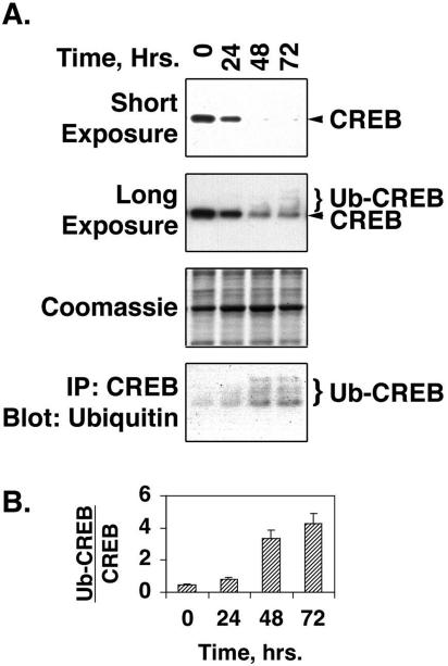
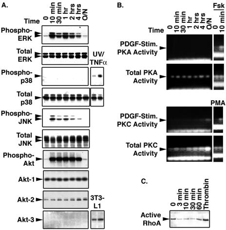
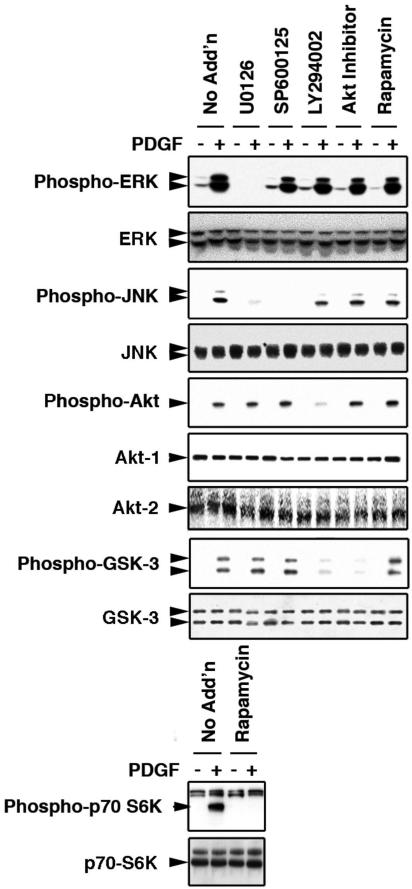
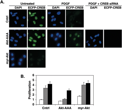
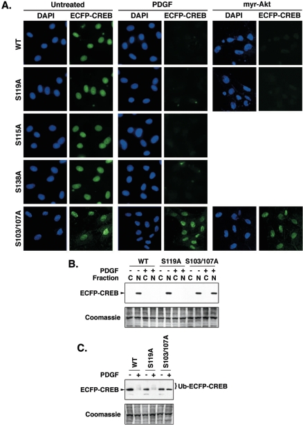
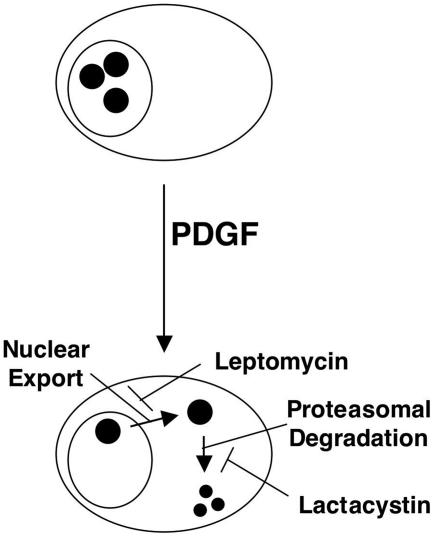
Cyclic AMP response element binding protein (CREB) content is diminished in smooth muscle cells (SMCs) in remodeled pulmonary arteries from animals with pulmonary hypertension and in the SMC layers of atherogenic systemic arteries and cardiomyocytes from hypertensive individuals. Loss of CREB can be induced in cultured SMCs by chronic exposure to hypoxia or platelet-derived growth factor BB (PDGF-BB). Here we investigated the signaling pathways and mechanisms by which PDGF elicits depletion of SMC CREB. Chronic PDGF treatment increased CREB ubiquitination in SMCs, while treatment of SMCs with the proteasome inhibitor lactacystin prevented decreases in CREB content. The nuclear export inhibitor leptomycin B also prevented depletion of SMC CREB alone or in combination with lactacystin. Subsequent studies showed that PDGF activated extracellular signal-regulated kinase, Jun N-terminal protein kinase, and phosphatidylinositol 3 (PI3)-kinase pathways in SMCs. Inhibition of these pathways blocked SMC proliferation in response to PDGF, but only inhibition of PI3-kinase or its effector, Akt, blocked PDGF-induced CREB loss. Finally, chimeric proteins containing enhanced cyan fluorescent protein linked to wild-type CREB or CREB molecules with mutations in several recognized phosphorylation sites were introduced into SMCs. PDGF treatment reduced the levels of each of these chimeric proteins except for one containing mutations in adjacent serine residues (serines 103 and 107), suggesting that CREB loss was dependent on CREB phosphorylation at these sites. We conclude that PDGF stimulates nuclear export and proteasomal degradation of CREB in SMCs via PI3-kinase/Akt signaling. These results indicate that in addition to direct phosphorylation, proteolysis and intracellular localization are key mechanisms regulating CREB content and activity in SMCs.


Figures

FIG. 1.
FIG. 1.
CREB depletion with siRNA induces a dedifferentiated, proliferative, and synthetic phenotype in PA SMCs. Rat PA SMCs were transfected with nonspecific (Cntrl) or CREB-specific (CREB) double-stranded siRNAs as indicated and incubated overnight in DMEM containing 15% FCS. Cells were then transferred to DMEM containing 0.2% FCS for 72 h. (A) Whole-cell lysates and RNA were prepared from the SMCs and subjected to Western blotting (left panels) or semiquantitative reverse transcriptase PCR (right panels) for the factors indicated to the left of each pair of panels. (B) BrdU (10 μM) was added to the siRNA-transfected cells for 12 h. Cells were then fixed in PBS containing 4% paraformaldehyde and subjected to immunostaining for BrdU incorporation as an index of cell proliferation. Representative bright-field images are shown. Magnification, ×200. (C) Eight randomly selected microscope fields were counted for BrdU-positive and total cell numbers by a blinded observer. Data shown are average percentages of BrdU-positive cells. (D) siRNA-treated cells and untreated or PDGF-treated (25 ng/ml for 72 h) cells were subjected to pentachrome staining. Representative bright-field images are shown. Magnification, ×400.
FIG. 2.
FIG. 2.
PDGF stimulates ubiquitination of CREB. (A) Rat PA SMCs maintained in complete medium were transferred to DMEM containing 0.2% FCS for 24 h. The cells were then treated with 25 ng/ml PDGF, and medium and PDGF were replaced every 24 h for 72 h. Nuclear extracts were prepared from the cells at the times indicated above the lanes. Portions (25 μg protein) of the extracts were resolved on 10% polyacrylamide-SDS gels and transferred to PVDF membranes. Another portion was incubated with CREB-specific antibodies linked to Sepharose beads conjugated with protein A. After a 1-h incubation, the beads were washed three times with PBS containing 0.1% Tween 20 and eluted with Laemmli loading buffer. The precipitated (IP) material was also resolved on SDS-polyacrylamide gels and transferred to PVDF membranes. The blots were subjected to Western blotting with antibodies to CREB or ubiquitin, as indicated to the right of each blot. Short exposure of the CREB Western blot shows loss of CREB with PDGF treatment. Long exposure of the CREB Western blot shows both CREB and ubiquitinated CREB (Ub-CREB) bands. A representative Coomassie blue-stained blot is shown as a loading control. (B) The bar graph shows the levels of ubiquitinated CREB relative to total CREB levels at each time point.
FIG. 3.
FIG. 3.
Leptomycin B and lactacystin inhibit PDGF-induced CREB depletion, loss of CREB transcriptional activity, and SMC proliferation. Rat PA SMCs were transfected with nonspecific (Cntrl) or CREB-specific double-stranded siRNAs as indicated and incubated overnight in DMEM containing 15% FCS. The cells were transferred to DMEM containing 0.2% FCS for 24 h. The cells were then treated with 100 nM leptomycin B (Lepto. B) and/or 500 nM lactacystin (Lacta.) for 30 min prior to the addition of 25 ng/ml PDGF. Fresh medium containing PDGF and inhibitors was added every 24 h for 72 h. (A) Cells were fixed, subjected to immunostaining with a CREB-specific antibody, and counterstained with DAPI to indicate nuclei. Magnification, ×400. (B) Cytosolic (“C”) and nuclear (“N”) extracts were prepared and separated on 10% polyacrylamide-SDS gels and transferred to PVDF membranes. Western blotting was performed with a CREB-specific antibody. The positions of CREB and ubiquitinated CREB (Ub-CREB) are indicated. A representative Coomassie blue-stained blot is shown as a loading control. (C) Prior to being treated with PDGF and inhibitors, cells were transfected with a plasmid containing a CREB-responsive promoter linked to the firefly luciferase gene (pCRESV-Luc). Cells were cotransfected with the internal control vector pRL-SV40 to correct for transfection efficiency. Following treatment with PDGF and inhibitors, firefly luciferase levels were determined as an index of CREB transcriptional activity. The figure shows transcription relative to levels measured in cells not treated with PDGF and inhibitors. Untreated, cross-hatched bars; PDGF treated, solid bars. (D) SMC proliferation was measured with CellTiter 96 AQ reagents as described in Materials and Methods. Proliferation is expressed relative to absorbances measured with cells not treated with PDGF or inhibitors. Untreated, cross-hatched bars; PDGF treated, solid bars.
FIG. 4.
FIG. 4.
PDGF stimulates ERK, JNK, and PI3-kinase intracellular signaling pathways. Rat PA SMCs maintained in complete medium were transferred to DMEM containing 0.2% FCS for 24 h. Cells were then treated with 25 ng/ml PDGF in the same medium for the times shown above each lane. (A) At each time point, whole-cell lysates were prepared and 25 μg of extract protein was resolved on 10% polyacrylamide-SDS gels and transferred to PVDF membranes. The blots were subjected to Western blot analysis with the antibodies to the proteins indicated on the left side of each panel. Where indicated, lysates from SMCs subjected to 20-min exposures to UV light and tumor necrosis factor alpha (TNFα) or lysates from 3T3-L1 cells were included as positive controls. (B) Total and PDGF-stimulated protein kinase A and C activities were measured with PepTag nonradioactive assay reagents as described in Materials and Methods. Cells treated with forskolin (Fsk) or phorbol myristate acetate (PMA) were included as positive controls as indicated. (C) Active RhoA was determined with RhoA activation assay reagents. Lysates from SMCs treated with thrombin for 30 min were included as a positive control.
FIG. 5.
FIG. 5.
Efficacies and specificities of intracellular signaling inhibitors. Rat PA SMCs were maintained in complete medium and transferred to DMEM containing 0.2% FCS 24 h before treatments. Cells were treated with 20 μM U0126, 40 μM SP600125, 40 μM LY294002, 10 μM Akt inhibitor, or 40 nM rapamycin for 30 min. “No Add'n” indicates cells not treated with an inhibitor. Duplicate wells either were left untreated or were subjected to the addition of PDGF to a final concentration of 25 ng/ml. After 30 min of incubation, the cells were lysed, and 25 μg of lysate protein was resolved on 10% polyacrylamide-SDS gels and transferred to PVDF membranes. The blots were subjected to Western blot analysis with antibodies to the phosphorylated and total signaling intermediates indicated to the left of each panel.
FIG. 6.
FIG. 6.
Inhibition of ERK, JNK, and PI3-kinase pathways blocks SMC proliferation. (A) Rat PA SMCs grown in complete medium on 96-well plates were transferred to DMEM containing 0.2% FCS for 24 h. The cells were then treated with the indicated signaling inhibitors at the concentrations specified in the legend to Fig. 5 for 30 min. PDGF was then added to a concentration of 25 ng/ml, and medium, PDGF, and inhibitors were replaced every 24 h for 72 h. Untreated, cross-hatched bars; PDGF treated, solid black bars. (B) Rat PA SMCs were transfected with nonspecific (control), CREB-specific, or Akt-specific double-stranded siRNAs as indicated and incubated overnight in DMEM containing 15% FCS. The cells were transferred to DMEM containing 0.2% FCS for 24 h. The cells were then treated with the indicated signaling inhibitors, and medium, PDGF, and inhibitors were replaced every 24 h for 72 h. Untreated, open bars; PDGF-treated, cross-hatched bars; CREB siRNA, shaded bars; CREB siRNA plus PDGF, solid black bars. Proliferation was measured with CellTiter 96 AQ reagents and is expressed relative to absorbances measured with untreated cells in both panel A and panel B.
FIG. 7.
FIG. 7.
Inhibition of PI3-kinase/Akt signaling blocks PDGF-induced CREB loss in SMCs. Rat PA SMCs were plated on eight-well slides at 20% confluence. The cells were transferred from complete medium to DMEM containing 0.2% FCS for 24 h and then treated with the signaling inhibitors indicated to the left of each row for 30 min at the concentrations indicated in the legend to Fig. 5. PDGF was then added to a concentration of 25 ng/ml. Duplicate wells were left untreated. Medium, PDGF, and inhibitors were replaced every 24 h for 72 h. (A) Cells were fixed and subjected to immunostaining for total CREB using an AlexaFluor 594-conjugated secondary antibody. The cells were also mounted with VectaShield with DAPI to visualize nuclei. Figure shows representative fluorescent deconvolution photomicrographs at a magnification of ×400. Rapa., rapamycin; Cntrl, control. (B) Cytosolic (“C”) and nuclear (“N”) fractions were resolved on 10% polyacrylamide-SDS gels and transferred to PVDF membranes. The figure shows a representative Western blot for total CREB and a Coomassie blue-stained gel as a loading control. (C) Whole-cell lysates (25 μg protein) from cells treated with and without PDGF and/or Akt inhibitor were resolved on polyacrylamide-SDS gels and transferred to PVDF membranes. The figure shows a representative long-exposure Western blot for total CREB and a Coomassie blue-stained gel as a loading control. Positions of CREB and ubiquitinated CREB (Ub-CREB) are indicated in the figure.
FIG. 8.
FIG. 8.
Regulation of Akt activity modulates SMC CREB content. Rat PA SMCs were grown in complete medium and stably transfected with a vector expressing wild-type CREB linked to ECFP. After selection, rapidly growing colonies exhibiting high levels of ECFP fluorescence were pooled and expanded. These cells were infected with adenoviruses expressing dominant negative Akt-AAA, constitutively active myr-Akt, or LacZ (Cntrl) at a multiplicity of infection of 100. Some cells also received CREB-specific siRNA as indicated. Twenty-four hours later, the cells were transferred to DMEM containing 0.2% FCS and incubated overnight before the addition of PDGF to a concentration of 25 ng/ml. Duplicate plates were left untreated as a control (Cntrl). Medium and PDGF were replaced every 24 h for 72 h. (A) Cells were fixed in PBS with 4% paraformaldehyde, and coverslips were affixed with VectaShield with DAPI to visualize nuclei. The figure shows representative fluorescence deconvolution images of DAPI or ECFP fluorescence at a magnification of ×416. (B) Cell proliferation was measured with CellTiter 96 AQ reagents. Open bars, untreated; cross-hatched bars, PDGF; solid bars, CREB siRNA.
FIG. 9.
FIG. 9.
PDGF-induced CREB depletion occurs via phosphorylation of CREB serines 103 and 107. Wild-type (WT) CREB-327 and CREB mutants containing serine-to-alanine mutations at serines 115, 119, and 138 and a dual mutation of serines 103 and 107 were linked in frame to ECFP in the plasmid pECFP-C1. Plasmids were stably transfected into rat PA SMCs as described in Materials and Methods. After selection in G418, rapidly growing colonies exhibiting bright ECFP fluorescence were pooled and expanded. The cells were plated on an eight-well microscope slide at 20% confluence. Some cells were infected with an adenovirus expressing constitutively active myr-Akt at a multiplicity of infection of 100 as indicated. The cells were transferred to DMEM containing 0.2% FCS for 24 h, and PDGF was added to a concentration of 25 ng/ml. Medium and PDGF was replaced every 24 h for 72 h. Duplicate wells were left untreated as a control. (A) Cells were fixed and coverslips were attached with VectaShield plus DAPI to visualize nuclei. The figure shows representative fluorescence deconvolution images of DAPI and ECFP fluorescence at a magnification of ×400. (B) Duplicate plates of cells harvested and separated into cytosolic (“C”) and nuclear (“N”) fractions, which were resolved on 10% polyacrylamide-SDS gels and transferred to PVDF membranes. The figure shows a representative Western blot for ECFP-CREB and a Coomassie blue-stained gel as a loading control. (C) Whole-cell lysates (25 μg protein) from cells expressing the indicated ECFP-CREB isoforms were resolved on polyacrylamide-SDS gels and transferred to PVDF membranes. The figure shows a representative long-exposure Western blot for ECFP-CREB and a Coomassie blue-stained gel as a loading control. The positions of ECFP-CREB and ubiquitinated ECFP-CREB (Ub-ECFP-CREB) are indicated in the figure.
FIG. 10.
FIG. 10.
Model of PDGF-induced SMC proliferation and CREB depletion. PDGF stimulates SMC proliferation via activation of ERK, JNK, and PI3-kinase signaling pathways. Inhibition of any one of these pathways blocks PDGF's mitogenic impact. PDGF-induced CREB loss is mediated by PI3-kinase and Akt signaling. Loss of CREB and other PI3-kinase/Akt-regulated events participate in SMC proliferation.
FIG. 11.
FIG. 11.
PDGF induces leptomycin-sensitive nuclear export and lactacystin-sensitive degradation of CREB in PA SMCs. Large oval, cell membrane; smaller circle, nuclear membrane; small, solid circles, CREB.

References

    1. Allen, R. T., K. D. Krueger, A. Dhume, and D. K. Agrawal. 2005. Sustained Akt/PKB activation and transient attenuation of c-Jun N-terminal kinase in the inhibition of apoptosis by IGF-1 in vascular smooth muscle cells. Apoptosis 10:525-535. - PubMed
    1. Atkins, J. B., J. Chlan-Fourney, H. E. Nye, N. Hiroi, W. A. Carlezon, and E. J. Nestler. 1999. Region-specific induction of dFosB by repeated administration of typical versus atypical antipsychotic drugs. Synapse 33:118-128. - PubMed
    1. Beals, C. R., C. M. Sheridan, C. W. Turck, P. Gardner, and G. R. Crabtree. 1997. Nuclear export of NF-ATc enhanced by glycogen synthase kinase-3. Science 275:1930-1933. - PubMed
    1. Biggs, W. H., III, J. Meisenhelder, T. Hunter, W. K. Cavenee, and K. C. Arden. 1999. Protein kinase B/Akt-mediated phosphorylation promotes nuclear exclusion of the winged helix transcription factor FKHR1. Proc. Natl. Acad. Sci. USA 96:7421-7426. - PMC - PubMed
    1. Bornfeldt, K. E., and E. G. Krebs. 1999. Crosstalk between protein kinase A and growth factor receptor signaling pathways in arterial smooth muscle. Cell. Signal. 11:465-477. - PubMed

Publication types

MeSH terms