. 2006 Jul;26(13):4982-97.
doi: 10.1128/MCB.00371-06.

Neuritic deposits of amyloid-beta peptide in a subpopulation of central nervous system-derived neuronal cells

Affiliations

Neuritic deposits of amyloid-beta peptide in a subpopulation of central nervous system-derived neuronal cells

Zoia Muresan et al. Mol Cell Biol. 2006 Jul.

Abstract

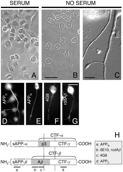
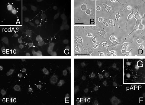
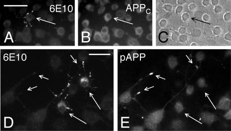
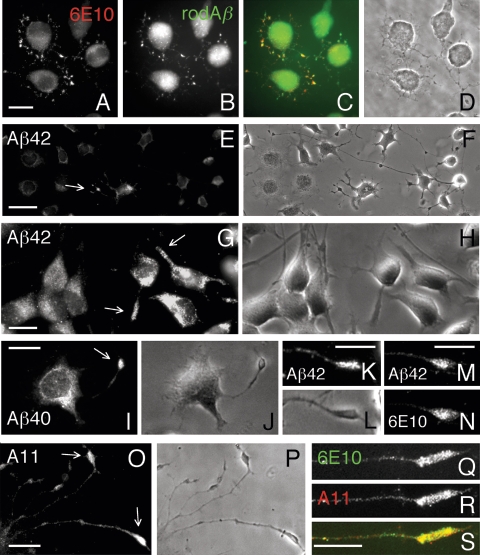
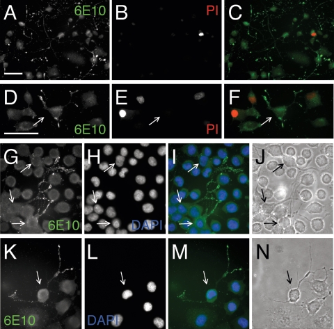
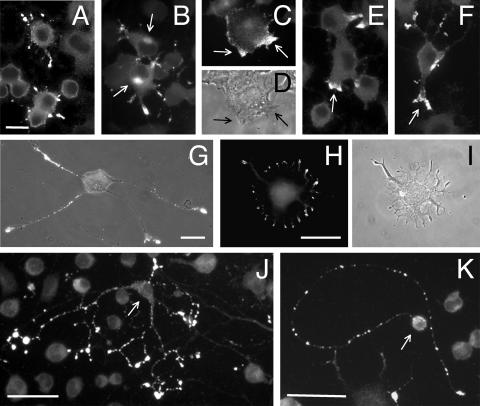
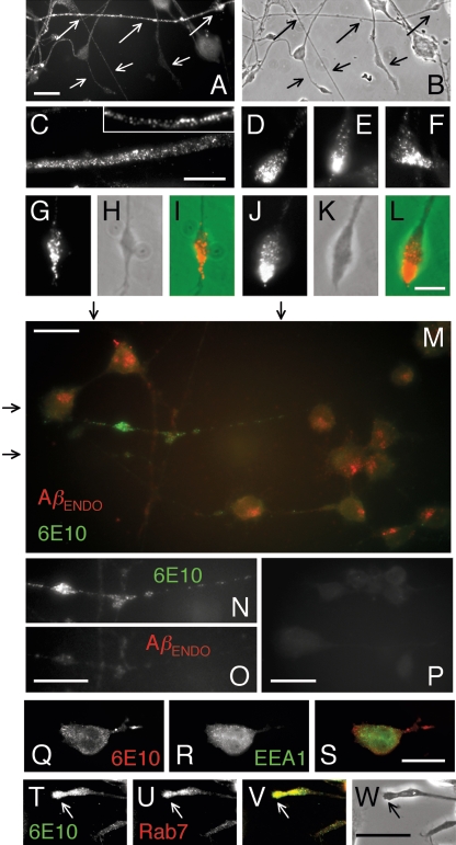
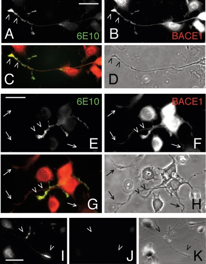
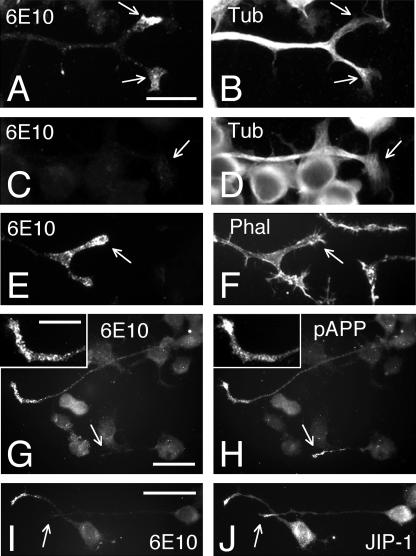
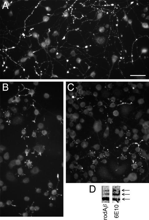
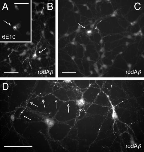
Our goal is to understand the pathogenesis of amyloid-beta (Abeta) deposition in the Alzheimer's disease (AD) brain. We established a cell culture system where central nervous system-derived neuronal cells (CAD cells) produce and accumulate within their processes large amounts of Abeta peptide, similar to what is believed to occur in brain neurons, in the initial phases of AD. Using this system, we show that accumulation of Abeta begins within neurites, prior to any detectable signs of neurodegeneration or abnormal vesicular transport. Neuritic accumulation of Abeta is restricted to a small population of neighboring cells that express normal levels of amyloid-beta precursor protein (APP) but show redistribution of BACE1 to the processes, where it colocalizes with Abeta and markers of late endosomes. Consistently, cells that accumulate Abeta appear in isolated islets, suggesting their clonal origin from a few cells that show a propensity to accumulate Abeta. These results suggest that Abeta accumulation is initiated in a small number of neurons by intracellular determinants that alter APP metabolism and lead to Abeta deposition and neurodegeneration. CAD cells appear to recapitulate the biochemical processes leading to Abeta deposition, thus providing an experimental in vitro system for studying the molecular pathobiology of AD.


Figures

FIG. 1.
FIG. 1.
Differentiated CAD cells express and transport APP. (A to C) Neuronal phenotype of differentiated CAD cells. (A) CAD cells cultured in the presence of serum. Note that upon serum withdrawal, CAD cells extend long processes (B). (C) Processes at high magnification. (D to G) APP is localized to cell bodies and processes of CAD cells, as detected with antibodies to carboxy-terminal (APPC) (D), amino-terminal (APPN) (E), and Aβ (4G8 [F] and anti-rodent Aβ [rodAβ] [G]) regions of APP. Note that no particular accumulation of anti-Aβ immunoreactive material at neurite terminals is seen (F and G). (H) Diagram of APP695 showing the polypeptides generated by secretase cleavage in the two main APP processing pathways. The positions of epitopes detected by antibodies used in this study are shown (a to d). The vertical lines indicate the transmembrane domain of APP. Bars = 50 μm (A and B), 10 μm (C), and 20 μm (D to G).
FIG. 2.
FIG. 2.
Clusters of differentiated CAD cells show accumulations of anti-Aβ immunoreactive material along neurites. CAD cells were labeled with an antibody to rodent Aβ (rodAβ) (A and B), antibody 6E10 (C to F), or an antibody to pAPP (G). Note that the 6E10 and anti-pAPP antibodies preferentially label the neurite terminals. (B and D) Phase-contrast micrographs. Bars = 20 μm (A and B) and 50 μm (C to G).
FIG. 3.
FIG. 3.
Neuritic deposits labeled by antibody 6E10 do not contain carboxy-terminal APP epitopes. The deposits detected with antibody 6E10 (A and D) do not cross-react with antibodies to the cytoplasmic domain of APP (APPC) (B) or to pAPP (E). Long arrows point to cells with neuritic deposits. Short arrows in panels D and E point to pAPP-containing terminals of adjacent cells. (C) Phase-contrast micrograph. Bars = 50 μm.
FIG. 4.
FIG. 4.
Differentiated CAD cells develop bona fide Aβ accumulations. (A to D) Antibody 6E10 and the antibody to rodent Aβ (rodAβ) costain deposits at neurite terminals in clusters of CAD cells. (E to L, O, and P) Neuritic deposits in CAD cells are labeled by antibodies to the carboxy- terminal end of Aβ (detecting Aβ40 and Aβ42 polypeptides) (E to L) and by an antioligomer antibody (A11) (O and P). Arrows point to labeled processes. Note that increased levels of Aβ42 polypeptides are often detected in clusters of cells (panels G and H, the three cells at right). (M, N, and Q to S) Antibody 6E10 and anti-Aβ42 (M and N) or antioligomer antibody (Q to S) costain deposits at neurite terminals. Note the significant colocalization in each case. (D, F, H, J, L, and P) Phase-contrast micrographs. Bars = 20 μm (A to D and G to S) and 40 μm (E and F).
FIG. 5.
FIG. 5.
CAD cells that accumulate Aβ are not necrotic or apoptotic. (A to F) No accumulation of propidium iodide (PI) is seen in cells that contain 6E10-immunoreactive material in their neurites. Cells were incubated with PI before fixation and immunolabeling with antibody 6E10. Note that few cells contain PI-stained nuclei (A to C), indicative of plasmalemmal damage and intracellular penetration of PI. A rare region containing several necrotic cells is shown in panels D to F. Note that the cell containing Aβ accumulations (arrow) is not necrotic. (G to N) Cells that contain neuritic Aβ accumulations do not show nuclear fragmentation (arrows). CAD cells were stained with antibody 6E10 and DAPI. (J and N) Phase-contrast micrographs. Bars = 50 μm (A to C and D to N).
FIG. 6.
FIG. 6.
Characterization of Aβ-accumulating CAD cells. (A to K) Gallery of images showing cells that contain deposits labeled with antibody 6E10. Brightness of images was increased to allow visualization of cell bodies. Note that deposits are present both in short (A and B) and long (G, J, and K) neurites and affect all neurites of a cell (G to J). Arrows in panel B point to immunostained material present in the perinuclear region. Arrows in panels C to F point to immunostained material present at the distal ends of emerging neurites. The cells indicated with arrows in panels J and K extend long processes, which contain large, 6E10-labeled varicosities. (D and I) Phase-contrast micrographs. (G) Combined phase-contrast-fluorescence micrograph. Bars = 20 μm (A to G) and 50 μm (H to K).
FIG. 7.
FIG. 7.
Aβ accumulations are intracellular but not in early endosomes. (A to L) “Anatomy” of particulate material detected with antibody 6E10 within neurites (A to C and inset in C) and at their terminals (D to L). Note that immunolabeled particles are intracellular throughout the neurites (A to C) and accumulate in the distal portion of the terminal (D to L). Note that most neurites do not show 6E10-immunoreactivity (small arrows in A and B). Long arrows in panels A and B point to a heavily labeled neurite. (M to P) Intracellular Aβ accumulations do not colocalize with early endocytic Aβ. CAD cells were cultured for 30 min in the presence of anti-rodent Aβ antibody, fixed, and immunolabeled for Aβ accumulations with antibody 6E10 (green [M and N]) and endocytosed anti-rodent Aβ with anti-rabbit IgG (AβENDO, red [M and O]). Note that following uptake, most anti-rodent Aβ (AβENDO) accumulates in perinuclear endosomes but not in regions labeled by the 6E10 antibody. (N and O) Enlargements of the marked area (arrows) in panel M. No labeling with anti-rabbit IgG is detected when cells are incubated in the absence of anti-rodent Aβ antibody (P). (Q to W) Neuritic Aβ accumulations, detected with antibody 6E10, colocalize with the late endosomal and autophagic vacuole marker Rab7 (T to W) but not with the early endosomal antigen 1 (EEA1) (Q to S). The arrows in panels T to W point to a terminal containing Aβ accumulations. Bars = 10 μm (C), 5 μm (D to L and inset in C), 20 μm (A, B, M to P, and T to W), 40 μm (Q to S).
FIG. 8.
FIG. 8.
Aβ accumulations colocalize with β-secretase. (A to H) CAD cells were double labeled for Aβ accumulations (antibody 6E10) and BACE1. Note that BACE1 is concentrated in the cell body. In addition, BACE1 is enriched within neurites of cells labeled by antibody 6E10, where it colocalizes with the Aβ accumulations (arrowheads). Arrows point to neurite terminals of cells that are not labeled by antibody 6E10, which contain little BACE1. (I to K) Control experiments, done in identical conditions as described above but in the absence of the primary antibody to BACE1. Note that the intense fluorescence at neurite endings stained with antibody 6E10 (I, arrowheads) does not bleed through into the red channel (J, arrowheads). Arrowheads point to neurite terminals labeled by antibody 6E10. (D, H, and K) Phase-contrast micrographs. Bars = 40 μm (A to D and I to K) and 20 μm (E to H).
FIG. 9.
FIG. 9.
Accumulation of Aβ within CAD cell neurites does not block vesicular transport of neuronal cargos. (A to F) Normal appearance of microtubules (A to D) and actin cytoskeleton (E and F) in neurites containing Aβ accumulations. CAD cells were costained with antibody 6E10 and either antitubulin antibody (Tub) or phalloidin-FITC (Phal; to stain actin filaments). Arrows in panels A, B, E, and F point to terminals containing Aβ deposits. The arrows in panels C and D point to the terminal of a control, normal process. (G to J) Transport and accumulation of pAPP- and JIP-1-containing vesicles is normal in cells that show Aβ deposits within neurites. CAD cells were double labeled with antibody 6E10 and either anti-pAPP (G and H) or anti-JIP-1 (I and J) antibody. Arrows point to terminals that do not contain Aβ accumulations. Insets in panels G and H are enlargements of the neurite terminal that contains Aβ and show that the 6E10 immunoreactive material does not colocalize with pAPP. Bars = 20 μm (A to F and insets in G and H) and 40 μm (G to J).
FIG. 10.
FIG. 10.
CAD cell population enriched in Aβ-accumulating cells. (A to C) Immunostaining of CAD cells with antibody 6E10. Cells are shown at different stages of differentiation (B and C, early stages; A, late stage). Note the increased proportion of cells contain 6E10-immunoreactive material. Bar = 50 μm. (D) Immunoblot of concentrated lysates from differentiated CAD cells done with antibodies to Aβ (rodAβ and 6E10). Arrows show the Aβ monomer (bottom arrow) and Aβ oligomers (upper two arrows). Higher oligomer species are likely present in the lysates but are below the detection limit of our Western blot procedure. Note that antibody 6E10 detects Aβ oligomers with higher sensitivity than the anti-rodent Aβ antibody.
FIG. 11.
FIG. 11.
Aβ accumulations in neurites of cortical neurons. Primary cultures of cortical neurons were immunolabeled with antibody 6E10 (A) or an antibody to rodent Aβ (rodAβ) (B to D). Note that a small number of cells show increased labeling in the cell body (arrows in A to C) and processes. (D) Cluster of intensely labeled neurons that show discrete accumulations of immunoreactive material along their processes. Arrows point to such a process, emanating from the cell marked with a star. Bars = 50 μm.

Similar articles

Cited by

References

    1. Ando, K., M. Oishi, S. Takeda, K. Iijima, T. Isohara, A. C. Nairn, Y. Kirino, P. Greengard, and T. Suzuki. 1999. Role of phosphorylation of Alzheimer's amyloid precursor protein during neuronal differentiation. J. Neurosci. 19:4421-4427. - PMC - PubMed
    1. Arboleda, G., C. Waters, and R. M. Gibson. 2005. Metabolic activity: a novel indicator of neuronal survival in the murine dopaminergic cell line CAD. J. Mol. Neurosci 27:65-77. - PubMed
    1. Aston-Jones, G., and J. D. Cohen. 2005. An integrative theory of locus coeruleus-norepinephrine function: adaptive gain and optimal performance. Annu. Rev. Neurosci. 28:403-450. - PubMed
    1. Bard, F., R. Barbour, C. Cannon, R. Carretto, M. Fox, D. Games, T. Guido, K. Hoenow, K. Hu, K. Johnson-Wood, K. Khan, D. Kholodenko, C. Lee, M. Lee, R. Motter, M. Nguyen, A. Reed, D. Schenk, P. Tang, N. Vasquez, P. Seubert, and T. Yednock. 2003. Epitope and isotype specificities of antibodies to beta-amyloid peptide for protection against Alzheimer's disease-like neuropathology. Proc. Natl. Acad. Sci. USA 100:2023-2028. - PMC - PubMed
    1. Billings, L. M., S. Oddo, K. N. Green, J. L. McGaugh, and F. M. Laferla. 2005. Intraneuronal Aβ causes the onset of early Alzheimer's disease-related cognitive deficits in transgenic mice. Neuron 45:675-688. - PubMed

Publication types

MeSH terms