. 2006 Jul;26(13):5070-85.
doi: 10.1128/MCB.02006-05.

Regulated activating Thr172 phosphorylation of cyclin-dependent kinase 4(CDK4): its relationship with cyclins and CDK "inhibitors"

Affiliations

Regulated activating Thr172 phosphorylation of cyclin-dependent kinase 4(CDK4): its relationship with cyclins and CDK "inhibitors"

Laurence Bockstaele et al. Mol Cell Biol. 2006 Jul.

Abstract

Cyclin-dependent kinase 4 (CDK4) is a master integrator of mitogenic and antimitogenic extracellular signals. It is also crucial for many oncogenic transformation processes. Various molecular features of CDK4 activation remain poorly known or debated, including the regulation of its association with D-type cyclins, its activating Thr172 phosphorylation, and the roles of Cip/Kip CDK "inhibitors" in these processes. Thr172 phosphorylation of CDK4 was reinvestigated using two-dimensional gel electrophoresis in various experimental systems, including human fibroblasts, canine thyroid epithelial cells stimulated by thyrotropin, and transfected mammalian and insect cells. Thr172 phosphorylation of CDK4 depended on prior D-type cyclin binding, but Thr172 phosphorylation was also found in p16-bound CDK4. Opposite effects of p27 on cyclin D3-CDK4 activity observed in different systems depended on its stoichiometry in this complex. Thr172-phosphorylated CDK4 was enriched in complexes containing p21 or p27, even at inhibitory levels of p27 that precluded CDK4 activity. Deletion of the p27 nuclear localization signal sequence relocalized cyclin D3-CDK4 in the cytoplasm but did not affect CDK4 phosphorylation. Within cyclin D3 complexes, T-loop phosphorylation of CDK4, but not of CDK6, was directly regulated, identifying it as a determining target for cell cycle control by extracellular factors. Collectively, these unexpected observations indicate that CDK4-activating kinase(s) should be reconsidered.


Figures

FIG. 1.
FIG. 1.
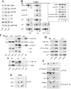
Two-dimensional gel electrophoresis identification of Thr172-phosphorylated CDK4. (A) 32P-metabolically labeled extracts from human IMR-90 fibroblasts stimulated for 16 h with serum (16 h) and from dog thyrocytes stimulated for 20 h with TSH plus EGF (ET) were immunoprecipitated with a CDK4 antibody, separated by 2D gel electrophoresis, and electroblotted. The membranes were exposed for autoradiographic detection of phosphorylated forms of CDK4 (32P). CDK4 phosphorylated on Thr172 or total CDK4 was then immunodetected from the same membranes by using enhanced chemiluminescence (P-T172-CDK4 and CDK4, respectively). As indicated, the polyclonal C-22 (Santa Cruz) or monoclonal DCS-156 antibodies were used. Unstimulated quiescent cells are shown for comparison (Cont). The different spots of CDK4 are numbered; only forms 3 and 4 are phosphorylated, including on Thr172. (B) Extracts of CHO cells transfected with vectors encoding CDK4-HA (K4), CDK4T172A-HA (K4T172A), CDK4T172E-HA (K4T172E), CDK4-HA plus cyclin D3 (K4+D3), CDK4T172A-HA plus cyclin D3 (K4T172A+D3), or CDK4T172E-HA plus cyclin D3 (K4T172E+D3) were immunoprecipitated (IP) with anti-HA or anti-cyclin D3 antibodies, separated by 2D gel electrophoresis, and electroblotted. K4T172A+D3 + K4+D3 is the 1:1 mixture of K4T172A+D3 and K4+D3 samples before 2D gel separation. CDK4 phosphorylated on Thr172 or total CDK4 was immunodetected from the same membranes (P-T172-CDK4 and CDK4, respectively). (C) The same CHO cell extracts were immunoprecipitated with anti-HA (for CDK4) or anti-cyclin D3 antibodies, assayed for pRb kinase activity, separated by SDS-PAGE, and immunoblotted. CDK4 was detected using anti-HA antibody, and the pRb fragment phosphorylated in vitro at Ser780 (P-Rb-780) were detected using a phospho-specific antibody.
FIG. 2.
FIG. 2.
Phospho-Thr172 CDK4 is associated with both D-type cyclins and CDK inhibitors. (A to C) IMR-90 fibroblasts; (D to F) dog thyrocytes. (A) Western blotting analyses of CDK4, cyclin D3, cyclin D1, p21, p27, and p16 from whole-cell extracts of quiescent unstimulated IMR-90 cells (Cont) or cells stimulated by 20% serum for 16 or 24 h. p38 mitogen-activated protein kinase (MAPK) was detected as a loading control. (B) CDK4 separated by 2D gel electrophoresis was detected by Western blotting with a CDK4 antibody from (co)immunoprecipitates (IP) of cyclin D1, cyclin D3, p21, p27, or p16 from quiescent unstimulated IMR-90 cells (Cont), cells stimulated with 20% serum for 16 h (16 h), or cells stimulated for 16 h and treated for 3 h with the protein synthesis inhibitor cycloheximide (100 μg/ml) in the presence of serum (16 h + 3 h Cx). In the inset, controls of cycloheximide impact on the presence of proteins in whole-cell extracts are shown; D1-bound CDK4 and p16-bound CDK4 are CDK4 coimmunoprecipitated using cyclin D1 and p16 antibodies, respectively. (C and D) Extracts from IMR-90 cells (C) stimulated or not (Cont) by 20% serum for 16 h or dog thyrocytes (D) stimulated or not (Cont) by forskolin (Forsk) were immunoprecipitated with anti-p16, anti-cyclin D1, anti-cyclin D3, anti-p21, or anti-p27 antibodies or a control immunoglobulin (IgG), assayed for pRb kinase activity, separated by SDS-PAGE, and immunoblotted. Cyclin D3, CDK4 and its activating Thr172 phosphorylation (P-T172-CDK4), cyclin D1, p16, p21, p27, and the pRb fragment phosphorylated in vitro at Ser780 (P-Rb-780) were detected using specific antibodies. (E) Western blotting analyses of CDK4 separated by 2D gel electrophoresis from coimmunoprecipitates of p27 from quiescent unstimulated dog thyrocytes (Cont) or cells stimulated with TSH (1 mU/ml) or TSH plus EGF plus 10% serum (ETS) for 20 h. (F) Extracts from dog thyrocytes stimulated for 20 h with TSH were immunoprecipitated with p27 antibodies (p27 IP) or a phospho-specific (Ser10) p27 antibody (P-S10-p27 IP), separated by 2D gel electrophoresis, and electroblotted. Immunodetections were performed using antibodies against total p27 or p27 phosphorylated on Ser10 (P-S10-p27) or with a mixture of CDK4 and p27 antibodies. In panels B, E, and F, arrows 3 indicate the main Thr172-phosphorylated form of CDK4.
FIG. 3.
FIG. 3.
Different impacts of p27 stoichiometry on activity and Thr172 phosphorylation of CDK4. (A) Extracts of CHO cells transfected with vectors encoding V5-tagged wild-type p27 or V5-tagged p27 lacking its C-terminal NLS region, or of insect Sf9 cells transduced with baculovirus encoding p27-V5, were immunoprecipitated (IP) with anti-V5 antibody, separated by 2D gel electrophoresis, and electroblotted. p27 phosphorylated on Ser10 or total p27 was immunodetected from the same membranes (P-S10-p27 and p27, respectively). (B) Extracts of Sf9 cells or CHO cells transduced/transfected with baculoviruses or plasmids encoding cyclin D3, CDK4-HA plus cyclin D3, or CDK4-HA plus cyclin D3 plus p27-V5 or untagged p27 (p27) (and dilutions of p27[-V5] vectors to 1/5, 1/7, 1/10, and 1/20) were immunoprecipitated with anti-cyclin D3, anti-p27, or anti-V5 antibodies, assayed for pRb kinase activity, separated by SDS-PAGE, and immunoblotted. Cyclin D3, p27, and the pRb fragment phosphorylated in vitro at Ser780 (P-Rb-780) were detected using their specific antibodies; CDK4-HA and p27-V5 were detected using anti-HA and anti-V5 antibodies, respectively. (C) Extracts of Sf9 or CHO cells transduced/transfected as for panel B were immunoprecipitated with anti-HA (for CDK4), anti-cyclin D3, or anti-V5 antibodies, separated by 2D gel electrophoresis, and electroblotted for detection of CDK4 with an anti-CDK4 antibody. Arrows indicate the Thr172-phosphorylated form of CDK4.
FIG. 4.
FIG. 4.
Various amounts of p27 differently affect cyclin D3-CDK4 activity in vitro. Cyclin D3-CDK4-HA complexes formed by their coexpression in infected Sf9 cells were incubated in vitro with decreasing amounts of p27-V5 (1, 1/10, 1/20, and 1/30 dilutions) produced in Sf9 cells. This mixture and cyclin D3-CDK4-HA-p27-V5 complexes formed by their coexpression in Sf9 cells were immunoprecipitated (IP) using anti-cyclin D3 or anti-V5 antibodies, assayed for pRb kinase activity, separated by SDS-PAGE, and immunoblotted. Cyclin D3 and the pRb fragment phosphorylated in vitro at Ser780 (P-Rb-780) were detected using specific antibodies; CDK4 and p27 were detected with anti-HA and anti-V5 antibodies.
FIG. 5.
FIG. 5.
p27 can associate with cyclin-free CDK4. (A) Extracts of CHO cells transfected with vectors encoding CDK4-HA or CDK4-HA plus p27-V5 or untagged p27 (p27), or Sf9 cells infected with baculoviruses encoding cyclin D3 plus CDK4-HA or p27-V5 plus CDK4-HA, were immunoprecipitated (IP) with anti-cyclin D3, anti-HA, anti-p27, or anti-V5 antibodies, assayed for pRb kinase activity, separated by SDS-PAGE, and immunoblotted. Cyclin D3 and the pRb fragment phosphorylated in vitro at Ser780 (P-Rb-780) were detected using specific antibodies. CDK4 and p27 were detected using, respectively, anti-HA and anti-V5 (left and middle panels) or anti-p27 (right panel) antibodies. (B) The same CHO or Sf9 cell extracts were immunoprecipitated with anti-HA or anti-V5 antibodies, separated by 2D gel electrophoresis, and electroblotted for detection of CDK4 with an anti-CDK4 antibody. Arrows indicate the form of CDK4 phosphorylated on Thr172.
FIG. 6.
FIG. 6.
The subcellular localization of CDK4 does not affect its Thr172 phosphorylation. CHO cells were transfected as indicated with different combinations of vectors encoding cyclin D3, HA-tagged CDK4 (K4), V5-tagged wild-type p27 (p27) or V5-tagged p27 lacking its C-terminal NLS region (p27-NLS). (A and B) Cells were fixed 48 h after transfection and processed for double indirect immunofluorescent staining with either mouse monoclonal anti-cyclin D3 and rabbit polyclonal anti-CDK4 antibodies or mouse monoclonal anti-V5 (p27) and rabbit polyclonal anti-CDK4 antibodies. Nuclei were counterstained with Hoechst dye. Images were recorded using a 100× immersion lens and the SPOT RT camera (Diagnostic Instrument, Inc.). To demonstrate the colocalizations of cyclin D3 and CDK4 or of CDK4 and p27 or p27-NLS, green and red fluorescent images were merged using the Adobe Photoshop program. (A) Single and double transfections. (B) Triple transfections. In some transfections, the p27 or p27-NLS expression plasmids were diluted. Only the 10-fold dilution of the p27 plasmid is illustrated (K4+D3 + 1/10p27). (C) Extracts from the same transfection experiment as in panels A and B were immunoprecipitated (IP) with anti-cyclin D3 or anti-V5 antibodies, separated by 2D gel electrophoresis, and electroblotted. CDK4 was detected using anti-CDK4 antibody. In some transfections, the plasmids encoding p27 or p27-NLS were diluted as indicated (1/5 and 1/10). Arrows indicate the form of CDK4 phosphorylated on Thr172.
FIG. 6.
FIG. 6.
The subcellular localization of CDK4 does not affect its Thr172 phosphorylation. CHO cells were transfected as indicated with different combinations of vectors encoding cyclin D3, HA-tagged CDK4 (K4), V5-tagged wild-type p27 (p27) or V5-tagged p27 lacking its C-terminal NLS region (p27-NLS). (A and B) Cells were fixed 48 h after transfection and processed for double indirect immunofluorescent staining with either mouse monoclonal anti-cyclin D3 and rabbit polyclonal anti-CDK4 antibodies or mouse monoclonal anti-V5 (p27) and rabbit polyclonal anti-CDK4 antibodies. Nuclei were counterstained with Hoechst dye. Images were recorded using a 100× immersion lens and the SPOT RT camera (Diagnostic Instrument, Inc.). To demonstrate the colocalizations of cyclin D3 and CDK4 or of CDK4 and p27 or p27-NLS, green and red fluorescent images were merged using the Adobe Photoshop program. (A) Single and double transfections. (B) Triple transfections. In some transfections, the p27 or p27-NLS expression plasmids were diluted. Only the 10-fold dilution of the p27 plasmid is illustrated (K4+D3 + 1/10p27). (C) Extracts from the same transfection experiment as in panels A and B were immunoprecipitated (IP) with anti-cyclin D3 or anti-V5 antibodies, separated by 2D gel electrophoresis, and electroblotted. CDK4 was detected using anti-CDK4 antibody. In some transfections, the plasmids encoding p27 or p27-NLS were diluted as indicated (1/5 and 1/10). Arrows indicate the form of CDK4 phosphorylated on Thr172.
FIG. 7.
FIG. 7.
Thr172 phosphorylation of CDK4 is regulated. (A) Extracts from dog thyrocytes stimulated for 24 h with forskolin (Forsk) or forskolin plus TGF-β (2 ng/ml) were immunoprecipitated (IP) with anti-cyclin D3 antibody, assayed for pRb kinase activity, separated by SDS-PAGE, and immunoblotted. Cyclin D3, CDK4 and its activating Thr172 phosphorylation (P-T172-CDK4), p27, and the pRb fragment phosphorylated in vitro at Ser780 (P-Rb-780) were detected using specific antibodies. In the right panel, similar coimmunoprecipitates from dog thyrocytes stimulated with TSH or TSH plus TGF-β (2 ng/ml) were separated by 2D gel electrophoresis and electroblotted for detection of CDK4 with an anti-CDK4 antibody. (B) Extracts from quiescent serum-starved T98G cells (Cont) or cells stimulated by 15% FBS for 10 h were immunoprecipitated with anti-cyclin D3 or anti-p27 antibodies, assayed for pRb kinase activity, separated by SDS-PAGE, and immunoblotted. Cyclin D3, CDK4, CDK6, CDK2, p27, and the pRb fragment phosphorylated in vitro at Ser780 (P-Rb-780) were detected using specific antibodies. In the right panel, the same coimmunoprecipitates were separated by 2D gel electrophoresis and electroblotted for detection of CDK4 and CDK6 with anti-CDK4 and anti-CDK6 antibodies. In the bottom panel, similar cyclin D3 coimmunoprecipitates from serum-starved (Cont) and stimulated (10 h) T98G cells were separated by 2D gel electrophoresis and electroblotted for detection using the phospho-CDK4(T172) antibody (PT-172). Total CDK4 and CDK6 were then detected from the same membrane by using anti-CDK4 and anti-CDK6 antibodies. Arrows in panels A and B indicate the regulated Thr172-phosphorylated form of CDK4 and the corresponding, but unregulated, phosphorylated (Thr177) form of CDK6. (C) CAK expression and activity in the same cultures of dog thyrocytes and T98G cells as in panels A and B. Cyclin H (Cyc H), CDK7, and Mat1 were detected from whole-cell extracts of T98G cells (serum starved and stimulated for 10 h) or dog thyrocytes treated as in panel A and from anti-cyclin H immunoprecipitates (IP Cyc H) of dog thyrocytes. In the right panel, CAK activity was assessed by the incubation of GST-CDK2 in the presence of [32P]ATP, with (+) or without (α) recombinant cyclin H-CDK7-Mat1 complex (CAK) or with crude extracts of T98G cells (serum starved [Cont] or stimulated for 10 or 26 h) or of dog thyrocytes (stimulated or not [Cont] with forskolin and TGF-β). GST-CDK2 was recovered using glutathione-Sepharose, put on SDS-polyacrylamide gels, and electroblotted, and its phosphorylation was detected by autoradiography (32P-GST-CDK2). GST-CDK2 was then immunodetected using the PSTAIRE antibody. The lower band of the 32P-GST-CDK2 doublet reflects the T-loop phosphorylation.
FIG. 8.
FIG. 8.
CAK phosphorylates CDK6 more readily than CDK4 in vitro. Inactive cyclin D3 complexes containing CDK4 or CDK6 were immunoprecipitated using the anti-cyclin D3 antibody (cyclin D3 IP) from serum-starved T98G cells. The immunoprecipitated complexes were incubated in the presence of 15 mM Mg2+ and 1 mM ATP or 50 μM [32P]ATP, without (−) or with (+) recombinant cyclin H-CDK7-Mat1 complex (CAK) or with CAK and 100 mM EDTA. The cyclin D3 coimmunoprecipitates were then separated by 2D gel electrophoresis and electroblotted for immunodetection using the phospho-CDK4(T172) antibody (PT-172). Total CDK4 and CDK6 were then detected from the same membrane using anti-CDK4 and anti-CDK6 antibodies. In the case of incubation with [32P]ATP, 32P labeling was revealed by autoradiography after electroblotting (P-CDK6 and P-CDK4 indicate the positions of phosphorylated forms of CDK6 and CDK4 as detected by CDK4 and CDK6 antibodies). Arrows indicate the Thr172-phosphorylated form of CDK4 and the corresponding phosphorylated (Thr177) form of CDK6. In the bottom panel, the cyclin D3 coimmunoprecipitates were assayed for pRb kinase activity (PRb-780) after incubation with or without (α) CAK.

References

    1. Alt, J. R., A. B. Gladden, and J. A. Diehl. 2002. p21(Cip1) promotes cyclin D1 nuclear accumulation via direct inhibition of nuclear export. J. Biol. Chem. 277:8517-8523. - PubMed
    1. Aprelikova, O., Y. Xiong, and E. T. Liu. 1995. Both p16 and p21 families of cyclin-dependent kinase (CDK) inhibitors block the phosphorylation of cyclin-dependent kinases by the CDK-activating kinase. J. Biol. Chem. 270:18195-18197. - PubMed
    1. Bagui, T. K., R. J. Jackson, D. Agrawal, and W. J. Pledger. 2000. Analysis of cyclin D3-cdk4 complexes in fibroblasts expressing and lacking p27(kip1) and p21(cip1). Mol. Cell. Biol. 20:8748-8757. - PMC - PubMed
    1. Bagui, T. K., S. Mohapatra, E. Haura, and W. J. Pledger. 2003. p27Kip1 and p21Cip1 are not required for the formation of active D cyclin-cdk4 complexes. Mol. Cell. Biol. 23:7285-7290. - PMC - PubMed
    1. Baptist, M., F. Lamy, J. Gannon, T. Hunt, J. E. Dumont, and P. P. Roger. 1996. Expression and subcellular localization of CDK2 and cdc2 kinases and their common partner cyclin A in thyroid epithelial cells: comparison of cyclic AMP-dependent and -independent cell cycles. J. Cell. Physiol. 166:256-273. - PubMed

Publication types

MeSH terms

LinkOut - more resources