. 2006 Jul;26(13):5131-45.
doi: 10.1128/MCB.02227-05.

Potential roles for ubiquitin and the proteasome during ribosome biogenesis

Affiliations

Potential roles for ubiquitin and the proteasome during ribosome biogenesis

Diana A Stavreva et al. Mol Cell Biol. 2006 Jul.

Erratum in

  • Mol Cell Biol. 2006 Aug;26(16):6308

Abstract

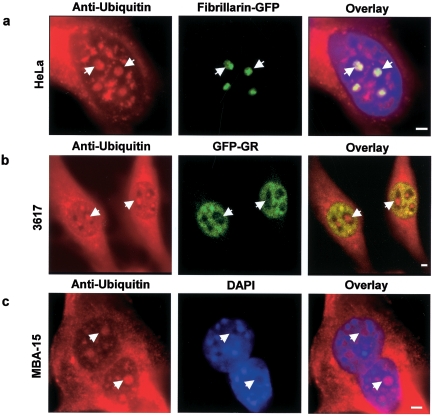
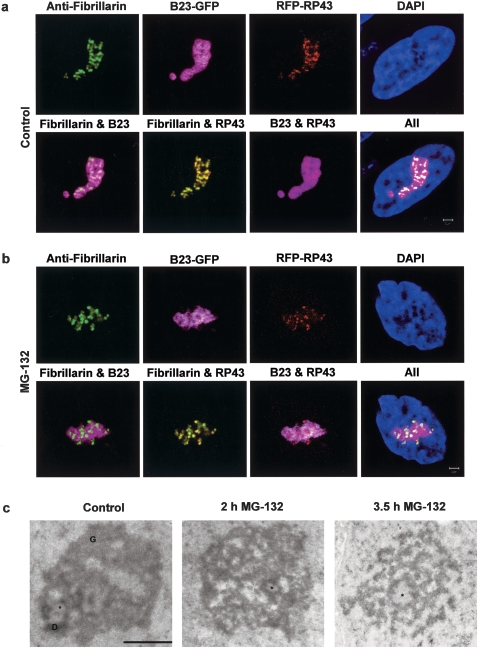
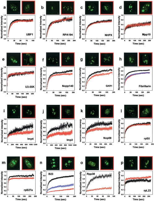
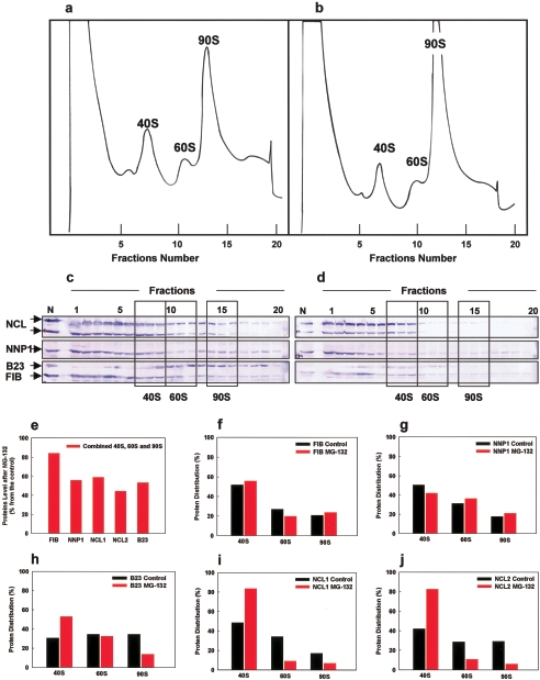
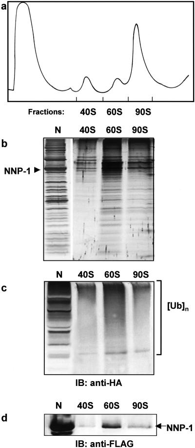
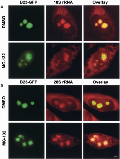
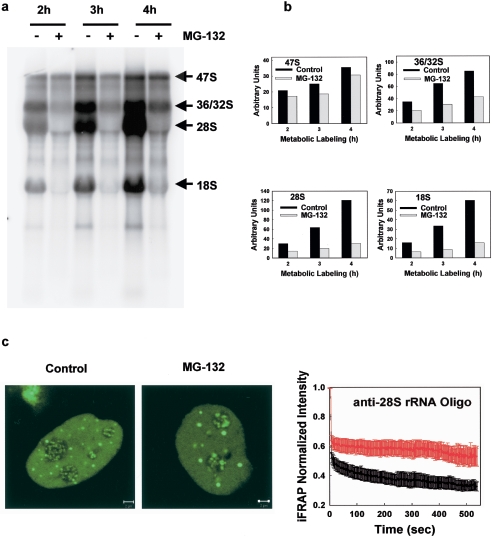
We have investigated the possible involvement of the ubiquitin-proteasome system (UPS) in ribosome biogenesis. We find by immunofluorescence that ubiquitin is present within nucleoli and also demonstrate by immunoprecipitation that complexes associated with pre-rRNA processing factors are ubiquitinated. Using short proteasome inhibition treatments, we show by fluorescence microscopy that nucleolar morphology is disrupted for some but not all factors involved in ribosome biogenesis. Interference with proteasome degradation also induces the accumulation of 90S preribosomes, alters the dynamic properties of a number of processing factors, slows the release of mature rRNA from the nucleolus, and leads to the depletion of 18S and 28S rRNAs. Together, these results suggest that the UPS is probably involved at many steps during ribosome biogenesis, including the maturation of the 90S preribosome.


Figures

FIG. 1.
FIG. 1.
Ubiquitin is present in the nucleolus, and the pre-rRNP complexes are ubiquitinated. (a) Immunofluorescence detection of ubiquitin in the nucleolus. Antibodies against ubiquitin stain the cytoplasm and nucleus as expected but also consistently colocalize with nucleoli, identified here in HeLa cells by fibrillarin expression. (b) 3617 cells, a derivative cell line from 3134, stably expressing GFP-tagged glucocorticoid receptor (GFP-GR) were also used (27). GFP-GR stains the nucleoplasm but is absent from nucleoli. These nucleoli are clearly stained with an anti-ubiquitin antibody. (c) In addition, MBA-15 cells also show a clear nuclear localization of ubiquitin as seen from the overlay with the DAPI DNA staining. Some of the nucleoli are indicated with arrows. Scale bar, 2 μm.
FIG. 2.
FIG. 2.
Proteasome inhibition by MG-132 and lactacystin induces morphological changes in the nucleolus. Treatment of B23-GFP transfected HeLa cells with 100 μM MG-132 or 50 μM lactacystin (data not shown) for 2 h changes the relative distributions of B23 and fibrillarin, as well as B23 and RP43 (compare images in the second rows of panels a and b). Scale bar, 2 μm. The normal ultrastructural organization of the nucleolus is affected after treatment with 100 μM MG-132 (c). Although structural counterparts of fibrillar centers (FC, asterisk) are observed both after 2 h and after 3.5 h of treatment, granular components (G) and dense fibrillar components (D) gradually disintegrate. Scale bar, 1 μm.
FIG. 3.
FIG. 3.
Effects of proteasome inhibition on the kinetics of selected nucleolar factors. Proteasome inhibition has minimal effects on rRNA gene transcription factors. A treatment with 100 μM MG-132 for up to 3 h does not affect the distribution or kinetics of UBF1 and RPA194, factors involved in Pol I transcription (a and b). Proteasome inhibition alters the mobility and cellular distribution of a subset of early pre-rRNA processing factors and small subunit ribosomal proteins. The kinetics of six (f to k) of nine (c to k) early processing factors were affected, whereas the remaining three did not show detectable changes. In addition, the cellular distribution of four of them (fibrillarin, Imp4, Imp3, and Nop58) was significantly altered (h to k). The mobility and distribution of one of the small subunit proteins (rpS5) was unaffected (l); however, the dynamics of another small subunit protein was reduced (m). Proteasome inhibition radically alters the mobility of two late pre-rRNA processing factors and increases the mobility of a large subunit protein (n to p). A 2- to 3-h treatment with either 100 μM MG-132 or 50 μM lactacystin leads to a dramatic change in the distribution of B23 and Rpp38 and drastic reduction in the mobility of both factors in nucleoli (n and o). In contrast the FRAPs of a large ribosomal subunit protein, rpL23, are faster than in control cells (p). Scale bars, 2 μm. The normal distribution of the proteins in HeLa cells is shown in black-framed pictures. The localization after proteasome inhibition is shown in red-framed pictures. Control FRAPs are shown in black, FRAPs after 100 μM MG-132 treatment are shown in red, and FRAPs after the addition of 50 μM lactacystin are shown in blue (error bars are ± the SE).
FIG. 4.
FIG. 4.
Effects of proteasome inhibition on pre-rRNP complexes. Equal amounts of nuclear extracts from control (DMSO) or MG-132-treated 293EBNA cells were fractionated on a 10 to 40% sucrose gradient (a and b). MG-132 treatment (b) induces an accumulation of the 90S pre-ribosome (143% ± 13%) compared to the control (a). The 40S and 60S levels were less affected (81% ± 6% and 92% ± 20%, respectively). The data are the means of three independent experiments (± the SE). Proteins in gradient fractions from control (a) and MG-132-treated 293EBNA cells (b) were further analyzed by immunoblotting with anti-nucleolin (NCL), anti-NNP-1, anti-B23, and anti-fibrillarin (FIB) antibodies (c and d). Three fractions from each peak representing the 40S, 60S, and 90S pre-ribosomes were selected, and the staining intensity of each of the protein bands present in the areas surrounded by boxes was measured. Shown are changes in the proteins' level after proteasome inhibition relative to the control (e), as well as changes in the proteins' distribution among the 40S, 60S, and 90S pre-ribosomal fractions (f to j).
FIG. 5.
FIG. 5.
Proteasome inhibition increases the ubiquitination of complexes associated with late processing factors. (a) Complexes isolated before (−) and after (+) MG-132 treatment. 293EBNA cells were cotransfected with HA-ubiquitin and FLAG-NNP-1, FLAG-B23, FLAG-fibrillarin (FIB), or FLAG-nucleolin (NCL) as affinity baits. Complexes were separated by SDS-PAGE and silver stained. A star indicates the estimated location of the bait protein, based on its molecular mass as determined by the molecular mass standards (in kilodaltons) shown at the left side (see also panel c, which demonstrates the presence of the bait protein in each extract). (b and c) Western blots with anti-HA (ubiquitin tag) and anti-FLAG (bait tag) antibodies are shown. Mock, extract from cells transfected with the expression plasmid pcDNA3.1 without an inserted gene. (d) As an input control, the GAPDH content in each of the cell extracts is indicated at the bottom.
FIG. 6.
FIG. 6.
Immunoprecipitated complexes are parts of the pre-rRNP particles. (a) Nuclear extract prepared from the FLAG-NNP-1 and HA-ubiquitin cotransfected 293 EBNA cells was fractionated on a 10 to 40% sucrose gradient with continuous monitoring of the absorbance at 254 nm. The FLAG-NNP-1-associated pre-rRNP complex was immunoisolated from each of the 40S, 60S, and 90S fractions; separated by SDS-PAGE (b); and analyzed by Western blotting with anti-HA (c) and anti-FLAG (d) antibodies, respectively. N, NNP-1-associated pre-rRNP complex isolated from FLAG-NNP-1-transfected cells.
FIG. 7.
FIG. 7.
Proteasomal inhibition alters the nucleolar distribution of both rRNA and rRNA precursors in HeLa cells. In the control (DHSO) both 18S and 28S RNA FISH signals colocalize to a large extent with the B23 GFP-tagged construct (first rows in panels a and b). However, after proteasome inhibition (MG-132), both 18S rRNA and 28S rRNA signals exhibit a mixed pattern, with some areas colocalizing with B23 and others not (second rows in panels a and b). Note that the degree of mislocalization varied from cell to cell. The second row in panel a shows an example of a significant separation of the two signals, while the second row in panel b shows an example of a more modest effect. Scale bar, 5 μm.
FIG. 8.
FIG. 8.
Proteasomal inhibition affects pre-rRNA processing, and impairs the production of mature 28S. (a) 32P metabolic labeling. Proteasome inhibition has modest effects on the levels of rRNA gene transcription (compare the levels of 47S in control and MG-132-treated cells), leads to reduced formation of 36S/32S pre-rRNA, and impaired production of mature 28S and 18S rRNA. (b) Quantification of the 32P-labeled 47S and 36/32S pre-rRNAs and 32P-labeled mature 28S and 18S rRNAs corresponding to the representative experiment shown in panel a. (c) iFRAP experiments on an anti-28S rRNA oligonucleotide microinjected in control and MG-132-treated HeLa cells reveal a slower loss of 28S rRNA fluorescence from the nucleolus after proteasome inhibition. This indicates impaired production and export of mature 28S rRNA. Curves: black, control iFRAP; red, iFRAP after MG-132 treatment (± the SE). Scale bars, 2 μm.

References

    1. Andersen, J. S., Y. W. Lam, A. K. Leung, S. E. Ong, C. E. Lyon, A. I. Lamond, and M. Mann. 2005. Nucleolar proteome dynamics. Nature 433:77-83. - PubMed
    1. Arabi, A., C. Rustum, E. Hallberg, and A. P. Wright. 2003. Accumulation of c-Myc and proteasomes at the nucleoli of cells containing elevated c-Myc protein levels. J. Cell Sci. 116:1707-1717. - PubMed
    1. Boulanger, J., A. Vezina, S. Mongrain, F. Boudreau, N. Perreault, B. A. Auclair, J. Laine, C. Asselin, and N. Rivard. 2005. Cdk2-dependent phosphorylation of homeobox transcription factor CDX2 regulates its nuclear translocation and proteasome-mediated degradation in human intestinal epithelial cells. J. Biol. Chem. 280:18095-18107. - PubMed
    1. Carmo-Fonseca, M., L. Mendes-Soares, and I. Campos. 2000. To be or not to be in the nucleolus. Nat. Cell Biol. 2:E107-E112. - PubMed
    1. Chan, Y. L., K. Suzuki, and I. G. Wool. 1995. The carboxyl extensions of two rat ubiquitin fusion proteins are ribosomal proteins S27a and L40. Biochem. Biophys. Res. Commun. 215:682-690. - PubMed

Publication types

MeSH terms

LinkOut - more resources