. 2006 Jul;26(13):5180-9.
doi: 10.1128/MCB.00966-05.

FAT10/diubiquitin-like protein-deficient mice exhibit minimal phenotypic differences

Affiliations

FAT10/diubiquitin-like protein-deficient mice exhibit minimal phenotypic differences

Allon Canaan et al. Mol Cell Biol. 2006 Jul.

Abstract

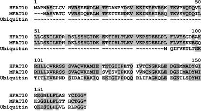
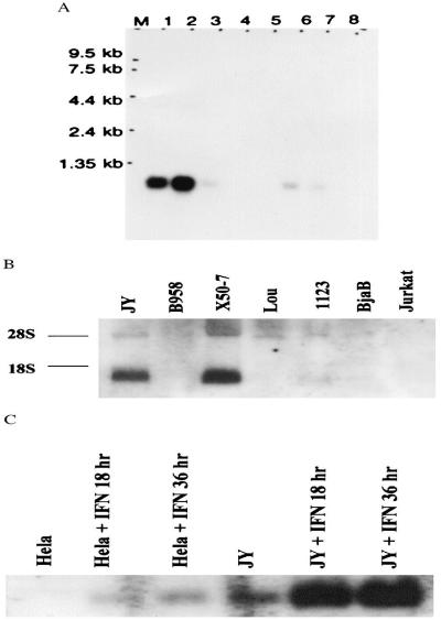
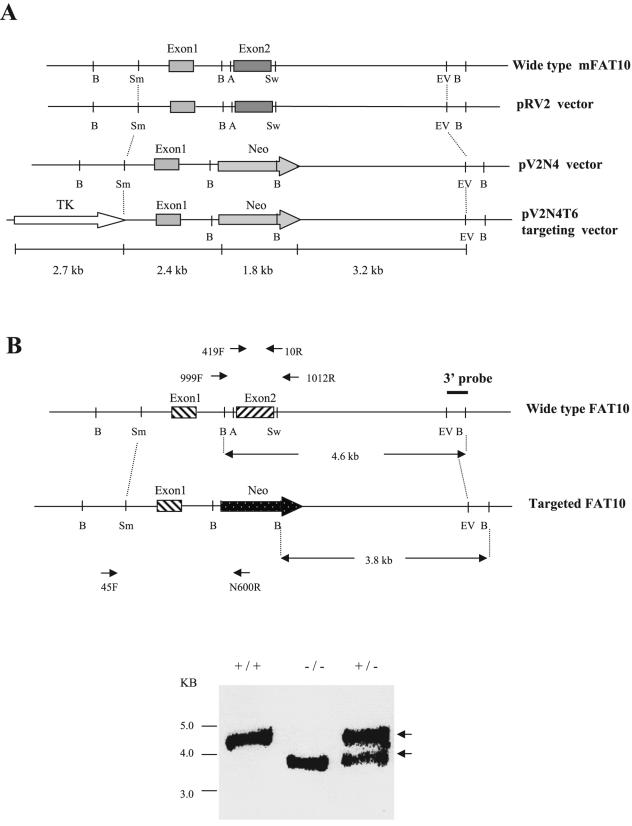
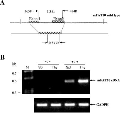
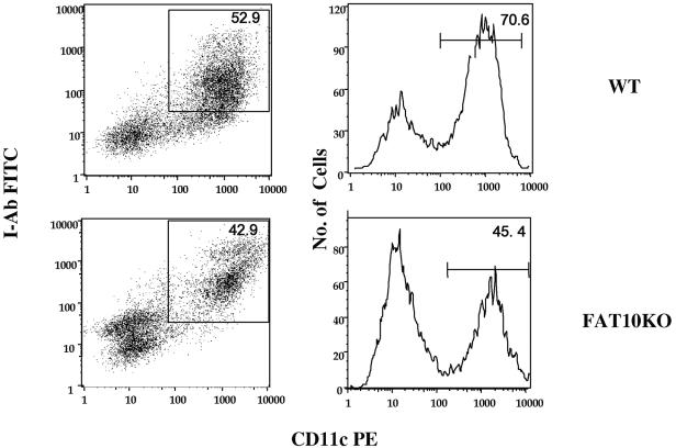
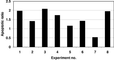
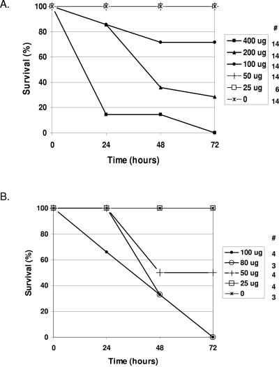
The FAT10 gene encodes a diubiquitin-like protein containing two tandem head-to-tail ubiquitin-like domains. There is a high degree of similarity between murine and human FAT10 sequences at both the mRNA and protein levels. In various cell lines, FAT10 expression was shown to be induced by gamma interferon or by tumor necrosis factor alpha. In addition, FAT10 expression was found to be up-regulated in some Epstein-Barr virus-infected B-cell lines, in activated dendritic cells, and in several epithelial tumors. However, forced expression of FAT10 in cultured cells was also found to produce apoptotic cell death. Overall, these findings suggest that FAT10 may modulate cellular growth or cellular viability. Here we describe the steps to generate, by genetic targeting, a FAT10 gene knockout mouse model. The FAT10 knockout homozygous mice are viable and fertile. No gross lesions or obvious histological differences were found in these mutated mice. Examination of lymphocyte populations from spleen, thymus, and bone marrow did not reveal any abnormalities. However, flow cytometry analysis demonstrated that the lymphocytes of FAT10 knockout mice were, on average, more prone to spontaneous apoptotic death. Physiologically, these mice demonstrated a high level of sensitivity toward endotoxin challenge. These findings indicate that FAT10 may function as a survival factor.


Figures

FIG. 1.
FIG. 1.
Amino acid sequence comparison of human and mouse FAT10 proteins with human ubiquitin protein. The human and mouse FAT10 amino acid sequences are indicated as HFAT10 and MFAT10, respectively, to the left of the sequences. The shaded amino acids are identical. Gaps introduced to maximize alignment (∼) are indicated. *, stop condon.
FIG. 2.
FIG. 2.
DNA sequence analysis reveals high degree of similarity between human and mouse FAT10 for expression regulation. The human and mouse FAT10 sequences that were compared encompassed the two exons, the intermediate intron, and a 2-kb upstream sequence that includes the 5′ untranslated region (5′UTR) and the promoter regions. TRANSFAC analysis (http://www.gene-regulation.com/pub/databases.html) was performed for the regulatory binding sites. NF-κB, IRF, and AP-1 are shown schematically for both strands.
FIG. 3.
FIG. 3.
RT-PCR analysis of FAT10 expression from total RNA from various mouse tissues. A. Schematic representation of the locations of RT-PCR primers on the mouse FAT10 gene. Intron-spanning primers were designed to eliminate the formation of genomic amplicons. B. Gel electrophoresis analysis of various mouse tissues for FAT10 gene expression. The origins of tissues are indicated above the gel, and the positions of the DNA markers are indicated to the left of the gel.
FIG. 4.
FIG. 4.
Northern blot analysis of FAT10 mRNA expression in different mouse tissues. A. The FAT10 mRNA expression pattern in different mouse tissues. The origins of tissues are indicated above the gel, and the positions of the kilobase markers are indicated to the left of the gel. B. GADPH probe hybridized to the same membrane as in panel A for RNA loading controls.
FIG. 5.
FIG. 5.
Northern blot analysis of human FAT10 mRNA. A. Northern blot of multiple tissues from healthy humans. Lane 1, spleen; 2, thymus; 3, prostate; 4, testis; 5, ovary; 6, small intestine; 7, colon mucosal lining; 8, peripheral blood leukocytes. Lane M contains molecular size markers. B. Analysis of total RNA blots from various cell lines. The origins of cell lines are indicated above the gel, and the two rRNA bands are indicated as 28S and 18S to the left of the gel. C. IFN-γ treatment for 0, 18, and 36 h induces progressive increases in FAT10 mRNA expression in HeLa and JY human cell lines.
FIG. 6.
FIG. 6.
Disruption of murine FAT10 expression by gene targeting. A. Schematic cascade of the constructs generated in the process of generating the targeting vector pV2N4T6. Neomycin (Neo) expression cassette replaced the second exon of FAT10, and a thymidine kinase (TK) gene was placed upstream of the first exon of FAT10. Restriction enzyme sites: A, AvrII; B, BamHI; EV, EcoRV; Sm, SmaI; Sw, SwaI. B. Screening of ES cells for FAT10 disruption was carried out by Southern blotting and PCR analysis. Following BamHI digestion, the three external probes hybridized to a 4.6-kb fragment in the wild-type mice (+/+) and to a 3.8-kb fragment in the FAT10 knockout mice (−/−). The two fragments appeared in the heterozygous ES cell (+/−). PCR screenings for homozygous mice (−/−) and wild-type mice (+/+) were done using the oligonucleotide pair: 999F/1012R generated amplicons of 2.1 and 1.2 kb, respectively. Both amplicons were generated for the heterozygote. However, the use of the oligonucleotide pair 45F/N600R resulted in a 2.9-kb amplicon that was shared only by FAT10 knockout mice (−/−) and heterozygous mice.
FIG. 7.
FIG. 7.
>RT-PCR of mouse FAT10 cDNA from total RNA from FAT10 knockout mice. A. Schematic representation of mouse FAT10 (mFAT10) 165F/424R primers used for RT-PCR. B. RT-PCR of mouse spleen (Spl) and thymus (Thy). The expected 530-bp RT-PCR products were found in the wild-type mouse (+/+) thymus and spleen but not in the FAT10 knockout mouse (−/−) thymus and spleen. Lane M contains molecular size markers. The 500-bp band of GADPH was used as an internal control.
FIG. 8.
FIG. 8.
FACS analysis of FAT10 knockout mouse dendritic cell population in vitro. FACS analysis of dendritic cells cultured from the bone marrow of wild-type (WT) and FAT10 knockout (FAT10KO) mice. For details of the procedure, see text and Materials and Methods. CD11c PE, phycoerythrin-labeled CD11c.
FIG. 9.
FIG. 9.
Leukocytes from FAT10-deficient mice demonstrate a higher level of apoptotic death. Bars represent the ratio of apoptotic leukocyte populations from FAT10-deficient mice to that of the FAT10-expressing control. Leukocytes from experiment 2 were obtained from bone marrow, while experiments 3 and 8 were performed on thymocytes. All other experiments were done on splenocytes. Analysis of variance analysis was performed on the raw data (two factors without replication, P = 0.042).
FIG. 10.
FIG. 10.
FAT10 knockout mice are highly susceptible to endotoxin administration. Mice were subjected to various doses of endotoxin (from 0 to 400 μg). Mortality was evaluated daily, and the initial numbers of mice in each group are indicated under the # sign. Survival of C57BL/6 mice (A) and FAT10 knockout mice (B) is shown.

References

    1. Amerik, A. Y., J. Nowak, S. Swaminathan, and M. Hochstrasser. 2000. The Doa4 deubiquitinating enzyme is functionally linked to the vacuolar protein-sorting and endocytic pathways. Mol. Biol. Cell 11:3365-3380. - PMC - PubMed
    1. Bates, E. E., O. Ravel, M. C. Dieu, S. Ho, C. Guret, J. M. Bridon, S. Ait-Yahia, F. Briere, C. Caux, J. Banchereau, and S. Lebecque. 1997. Identification and analysis of a novel member of the ubiquitin family expressed in dendritic cells and mature B cells. Eur. J. Immunol. 27:2471-2477. - PubMed
    1. Biggins, S., I. Ivanovska, and M. D. Rose. 1996. Yeast ubiquitin-like genes are involved in duplication of the microtubule organizing center. J. Cell Biol. 133:1331-1346. - PMC - PubMed
    1. Boddy, M. N., K. Howe, L. D. Etkin, E. Solomon, and P. S. Freemont. 1996. PIC 1, a novel ubiquitin-like protein which interacts with the PML component of a multiprotein complex that is disrupted in acute promyelocytic leukaemia. Oncogene 13:971-982. - PubMed
    1. Bryant, N. J., and T. H. Stevens. 1998. Vacuole biogenesis in Saccharomyces cerevisiae: protein transport pathways to the yeast vacuole. Microbiol. Mol. Biol. Rev. 62:230-247. - PMC - PubMed