. 2006 Aug;148(8):1099-107.
doi: 10.1038/sj.bjp.0706817. Epub 2006 Jun 19.

Acetyl-keto-beta-boswellic acid inhibits cellular proliferation through a p21-dependent pathway in colon cancer cells

Affiliations

Acetyl-keto-beta-boswellic acid inhibits cellular proliferation through a p21-dependent pathway in colon cancer cells

Jian-Jun Liu et al. Br J Pharmacol. 2006 Aug.

Abstract

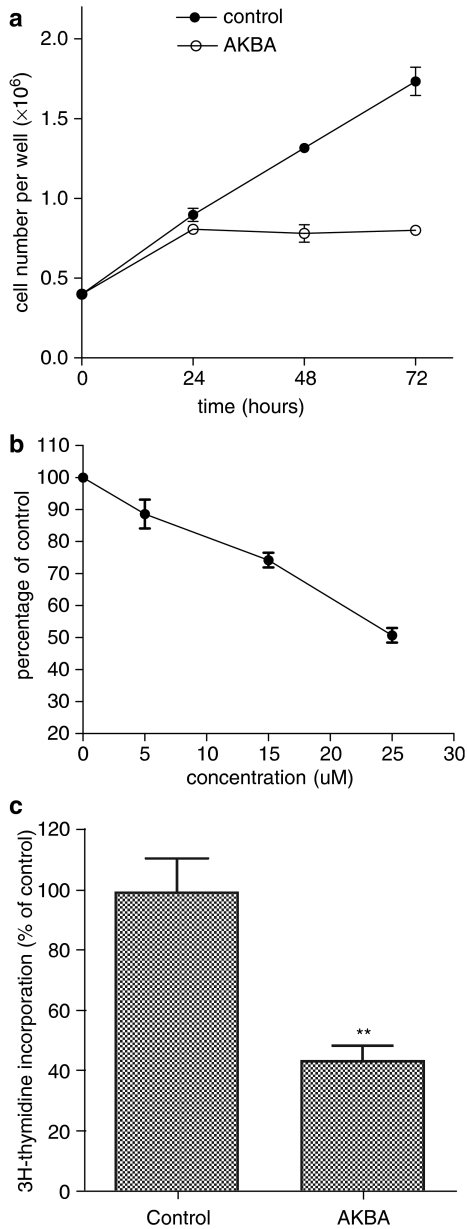
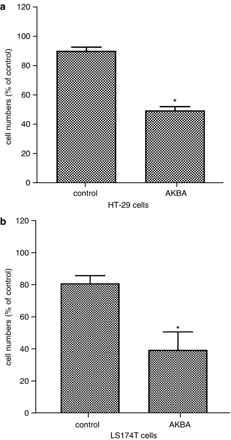
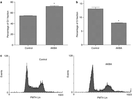
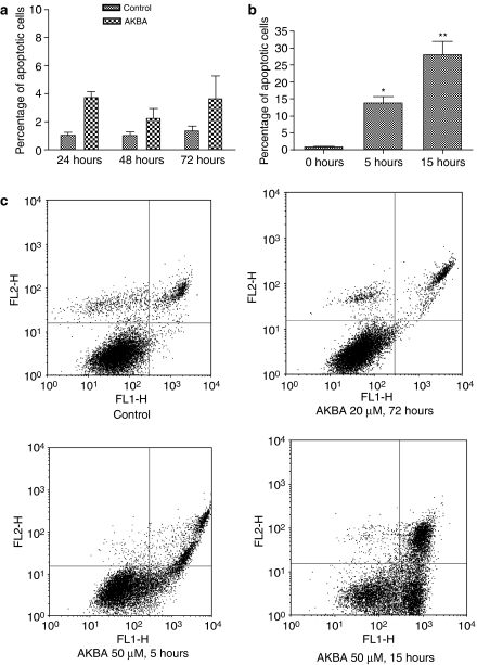
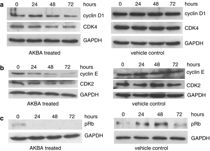
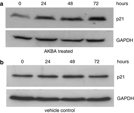
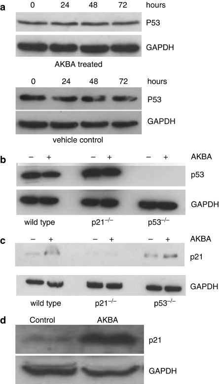
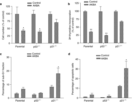
1. Although there is increasing evidence showing that boswellic acid might be a potential anticancer agent, the mechanisms involved in its action are unclear. 2. In the present study, we showed that acetyl-keto-beta-boswellic acid (AKBA) inhibited cellular growth in several colon cancer cell lines. Cell cycle analysis by flow cytometry showed that cells were arrested at the G1 phase after AKBA treatment. 3. Further analysis showed that cyclin D1 and E, CDK 2 and 4 and phosphorylated Rb were decreased in AKBA-treated cells while p21 expression was increased. 4. The growth inhibitory effect of AKBA was dependent on p21 but not p53. HCT-116 p53(-/-) cells were sensitized to the apoptotic effect of AKBA, suggesting that p21 may have protected cells against apoptosis by inducing a G1 arrest.5. In conclusion, we have demonstrated that AKBA inhibited cellular growth in colon cancer cells. These findings may have implications to the use of boswellic acids as potential anticancer agents in colon cancer.


Figures

Figure 1
Figure 1
AKBA inhibited growth of HCT-116 cells. (a) Changes in cell numbers after treatment with AKBA. HCT-116 ls were treated with 20 μM AKBA for 24, 48 and 72 h and cell numbers was counted using a hemacytometer; (b) changes in cellular viability after treatment with different concentrations of AKBA. HCT-116 cells were treated with 0, 5, 15 and 25 μM AKBA for 48 h. Cell viability was assayed using WST-1; (c) Changes in DNA synthesis after treatment with AKBA. HCT-116 cells were treated with 20 μM AKBA for 48 h. DNA synthesis was assayed using the 3H-thymidine incorporation assay. Control cells were treated with 0.2% ethanol. Results are expressed as mean±s.d. and are representative of at least three separate experiments. **P<0.01 compared to control.
Figure 2
Figure 2
AKBA inhibited growth of HT-29 and LS174T cells. HT-29 and LS174T cells were treated with 30 and 20 μM AKBA for 48 h, respectively. Control cells were treated with 0.2% ethanol. Cell numbers were counted using a hemacytometer. (a) Changes in cell numbers after treatment with AKBA in HT-29 cells; (b) changes in cell numbers after treatment with AKBA in LS174T cells. Results are shown as mean±s.d. *P<0.05 compared to control.
Figure 3
Figure 3
AKBA caused a G1 arrest in HCT-116 cells. HCT1-116 cells were treated with 20 μM AKBA for 48 h. DNA was stained with propidium iodide and analyzed by flow cytometry. (a) Percentage of G1 phase cells in AKBA treated and control cells; (b) percentage of S phase cells in AKBA treated and control cells; (c) representative flow cytometric histogram in AKBA treated and control cells. Experiments were performed in triplicates. Results are expressed as mean±s.d. and are representative of at least three separate experiments. *P<0.05 compared to control.
Figure 4
Figure 4
Apoptosis assayed by annexin V and propidium iodide staining. (a) Percentage of apoptotic HCT-116 cells after treatment with 20 μM AKBA for 24, 48 and 72 h; (b) percentage of apoptotic HCT-116 cells after treatment with 50 μM AKBA for 5 and 15 h; (c) representative density plots of flow cytometric data. Results are expressed as mean±s.d. and are representative of at least three separate experiments. *P<0.05 compared to control. **P<0.01 compared to control.
Figure 5
Figure 5
AKBA regulated the expression of G1 phase cyclins and CKDs in HCT-116 cells. HCT-116 cells were treated with 20 μM AKBA for 0, 24, 48 and 72 h. The expression of cyclin D1, cyclin E, CDK2, CDK4 and phophorylated Rb (pRb) were assayed by Western blot analysis. GAPDH was used as a housekeeping control. (a) Expression of cyclin D1 and CDK4 after treatment with AKBA; (b) expression of cyclin E and CDK2 after treatment with AKBA; (c) expression of pRb after treatment with AKBA. Results shown are representative of at least three separate experiments.
Figure 6
Figure 6
Expression of p21 in AKBA treated cells. HCT-116 cells were treated with 20 μM AKBA for 0, 24, 48 and 72 h. The expression of p21 was assayed by Western blot analysis. GAPDH was used as a housekeeping control. (a) Expression of p21 in HCT-116 cells after treatment with AKBA; (b) expression of p21 in HCT-116 cells treated with vehicle control. Results shown are representative of at least three separate experiments.
Figure 7
Figure 7
Expression of p21 and p53 in colon cancer cells after AKBA treatment. (a) Expression of p53 in HCT-116 cells treated with AKBA and vehicle control. HCT-116 cells were treated with 20 μM AKBA for 0, 24, 48 and 72 h. Expression of p53 was assayed by Western blot analysis; (b) expression of p53 in parental, p21−/− and p53−/− HCT-116 cells after treatment with AKBA. Cells were treated with 20 μM AKBA for 48 h. Expression of p53 was assayed by Western blot analysis. Control cells were treated with 0.2% ethanol; (c) changes in p21 expression in parental, p21−/− and p53−/− HCT-116 cells after treatment with AKBA. Cells were treated with 20 μM AKBA for 48 h. Expression of p21 was assayed by Western blot analysis; (d) changes in p21 expression in HT-29 cells after treatment with AKBA. HT-29 cells were treated with 30 μM AKBA for 72 h. Expression of p21 was assayed by Western blot analysis. Control cells were treated with 0.2% ethanol. Results shown are representative of three separate experiments. Expression of GAPDH was used as a housekeeping control.
Figure 8
Figure 8
Effect of AKBA on growth and apoptosis in parental, p21−/− and p53−/− HCT-116 cells. Cells were treated with 20 μM AKBA for 48 h. Control cells were treated with 0.1% ethanol. The number of cells was counted using a hemacytometer. DNA synthesis was assayed by 3H-thymidine incorporation. Apoptosis was assayed by both cell cycle analysis and annexin V staining. (a) Changes in cell numbers after treatment with AKBA in parental, p21−/− and p53−/− HCT-116 cells; (b) changes in DNA synthesis in parental, p21−/− and p53−/− HCT-116 cells after treatment with AKBA; (c) changes in sub-G1 fraction after treatment with AKBA in parental, p21−/− and p53−/− HCT-116 cells; (d) changes in apoptotic rate assayed by annexin V and propidium iodide double staining in parental, p21−/− and p53−/− HCT-116 cells. Results shown are representative of three separate experiments. *P<0.05 compared to control. **P<0.01 compared to control.

Similar articles

Cited by

References

    1. CAPUTI M., RUSSO G., ESPOSITO V., MANCINI A., GIORDANO A. Role of cell-cycle regulators in lung cancer. J. Cell Physiol. 2005;205:319–327. - PubMed
    1. GARTEL A.L., RADHAKRISHNAN S.K. Lost in transcription: p21 repression, mechanisms, and consequences. Cancer Res. 2005;65:3980–3985. - PubMed
    1. GARTEL A.L., TYNER A.L. The role of the cyclin-dependent kinase inhibitor p21 in apoptosis. Mol. Cancer Ther. 2002;1:639–649. - PubMed
    1. GERHARDT H., SEIFERT F., BUVARI P., VOGELSANG H., REPGES R. Therapy of active Crohn disease with Boswellia serrata extract H 15] Z. Gastroenterol. 2001;39:11–17. - PubMed
    1. GLASER T., WINTER S., GROSCURTH P., SAFAYHI H., SAILER E.R., AMMON H.P., SCHABET M., WELLER M. Boswellic acids and malignant glioma: induction of apoptosis but no modulation of drug sensitivity. Br. J. Cancer. 1999;80:756–765. - PMC - PubMed

Publication types