. 2006 Jun 27;45(25):7872-81.
doi: 10.1021/bi060683g.

Activation of the global gene regulator PrrA (RegA) from Rhodobacter sphaeroides

Affiliations

Activation of the global gene regulator PrrA (RegA) from Rhodobacter sphaeroides

Cédric Laguri et al. Biochemistry. .

Abstract

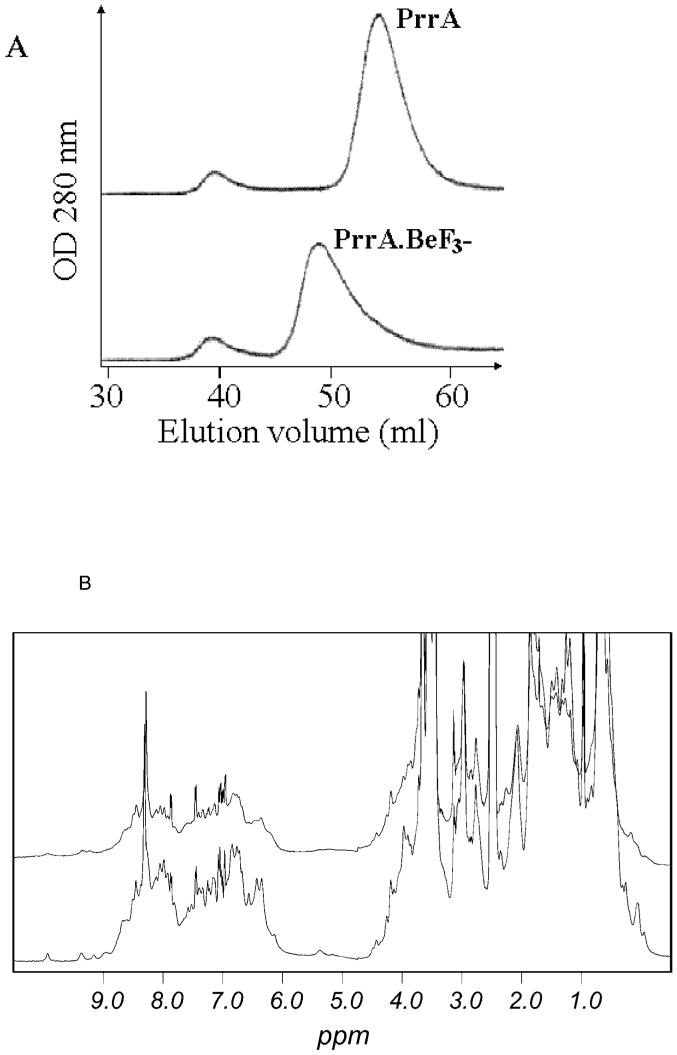
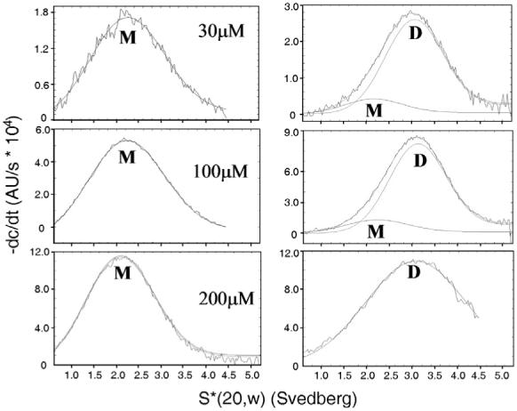
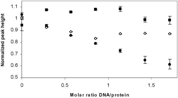
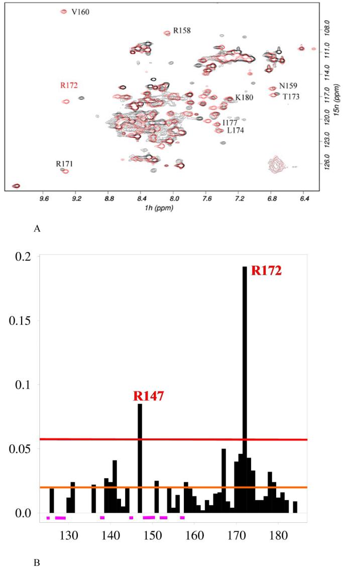
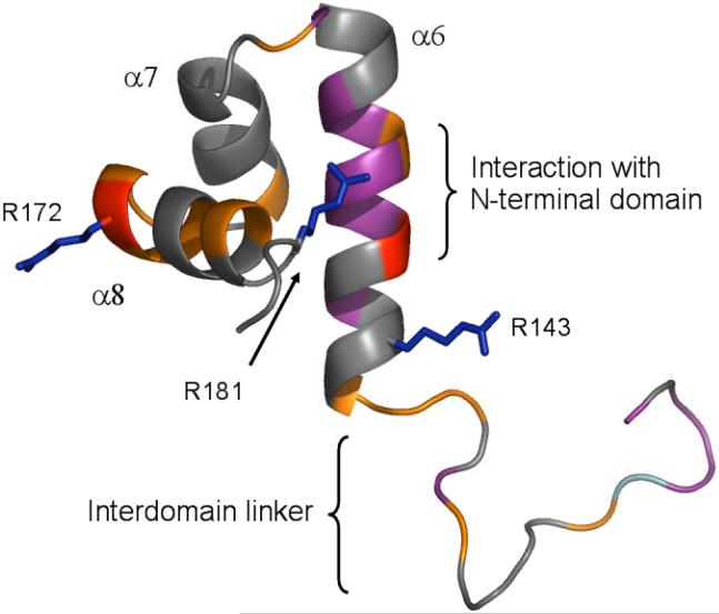
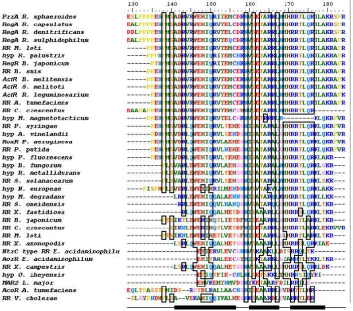
PrrA is a global transcription regulator activated upon phosphorylation by its cognate kinase PrrB in response to low oxygen levels in Rhodobacter sphaeroides. Here we show by gel filtration, analytical ultracentrifugation, and NMR diffusion measurements that treatment of PrrA with a phosphate analogue, BeF(3)(-), results in dimerization of the protein, producing a protein that binds DNA. No dimeric species was observed in the absence of BeF(3)(-). Upon addition of BeF(3)(-), the inhibitory activity of the N-terminal domain on the C-terminal DNA-binding domain is relieved, after which PrrA becomes capable of binding DNA as a dimer. The interaction surface of the DNA-binding domain with the regulatory domain of PrrA is identified by NMR as being a well-conserved region centered on helix alpha6, which is on the face opposite from the DNA recognition helix. This suggests that there is no direct blockage of DNA binding in the inactive state but rather that PrrA dimerization promotes a correct arrangement of two adjacent DNA-binding domains that recognizes specific DNA binding sequences.


Figures

Figure 1
Figure 1
Dimerisation of PrrA upon addition of BeF3-. A: Superdex G75 gel filtration profile of PrrA and PrrA.BeF3-. PrrA (purified as the monomer) and PrrA.BeF3- were injected onto the column. Monomer and dimer are eluted at their expected molecular weight; the dimer elutes at the same volume as the minor species from the E. coli growth. Some protein aggregates are formed during concentration and are eluted in the void volume (39 ml). The dimer peak has a “tail” suggesting the presence of a low quantity of monomer (<10 %) or an exchange during gel filtration between monomeric and dimeric species. The column was calibrated using standards of known molecular weights: bovine serum albumin (66 kDa), ovalbumin (45 kDa), carbonic anhydrase (26 kDa), lysozyme (14.3 kDa) and insulin (5.8 kDa). B: 1H 1D spectra of PrrA and PrrA.BeF3-. Spectra of PrrA (bottom) and PrrA.BeF3- (top) (500 μM) were recorded at 2°C under identical conditions and overlaid. The spectrum of PrrA.BeF3- shows a large broadening of the NMR signal resulting in an important decrease in signal intensity. Dimer purified from E. coli has a very similar NMR spectrum (not shown).
Figure 2
Figure 2
Velocity analytical centrifugation of PrrA and PrrA.BeF3- at different protein concentrations. Fitting of sedimentation coefficients was calculated with dcdt+ at the different protein concentrations indicated. Left: PrrA. Right: PrrA.BeF3-. The overall fitted functions and the individual functions corresponding to monomeric and dimeric species are indicated. The difference between the fitted function and original data (residual) showed good fits in all cases.
Figure 3
Figure 3
Changes in peak height in the methyl region of the NMR spectrum (0.5 - 0.9 ppm) of full-length PrrA (squares), PrrA.BeF-3 (open diamonds) and PrrAC (circles) on binding to the cycAP2 promoter. The changes are a result of line broadening, and have been normalized to an approximately equal starting height. Because free PrrAC has narrower lines than does PrrA.BeF3-, its changes in height on DNA binding are expected to be greater for the same degree of binding. Thus, the absolute changes in height are not readily interpretable, and the apparent similarity of the slopes for PrrAC and PrrA.BeF3- is coincidental.
Figure 4
Figure 4
Comparison of PrrA C-terminal domain spectra in the full-length protein and in PrrAC. A: Overlaid 15N-HSQC spectra of PrrA (black) and PrrAC(red), obtained in identical solution conditions. Signals from the C-terminal domain comprise most of the signals observed in full-length PrrA NMR spectra. PrrAC residue differences in NH peak positions from the backbone but also sidechain resonances from asparagines, glutamines and arginines can be observed on the 15N HSQCs. Most PrrAC resonances are easily identifiable in full-length PrrA and suggest the C-terminal domain is in a very similar conformation to that in the structure of PrrAC determined previously. A few examples are shown on the figure. Several peaks from the C-terminal domain could not be found without ambiguity, presumably because they experience large chemical shift variation. B: Backbone NH chemical shift differences of PrrAC between the isolated domain and full-length PrrA. Chemical shift differences are represented, as a function of residue, from the overlaid 15N-HSQC in A. Differences are weighted as [(δH)2 + (δN/10)2]1/2. In purple are represented PrrAC signals that unambiguously could not be found in the PrrA spectrum. The red and orange lines represent the lower limits defined for large and medium chemical shift differences respectively. These differences are represented in Fig. 5 on the PrrAC structure.
Figure 5
Figure 5
Chemical shift differences between PrrAC in isolation and in full length PrrA. In red are represented large HN backbone chemical shift changes and in purple peaks that could not be found in the PrrA spectrum. Medium backbone chemical shift changes are shown in orange. R143, R172 and R181 Hε experience large chemical shift changes and their sidechains are represented in blue. The end of the PrrA N-terminal domain (end of helix α5) is shown in cyan. The W146 peak was too weak to follow its variation.
Figure 6
Figure 6
Alignment of PrrAC with homologous RR effector domains and hypothetical proteins. Conservation >70% is outlined by boxes. PrrAC secondary structure (helices α6-8) is indicated at the bottom of the figure. The conservation occurs mainly in the first loop and the helix-turn-helix motif. Residues shown to be involved in DNA binding in the first loop (binding to phosphate backbone) and at the beginning of α8 (specific binding to bases) are highly conserved. Hyp: hypothetical protein. Species not otherwise given in the text are: Mesorhizobium loti, Rhodopseudomonas loti, Brucella suis, Brucella melitensis, Rhizobium leguminosarium, Agrobacterium tumefaciens, Caulobacter crescentus, Magnetospirillum magnetotacticum, Azotobacter vinelandii, Ralstonia metallidurans, Ralstonia solanacearum, Microbulbifer degradans, Shewanella oneidensis, Xylella fastidiosa, Eubacterium acidaminophilum, Xanthomonas campestris, Oceanobacillus iheyensis, Leishmania major.

Similar articles

Cited by

References

    1. Swem LR, Elsen S, Bird TH, Swem DL, Koch HG, Myllykallio H, Daldal F, Bauer CE. The RegB/RegA two-component regulatory system controls synthesis of photosynthesis and respiratory electron transfer components in Rhodobacter capsulatus. J. Mol. Biol. 2001;309:121–138. - PubMed
    1. Oh JI, Kaplan S. Redox signaling: globalization of gene expression. EMBO J. 2000;19:4237–4247. - PMC - PubMed
    1. Elsen S, Swem LR, Swem DL, Bauer CE. RegB/RegA, a highly conserved redox-responding global two- component regulatory system. Microbiol. Mol. Biol. Rev. 2004;68:263–279. - PMC - PubMed
    1. Elsen S, Dischert W, Colbeau A, Bauer CE. Expression of uptake hydrogenase and molybdenum nitrogenase in Rhodobacter capsulatus is coregulated by the RegB-RegA two- component regulatory system. J. Bact. 2000;182:2831–2837. - PMC - PubMed
    1. Laratta WP, Choi PS, Tosques IE, Shapleigh JP. Involvement of the PrrB/PrrA two-component system in nitrite respiration in Rhodobacter sphaeroides 2.4.3: evidence for transcriptional regulation. J. Bact. 2002;184:3521–3529. - PMC - PubMed

Publication types

MeSH terms

LinkOut - more resources