. 2006;8(4):R98.
doi: 10.1186/ar1978.

Relaxin and beta-estradiol modulate targeted matrix degradation in specific synovial joint fibrocartilages: progesterone prevents matrix loss

Affiliations

Relaxin and beta-estradiol modulate targeted matrix degradation in specific synovial joint fibrocartilages: progesterone prevents matrix loss

Gihan Hashem et al. Arthritis Res Ther. 2006.

Abstract

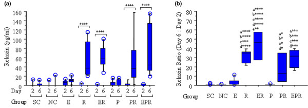
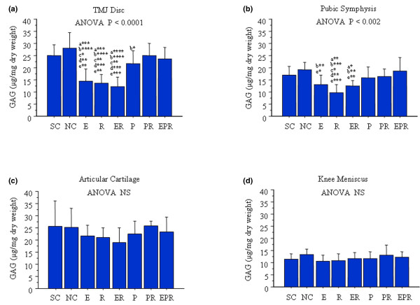
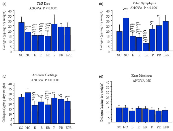
Relaxin, a 6-kDa polypeptide hormone, is a potent mediator of matrix turnover and contributes to the loss of collagen and glycosaminoglycans (GAGs) from reproductive tissues, including the fibrocartilaginous pubic symphysis of several species. This effect is often potentiated by beta-estradiol. We postulated that relaxin and beta-estradiol might similarly contribute to the enhanced degradation of matrices in fibrocartilaginous tissues from synovial joints, which may help explain the preponderance of diseases of specific fibrocartilaginous joints in women of reproductive age. The objective of this study was to compare the in vivo effects of relaxin, beta-estradiol, and progesterone alone or in various combinations on GAG and collagen content of the rabbit temporomandibular joint (TMJ) disc fibrocartilage, knee meniscus fibrocartilage, knee articular cartilage, and the pubic symphysis. Sham-operated or ovariectomized female rabbits were administered beta-estradiol (20 ng/kg body weight), progesterone (5 mg/kg), or saline intramuscularly. This was repeated 2 days later and followed by subcutaneous implantation of osmotic pumps containing relaxin (23.3 microg/kg) or saline. Tissues were retrieved 4 days later and analyzed for GAG and collagen. Serum relaxin levels were assayed using enzyme-linked immunosorbent assay. Relaxin administration resulted in a 30-fold significant (p < 0.0001) increase in median levels (range, approximately 38 to 58 pg/ml) of systemic relaxin. Beta-estradiol, relaxin, or beta-estradiol + relaxin caused a significant loss of GAGs and collagen from the pubic symphysis and TMJ disc and of collagen from articular cartilage but not from the knee meniscus. Progesterone prevented relaxin- or beta-estradiol-mediated loss of these molecules. The loss of GAGs and collagen caused by beta-estradiol, relaxin, or beta-estradiol + relaxin varied between tissues and was most prominent in pubic symphysis and TMJ disc fibrocartilages. The findings suggest that this targeted modulation of matrix loss by hormones may contribute selectively to degeneration of specific synovial joints.


Figures

Figure 1
Figure 1
Relaxin (R) administration increases serum R concentrations, which is enhanced by β-estradiol (E) and attenuated by progesterone (P). Systemic R concentrations were assayed in serum from sham-operated controls (SC), ovariectomized normal controls (NC), and rabbits treated with E, R, E + R (ER), P, P + R (PR), and E + P + R (EPR) groups on day 2 and day 6, respectively, using an enzyme-linked immunosorbent assay as described. (a) Histograms of the median with inter-quartile value showed that there was no statistically significant difference in the basal levels of R on day 2 between any of the groups. Additionally, the SC, NC, E, and P groups showed no statistically significant differences in R concentrations between day 2 and day 6. R administration resulted in a significant increase of systemic R concentration on day 6 in R, ER, PR, and EPR groups compared with the corresponding basal levels of R on day 2. (b) The fold change in R was determined by normalizing the day-6 concentration to the corresponding day-2 concentration of R for each animal, and the median with inter-quartile value was plotted for each group of animals. The fold increase in R was significantly greater in R (35-fold), ER (46-fold), PR (13-fold), and EPR (32-fold) groups compared with that in SC, NC, E, and P groups. No statistically significant difference of R ratio was found between any of the R-treated groups, with the exception of ER versus PR groups (p < 0.01). Data were collected from a minimum of six rabbits in each group and expressed as median with inter-quartile value. (* < 0.05; ** < 0.01; *** < 0.001; **** < 0.0001; a, versus SC; b, versus NC; c, versus E; d, versus P; e, versus PR.)
Figure 2
Figure 2
β-estradiol (E) or relaxin (R) treatment causes loss of GAGs from specific fibrocartilages while progesterone (P) inhibits this effect. Temporomandibular joint (TMJ) discs, pubic symphysis, knee meniscus, and articular cartilage were retrieved from control and hormone-treated rabbits, and total GAG content was determined by dimethylmethylene blue assay and normalized to the total dry weight for each sample. Histograms of the mean (± standard deviation) GAG concentration in sham-operated controls (SC), ovariectomized normal controls (NC), and rabbits treated with E, R, ER, P, P + R (PR), and E + P + R (EPR) groups were plotted. Ovariectomy had minimal effect on total GAGs present in any of these four tissues. E, R, and ER produced a significant reduction of GAGs in TMJ disc (a) and pubic symphysis (b) relative to SC and NC groups. P alone contributed to the maintenance of GAGs in all these tissues or prevented E-, R-, or ER-mediated loss of GAGs from both TMJ disc and pubic symphysis. None of these hormone treatments caused a significant change of GAGs in knee articular cartilage (c) and meniscus (d). Data were collected from a minimum of six rabbits in each group. (* < 0.05; ** < 0.01; *** < 0.001; **** < 0.0001; a, versus SC; b, versus NC; c, versus P; d, versus PR; e, versus EPR.) ANOVA, analysis of variance; NS, not significant.
Figure 3
Figure 3
β-estradiol (E), relaxin (R), and progesterone (P) treatment have differential effects on the loss of collagen in fibrocartilages. Temporomandibular joint (TMJ) discs, pubic symphysis, knee meniscus, and articular cartilage were retrieved from control and hormone-treated rabbits, and the total collagen content was determined by the Sircol assay and normalized to the total dry weight for each sample. Histograms of the mean (± standard deviation) collagen concentration in sham-operated controls (SC), ovariectomized normal controls (NC), and rabbits treated with E, R, E + R (ER), P, P + R (PR), and E + P + R (EPR) groups were plotted. Ovariectomy resulted in a significant reduction of collagen content in TMJ disc (a) and increase collage in the pubic symphysis (b) but had no effect on collagen in knee articular cartilage (c) and meniscus (d). Administration of E, R, and ER maintained the reduction of collagen content in TMJ disc produced by ovariectomy and caused a significant reduction in collagen in pubic symphysis relative to NC and in knee articular cartilage relative to SC and NC. In contrast, progesterone antagonized the effects of E, R, and ER on collagen content in TMJ disc, pubic symphysis, and the knee articular cartilage. Collagen content in knee meniscus was not affected by treatment with any of these hormones. Data were collected from a minimum of six rabbits in each group. (* < 0.05; ** < 0.01; *** < 0.001; **** < 0.0001; a, versus SC; b, versus NC; c, versus P; d, versus PR; e, versus EPR; NS, not significant.)

References

    1. Buckwalter JA, Mankin HJ, Grodzinsky AJ. Articular cartilage and osteoarthritis. Instructional Course Lectures. 2005;54:465–480. - PubMed
    1. Bramono DS, Richmond JC, Weitzel PP, Kaplan DL, Altman GH. Matrix metalloproteinases and their clinical applications in orthopaedics. Clin Orthop Relat Res. 2004;428:272–285. doi: 10.1097/01.blo.0000144166.66737.3a. - DOI - PubMed
    1. Ushiyama T, Inoue K, Nishioka J. Expression of estrogen receptor related protein (p29) and estradiol binding in human arthritic synovium. J Rheumatol. 1995;22:421–426. - PubMed
    1. Khalkhali-Ellis Z, Seftor EA, Nieva DR, Handa RJ, Price RH, Jr, Kirschmann DA, Baragi VM, Sharma RV, Bhalla RC, Moore TL, Hendrix MJ. Estrogen and progesterone regulation of human fibroblast-like synoviocytes function in vitro: implications in rheumatoid arthritis. J Rheumatol. 2000;27:1622–1631. - PubMed
    1. Kapila S, Xie YQ. Targeted induction of collagenase and stromelysin by relaxin in unprimed and β-estradiol-primed diarthrodial joint fibrocartilaginous cells but not in synoviocytes. Lab Invest. 1998;78:925–938. - PubMed

Publication types

MeSH terms