. 2006 Jul 10;203(7):1733-44.
doi: 10.1084/jem.20051715. Epub 2006 Jun 19.

Phosphoinositide-dependent kinase 1 targets protein kinase A in a pathway that regulates interleukin 4

Affiliations

Phosphoinositide-dependent kinase 1 targets protein kinase A in a pathway that regulates interleukin 4

Ajay Nirula et al. J Exp Med. .

Abstract

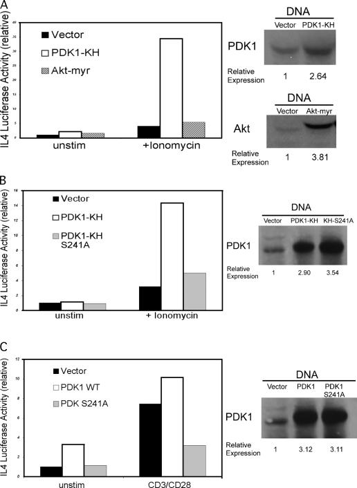
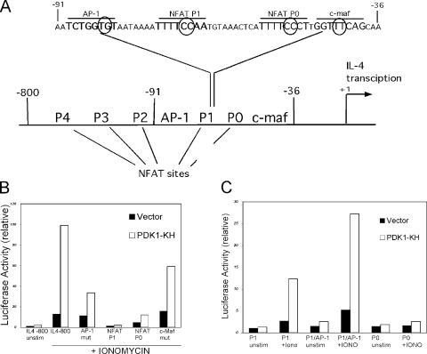
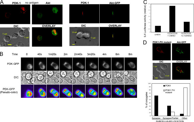
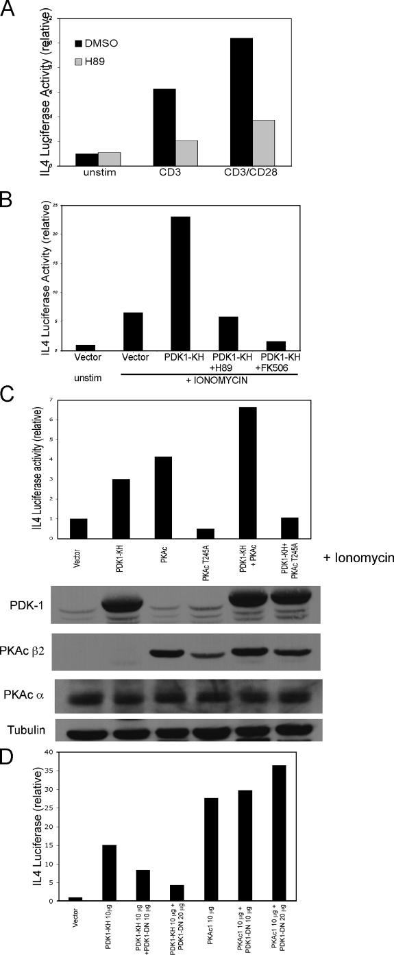
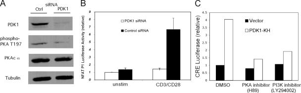
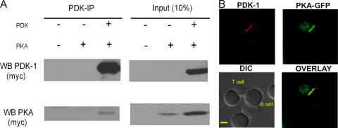
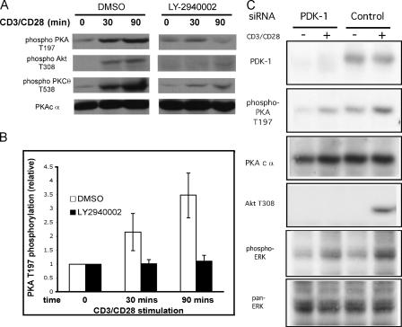
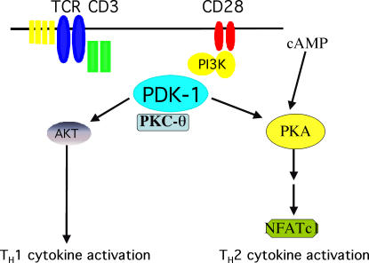
CD28 plays a critical role in T cell immune responses. Although the kinase Akt has been shown to act downstream of CD28 in T helper (Th)1 cytokine induction, it does not induce Th2 cytokines such as interleukin 4 (IL-4). We recently reported that phosphoinositide-dependent kinase 1 (PDK1) partially corrects the defect in IL-4 production present in CD28-deficient T cells, suggesting that PDK1 regulates IL-4 independently of Akt. We now describe a signaling pathway in which PDK1 targets IL-4 in the murine Th2 cell line D10. PDK1-mediated activation of this pathway is dependent on protein kinase A (PKA) and the nuclear factor of activated T cells (NFAT) P1 transcriptional element in the IL-4 promoter. PDK1 localizes to the immune synapse in a phosphatidylinositol 3-kinase-dependent manner, partially colocalizes with PKA at the synapse, and physically interacts with PKA. In RNA interference knockdown experiments, PDK1 is necessary for phosphorylation of PKA in T cells, as well as for activation of the IL-4 NFAT P1 element by the T cell receptor (TCR) and CD28. Phosphorylation of the critical PKA threonine residue is stimulated by engagement of TCR/CD28 via a PDK1-dependent mechanism. These findings together define a pathway linking the kinases PDK1 and PKA in the induction of the Th2 cytokine IL-4.


Figures

Figure 1.
Figure 1.
PDK1 but not Akt activates the IL-4 promoter. (A) PDK1-KH or a myristoylated membrane targeted form of Akt (Akt myr) was cotransfected with an IL-4 −800 Luciferase transcriptional reporter in the D10 cell line. 16 h later, 105 cells were either left unstimulated or were treated with ionomycin (0.5 μM) and were assayed for luciferase activity 6 h later. Luciferase data are shown relative to unstimulated IL-4 (or other reporter) luciferase activity (relative activity = 1) throughout the paper unless otherwise stated. A representative experiment is shown with all such experiments shown in this manuscript repeated at least three times. The mean result for each sample (performed in duplicate) is shown. Western blot expression controls were performed using 106 remaining cells with transfected PDK1-KH and Akt-myr compared with endogenous protein with quantification of fold-overexpression. (B) PDK1 kinase activity is required for IL-4 promoter activation. PDK1-KH and a PDK1-KH construct with a kinase-inactivating S241A mutation were coexpressed with IL-4 Luciferase. Western blot expression controls are shown. (C) PDK1-DN (S241A) blocks CD3/CD28-mediated activation of the IL-4 promoter. Wild-type PDK1 or PDK1-DN were coexpressed with the IL-4 Luciferase reporter. After 16 h, cells were stimulated with plate-coated anti–mouse CD3 antibody with soluble mouse anti-CD28 (1:500 in media) or were placed in a control plate (unstim). Protein expression controls are shown.
Figure 2.
Figure 2.
PDK1 targets the NFAT P1 site in the IL-4 promoter. (A) Schematic of the IL-4 promoter showing selected promoter elements and the sequence of the proximal promoter from −91 to −36. The elements studied in this analysis are highlighted with larger letters with mutated nucleotides circled. (B) PDK1-KH activation of IL-4 requires the NFAT P1 site. PDK1-KH was coexpressed with wild type or mutant IL-4 promoter constructs. Transfected D10 cells were treated with ionomycin 6 h before measurement of luciferase activity (except for samples labeled unstim). (C) PDK1-KH was coexpressed with multimerized 2X-reporter constructs for IL-4 NFAT P1, NFAT P1/AP-1, and NFAT P0. Cells were treated with ionomycin or left unstimulated as labeled and assayed for luciferase activity.
Figure 3.
Figure 3.
(A) PDK1 colocalizes with Akt at the immune synapse. D10 cells were transfected with myc-tagged wild-type PDK1 and with an Akt-GFP fusion protein. Transfected cells were allowed to form conjugates with CH27 B cells in the presence or absence of antigen. Cells were subsequently fixed and stained with an anti-myc Cy3-conjugated antibody and visualized using a Zeiss deconvolution microscope. Bar, 5 μm. (B) Kinetics of recruitment of PDK1 to the immune synapse. PDK1- GFP localization after conjugate formation between D10 T cells and conalbumin-loaded CH27 APCs. Green fluorescence (top), DIC (middle), and pseudo-colored images (bottom) were obtained every 20 s for 20 min with time-lapse fluorescence microscopy. Images are representative of several conjugates. *, APCs. (C) IL-4 promoter activation is sensitive to the PI3K inhibitor LY2940002. Cells were transfected with the IL-4 luciferase construct and either left unstimulated or stimulated with plate-bound anti-CD3ɛ antibody and soluble anti-CD28 antibody in the presence of LY294002 (10 μM) or DMSO control. (D) A PDK1 PH mutant (L472/474E) does not localize to the immune synapse. A representative micrograph is shown (top) for the PDK1 PH mutant in a D10-CH27 conjugate in the presence of antigen. Bar, 5 μm. Blinded analysis of 20 conjugates (in two separate experiments) using wild-type PDK1 or the PDK1 PH mutant was performed. The mean number of conjugates in which the respective PDK1 protein localizes predominantly to the immune synapse, partially to the synapse, or to other subcellular locations is shown (bottom).
Figure 4.
Figure 4.
Activation of the IL-4 promoter by PDK1-KH is inhibited by the PKA inhibitor H89. (A) Activation of the IL-4 promoter by CD3/CD28 is inhibited by the PKA inhibitor H89. D10 cells were transfected with IL-4 luciferase and were either unstimulated or stimulated with anti-CD3 antibody or with anti-CD3/CD28 antibodies in thepresence of H89 (10 μM) or a DMSO control. (B) PDK1-KH was cotransfected with IL-4 luciferase into cells that were subsequently stimulated with ionomycin in the presence of vehicle (DMSO), PKA inhibitor H89 (10 μM), or calcineurin inhibitor FK506 (200 ng/ml). (C) PKAc activates the IL-4 promoter with PKAc T245A defective in activation and capable of blocking PDK1-KH–mediated IL-4 activation. PDK1-KH, PKAc β2, and the PKAc β2 T245A mutant were cotransfected with IL-4 luciferase alone or in combination as labeled, with all samples undergoing ionomycin stimulation. Luciferase results are expressed relative to IL-4 Luciferase + vector in the presence of ionomycin (relative activity = 1). Protein expression controls are shown. Transfected PKA is visualized in the second row (PKAc β2) via anti-myc immunoblotting, whereas endogenous PKA is visualized in the third row (PKAc α) using a PKA catalytic α-specific antibody. (D) 10 μg of PDK1-KH or PKAc encoding plasmid vector was expressed alone or with 10 or 20 μg of PDK1-DN and IL-4 Luciferase. All samples were stimulated with ionomycin with results expressed relative to vector control + ionomycin (relative activity = 1).
Figure 5.
Figure 5.
(A) Knockdown of PDK1 expression using RNAi oligos in D10 cells resulted in decreased PKAc α phosphorylation at threonine 197 in the kinase domain. D10 cells were transfected with pooled mouse-PDK1–specific or control pooled RNAi oligos and a nonsignaling human CD16/CD7 fusion construct. On day 2, transfected cells were selected using magnetic bead selection targeting human CD16. Lysates were prepared from 106 cells and resolved by SDS-PAGE followed by subsequent immunoblotting. (B) Knockdown of PDK1 expression blocks CD3/CD28-mediated activation of an IL-4 NFAT P1 reporter. D10 cells were contransfected with an NFAT P1/AP-1 luciferase construct, a nonsignaling human CD16/CD7 fusion construct, and either pooled mouse-PDK1–specific or control pooled RNAi oligos. At day 2, transfected cells were purified. 105 cells were aliquoted in triplicate on a 96-well plate either unstimulated or stimulated with plate-bound anti-CD3ɛ/soluble anti-CD28 antibody. Results of a representative experiment are expressed as mean luciferase signal with error bars representing standard deviations from the mean. (C) PDK1-KH activates a CRE in a PKA-dependent manner. PDK1-KH or a control vector were cotransfected with a CRE Luciferase reporter in D10 cells. Luciferase activity was subsequently measured from cells treated with DMSO control, H89, or LY294002.
Figure 6.
Figure 6.
(A) PDK1 interacts with the PKA catalytic subunit. PKAc was expressed alone or in the presence of PDK1. NP-40 (1%) extracts were prepared from 25 × 106 transfected cells. Both proteins were tagged with a myc-epitope. PDK1 was immunoprecipitated with an antibody against the PDK1 carboxy-terminal region and was subsequently immunoblotted for transfected PDK1 and PKA using an anti-myc antibody. 10% of the amount of extract used for immunoprecipitation was immunoblotted as shown (right). (B) PDK1 and PKA partially colocalize at the immune synapse. D10 cells were transfected with myc-tagged PDK1 and with a PKAc-GFP construct. Cells were allowed to form conjugates with antigen-loaded CH27 cells 16 h later. Cells were fixed and stained with a myc-Cy3 antibody, with microscopy performed as described previously. Bar, 5 μm.
Figure 7.
Figure 7.
(A) PKA T197 phosphorylation is induced by CD3/CD28 stimulation of D10 T cells. 2 × 10⁶ D10 cells were aliquoted into individual wells on a 24-well plate either unstimulated or stimulated with plate-bound anti-CD3ɛ/soluble anti-CD28 for the indicated times, in the presence of DMSO or LY2940002 (10 μM). NP-40 extracts (106 cells) were loaded in each lane followed by serial immunoblotting. (B) Quantitation of PKA T197 phosphorylation. PKAc α T197 phosphorylation and total PKAc α signal were quantitated using Image J software. The phospho-T197 signal was divided by total PKA with the baseline T197 index (t = 0 min) expressed as 1.0 for both DMSO/LY2940002 samples. Phospho-T197/total PKA index was calculated for three separate experiments with the graph showing mean result and error bars representing standard deviations. (C) Knockdown of PDK1 expression blocks CD3/CD28-mediated induction of PKA T197 phosphorylation. D10 cells were contransfected with the CD16/CD7 fusion construct and either pooled mouse-PDK1–specific or control pooled RNAi oligos. At day 2, transfected cells were purified with 106 cells from each sample aliquoted on a 24-well plate either unstimulated or stimulated with plate-bound anti-CD3ɛ/soluble anti-CD28 for 90 min. NP-40 extracts (106 cells) were loaded in each lane followed by serial immunoblotting.
Figure 8.
Figure 8.
Model for activation of the IL-4 promoter by PDK1 and PKA in Th2 cells.

References

    1. Salomon, B., and J.A. Bluestone. 2001. Complexities of CD28/B7: CTLA-4 costimulatory pathways in autoimmunity and transplantation. Annu. Rev. Immunol. 19:225–252. - PubMed
    1. Fraser, J.D., B.A. Irving, G.R. Crabtree, and A. Weiss. 1991. Regulation of interleukin-2 gene enhancer activity by the T cell accessory molecule CD28. Science. 251:313–316. - PubMed
    1. Rulifson, I.C., A.I. Sperling, P.E. Fields, F.W. Fitch, and J.A. Bluestone. 1997. CD28 costimulation promotes the production of Th2 cytokines. J. Immunol. 158:658–665. - PubMed
    1. Shahinian, A., K. Pfeffer, K.P. Lee, T.M. Kundig, K. Kishihara, A. Wakeham, K. Kawai, P.S. Ohashi, C.B. Thompson, and T.W. Mak. 1993. Differential T cell costimulatory requirements in CD28-deficient mice. Science. 261:609–612. - PubMed
    1. Kane, L.P., P.G. Andres, K.C. Howland, A.K. Abbas, and A. Weiss. 2001. Akt provides the CD28 costimulatory signal for up-regulation of IL-2 and IFN-γ but not TH2 cytokines. Nat. Immunol. 2:37–44. - PubMed

Publication types

MeSH terms