

Cubic membranes: a legend beyond the Flatland* of cell membrane organization
- PMID: 16785319
- PMCID: PMC2063909
- DOI: 10.1083/jcb.200603055
Cubic membranes: a legend beyond the Flatland* of cell membrane organization
Abstract
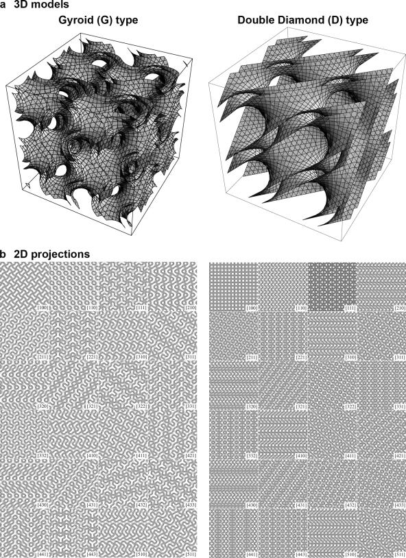
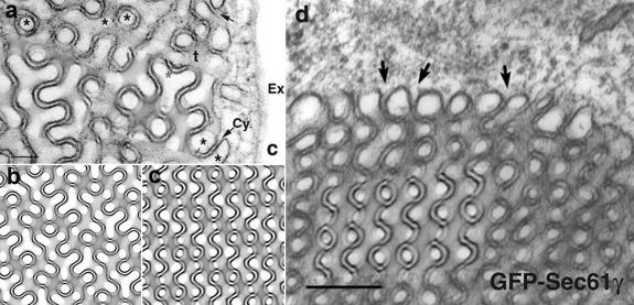
Cubic membranes represent highly curved, three-dimensional nanoperiodic structures that correspond to mathematically well defined triply periodic minimal surfaces. Although they have been observed in numerous cell types and under different conditions, particularly in stressed, diseased, or virally infected cells, knowledge about the formation and function of nonlamellar, cubic structures in biological systems is scarce, and research so far is restricted to the descriptive level. We show that the "organized smooth endoplasmic reticulum" (OSER; Snapp, E.L., R.S. Hegde, M. Francolini, F. Lombardo, S. Colombo, E. Pedrazzini, N. Borgese, and J. Lippincott-Schwartz. 2003. J. Cell Biol. 163:257-269), which is formed in response to elevated levels of specific membrane-resident proteins, is actually the two-dimensional representation of two subtypes of cubic membrane morphology. Controlled OSER induction may thus provide, for the first time, a valuable tool to study cubic membrane formation and function at the molecular level.
Figures
References
-
- Almsherqi, Z.A., C.S. McLachlan, P. Mossop, K. Knoops, and Y. Deng. 2005. Direct template matching reveals a host subcellular membrane gyroid cubic structure that is associated with SARS virus. Redox Rep. 10:167–171. - PubMed
-
- Anderson, R.G., L. Orci, M.S. Brown, L.M. Garcia-Segura, and J.L. Goldstein. 1983. Ultrastructural analysis of crystalloid endoplasmic reticulum in UT-1 cells and its disappearance in response to cholesterol. J. Cell Sci. 63:1–20. - PubMed
-
- Awad, T.S., Y. Okamoto, S.M. Masum, and M. Yamazaki. 2005. Formation of cubic phases from large unilamellar vesicles of dioleoylphosphatidylglycerol/monoolein membranes induced by low concentrations of Ca2+. Langmuir. 21:11556–11561. - PubMed
-
- Barauskas, J., M. Johnsson, and F. Tiberg. 2005. Self-assembled lipid superstructures: beyond vesicles and liposomes. Nano Lett. 5:1615–1619. - PubMed
-
- Black, V.H. 1972. The development of smooth-surfaced endoplasmic reticulum in adrenal cortical cells of fetal guinea pigs. Am. J. Anat. 135:381–417. - PubMed
Publication types
MeSH terms
Substances
LinkOut - more resources
Full Text Sources
Other Literature Sources
