Comparative Study
. 2006 Jul 1;177(1):694-701.
doi: 10.4049/jimmunol.177.1.694.

Fc gamma RIIa, not Fc gamma RIIb, is constitutively and functionally expressed on skin-derived human mast cells

Affiliations
Comparative Study

Fc gamma RIIa, not Fc gamma RIIb, is constitutively and functionally expressed on skin-derived human mast cells

Wei Zhao et al. J Immunol. .

Abstract

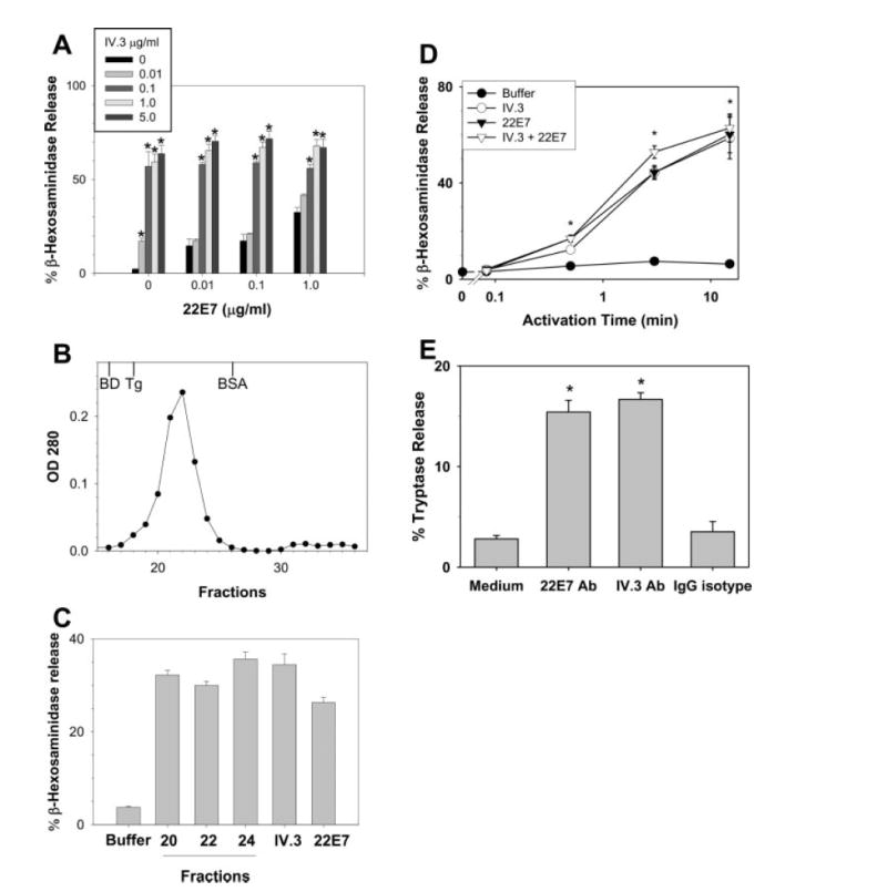
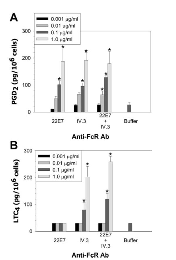
The expression of FcgammaR by human skin-derived mast cells of the MC(TC) type was determined in the current study. Expression of mRNA was analyzed with microarray gene chips and RT-PCR; protein by Western blotting and flow cytometry; function by release of beta-hexosaminidase, PGD(2), leukotriene C(4) (LTC(4)), IL-5, IL-6, IL-13, GM-CSF, and TNF-alpha. FcgammaRIIa was consistently detected along with FcepsilonRI at the mRNA and protein levels; FcgammaRIIc was sometimes detected only by RT-PCR; but FcgammaRIIb, FcgammaRI, and FcgammaRIII mRNA and protein were not detected. FcgammaRIIa-specific mAb caused skin MC(TC) cells to degranulate and secrete PGD(2), LTC(4), GM-CSF, IL-5, IL-6, IL-13, and TNF-alpha in a dose-dependent fashion. FcepsilonRI-specific mAb caused similar amounts of each mediator to be released with the exception of LTC(4), which was not released by this agonist. Simultaneous but independent cross-linking of FcepsilonRI and FcgammaRIIa did not substantially alter mediator release above or below levels observed with each agent alone. Skin MC(TC) cells sensitized with dust-mite-specific IgE and IgG, when coaggregated by Der p2, exhibited enhanced degranulation compared with sensitization with either IgE or IgG alone. These results extend the known capabilities of human skin mast cells to respond to IgG as well as IgE-mediated signals.


Figures

FIGURE 1
FIGURE 1
FcγRII mRNA and protein expression in human skin mast cells. A, RT-PCR. RNA preparations from skin mast cells and peripheral blood leukocytes (+, positive control) were reverse-transcribed and FcγRIIa, FcγRIIb, and FcγRIIc transcripts were selectively amplified. PCR products were separated by electrophoresis on 2% agarose gels and detected by ethidium bromide staining (upper panel). Reaction mixtures with primers only (no RNA/DNA template) served as negative controls (data not shown). These results were representative of three independent experiments, meaning that cells were obtained from three separate donors. Southern blotting performed with a digoxygenin-UTP-labeled probe recognizing all FcγRII isoforms was used to confirm amplification of this gene. The results are representative of studies performed on two separate skin mast cell cultures. The two bands in FcγRIIC blots represent donor-dependent allelic polymorphisms (57). B, Western blot. Lysates from two separate mast cell cultures (SMC1 and SMC2) of 6–8 wk were separated by SDS-PAGE under reducing conditions, and analyzed by immunoblotting with rabbit anti-FcγRIIa (Ab260) (lower panel) or rabbit anti-FcγRIIb (Ab163) (upper panel). As a control, the FcγRIIa-expressing cell line U937 and FcγRIIb-expressing peripheral blood leukocytes (PBL) were included. Each lane contains 2 × 10⁵ cell equivalents. Molecular mass markers are indicated to the left of these gels. C, Western blot of freshly dispersed mast cells. Lysates from freshly dipersed mast cells were tested as in B. These mast cells had been purified by sorting using FITC-labeled anti-Kit and anti-FcεRI mAbs within 2 days after their dispersal from skin. Peripheral blood basophils (PBB) and cord blood-derived mast cells (CBMC), obtained as described (24, 36), served as positive controls for CD32b, while Raji cells served as a positive control for CD32a. Each lane contains 5 × 10⁵ cell equivalents.
FIGURE 2
FIGURE 2
Surface expression of mast cell FcγRII. Mast cells were incubated at 4°C with mAbs against FcγR, FcεRI, and Kit as described, followed by FITC-labeled goat IgG anti-mouse IgG. Each primary mAb was used at 10 μg/ml. The isotype-matched control mAb (MOPC31C) is shown as the dotted line. Results are representative of a total of five separate experiments.
FIGURE 3
FIGURE 3
Degranulation by skin-derived mast cells after FcγRIIa and/or FcεRI cross-linking. A, Degranulation dose response. β-Hexosaminidase release was determined after 30 min of incubation with the mAbs as indicated. Release values were greater than the buffer control (p < 0.05) at all mAb concentrations. B, Gel filtration of IV.3 mAb. IV.3 mAb (0.5 mg/180 μl) was loaded onto a Superose 12 HR 10/30 column (Pharmacia) using a Shimadzu LC-10Avp HPLC system (Shimadzu) at a flow rate of 1 ml/min. The column was equilibrated and run with PBS. Elution fractions were collected at 0.5 ml/tube at 1 ml/min. The OD of each fraction was monitored at 280 nm and plotted. Gel filtration standards are shown at their elution positions for Blue Dextran (BD, ~2 × 10⁶ Da), thyroglobulin (Tg, 669,000 Da), and BSA (66,000 Da). C, Activation of skin mast cells with unaggregated IV.3 mAb. IV.3 in fractions from the ascending (#20), peak (#22), and descending (#24) portions of the elution profile along with prechromatography IV.3 and 22E7 were each used at 1 μg/ml to stimulate skin mast cells. β-Hexosaminidase was measured as a marker of degranulation. D, Time-course. β-Hexosaminidase release was determined after mast cells were incubated with 22E7 (1 μg/ml) and/or IV.3 (0.1 μg/ml) mAbs for 0, 5 and 30 s, and for 3 and 15 min. E, Degranulation of freshly dispersed skin mast cells. Mast cells were dispersed from skin, enriched to ~5% purity by Percoll density-dependent sedimentation and then stimulated with 22E7 (1 μg/ml) or IV.3 (1 μg/ml) mAbs for 30 min. Degranulation was assessed by measuring release of tryptase. Spontaneous release and release with a nonimmune IgG-isotype control were each <4%. Mean ± SE values from three independent experiments are shown. *, p < 0.05 by ANOVA when experimental values are compared with 0 μg/ml of IV.3 within each 22E7 group in A, and to the buffer control in D and E.
FIGURE 4
FIGURE 4
Release of PGD2 (A) and LTC4 (B) by skin mast cells stimulated by cross-linking FcεRI (22E7), FcγRIIa (IV.3) or both receptors simultaneously. PGD2 and LTC4 release were determined after skin mast cells (106 cells/ml) had been incubated with the indicated mAbs for 45 min in AIM-V medium. Results from three independent experiments, each single experiment performed in triplicate, are presented. *, p < 0.05 by ANOVA when experimental values within each group are compared with the buffer control.
FIGURE 5
FIGURE 5
Cytokine production by skin-derived mast cells challenged with anti-FcγRIIa and/or anti-FcεRI mAbs. A, IL-5. B, IL-6. C, IL-13. D, GM-CSF. E, TNF-α. Mast cells (106 cells/ml) were incubated with the indicated mAbs for 24 h in AIM-V medium containing SBTI and SCF. Results from three independent experiments, each single experiment performed in triplicate, are presented as box plots (because not all data followed a normal distribution) where the hatch mark is the median and the upper and lower ends of the box are the 25th and 75th percentiles. *, p < 0.05 by ANOVA when values for each cytokine were compared with those for the buffer control; †, p < 0.05 against IV.3 within the same mAb dose group.
FIGURE 6
FIGURE 6
Activation of skin-derived human mast cells by Ag through FcγRIIa and FcεRI pathways. A, Activation by preformed IgG anti-NP–NP-BSA and IgE anti-NP–NP-BSA immune complexes. Immune complexes containing 8.7 μg/ml Ab with 0.13 μg/ml NP-BSA, 4.4 μg/ml Ab with 0.7 μg/ml NP-BSA, and 2.2 μg/ml Ab with 0.3 μg/ml NP-BSA were incubated with skin mast cells for 30 min. Supernatants and cell lysates were prepared for β-hexosaminidase measurements. Data are expressed as mean ± SE from three independent experiments, each performed in triplicate. B, Co-crosslinking FcγRIIa and FcεRI by Ag. Skin-derived mast cells that had been sensitized with Der p2-specific IgE, Der p2-specific IgG, or both mAbs were challenged with aggregated Der p2. Releasates were collected after 30 min to measure mediator release as described in materials and methods. Data expressed as mean ± SE from three individual experiments. *, p < 0.05 by ANOVA when experimental values are compared with the Ab-only control in A.

References

    1. Unkeless JC, Jin J. Inhibitory receptors, ITIM sequences and phosphatases. Curr Opin Immunol. 1997;9:338–343. - PubMed
    1. Malaviya R, Georges A. Regulation of mast cell-mediated innate immunity during early response to bacterial infection. Clin Rev Allergy Immunol. 2002;22:189–204. - PubMed
    1. Marone G, Florio G, Petraroli A, Triggiani M, De Paulis A. Role of human Fcε RI+ cells in HIV-1 infection. Immunological Reviews. 2001;179:128–138. - PubMed
    1. Marshall JS, King CA, McCurdy JD. Mast cell cytokine and chemokine responses to bacterial and viral infection. Curr Pharm Des. 2003;9:11–24. - PubMed
    1. Urban JF, Schopf L, Morris SC, Orekhova T, Madden KB, Betts CJ, Gamble HR, Byrd C, Donaldson D, Else K, Finkelman FD. Stat6 signaling promotes protective immunity against Trichinella spiralis through a mast cell- and T cell-dependent mechanism. J Immunol. 2000;164:2046–2052. - PubMed

Publication types

MeSH terms