Comparative Study
. 2006 Aug 15;84(3):553-67.
doi: 10.1002/jnr.20968.

Gene delivery to the spinal cord: comparison between lentiviral, adenoviral, and retroviral vector delivery systems

Affiliations
Comparative Study

Gene delivery to the spinal cord: comparison between lentiviral, adenoviral, and retroviral vector delivery systems

Ahmed A Abdellatif et al. J Neurosci Res. .

Abstract

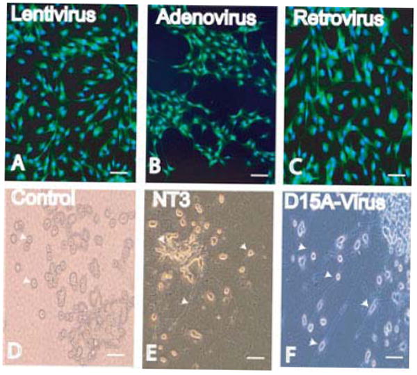
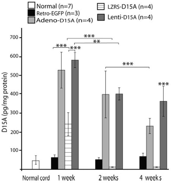
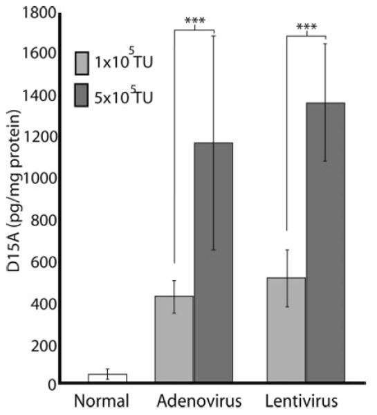
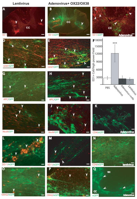
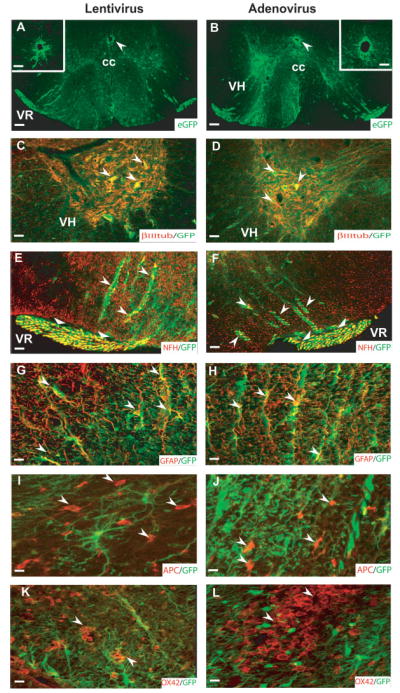
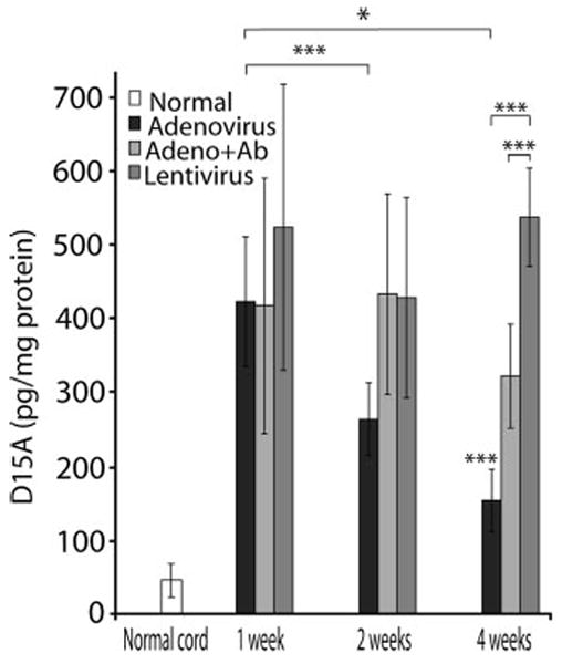
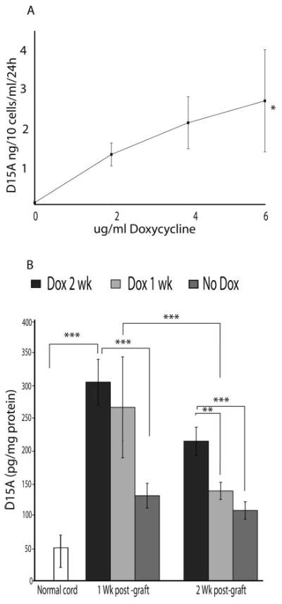
Viral gene delivery for spinal cord injury (SCI) is a promising approach for enhancing axonal regeneration and neuroprotection. An understanding of spatio-temporal transgene expression in the spinal cord is essential for future studies of SCI therapies. Commonly, intracellular marker proteins (e.g., EGFP) were used as indicators of transgene levels after viral delivery, which may not accurately reflect levels of secreted transgene. This study examined transgene expression using ELISA after viral delivery of D15A, a neurotrophin with BDNF and NT-3 activities, at 1, 2, and 4weeks after in vivo and ex vivo delivery using lentiviral, adenoviral, and retroviral vectors. Further, the inflammatory responses and viral infection patterns after in vivo delivery were examined. Lentiviral vectors had the most stable pattern of gene expression, with D15A levels of 536 +/- 38 and 363 +/- 47 pg/mg protein seen at 4 weeks after the in vivo and ex vivo delivery, respectively. Our results show that protein levels downregulate disproportionately to levels of EGFP after adenoviral vectors both in vivo and ex vivo. D15A dropped from initial levels of 422 +/- 87 to 153 +/- 18 pg/mg protein at 4 weeks after in vivo administration. Similarly, ex vivo retrovirus-mediated transgene expression exhibited rapid downregulation by 2 weeks post-grafting. Compared to adenoviral infection, macrophage activation was attenuated after lentiviral infection. These results suggest that lentiviral vectors are most suitable in situations where stable long-term transgene expression is needed. Retroviral ex vivo delivery is optional when transient expression within targeted spinal tissue is desired, with adenoviral vectors in between.


Figures

Fig. 1
Fig. 1
Schwann cells were infected in vitro with either lentivirus (A), adenovirus (B), or retrovirus (C) (MOI = 20). Cells were stained for EGFP and counterstained with Hoechst 33342. TrkC-expressing PC12 cells (arrow heads in D) were used to determine the bioactivity of D15A. Recombinant human (rh) NT-3 was added (100 pg/ml–1 ng/ml) to the media of PC12 cells for 3–4 days, neurite outgrowth from PC12 cells (arrow heads in E) was compared to that of conditioned media of infected NIH3T3 cells added at a concentration range of 10–100%, bioactive D15A in media was identified by examining neurite outgrowth (arrowheads in F) from PC12 cells. Conditioned media at a concentration of 75–100% were similar to an NT-3 concentration of 500 pg/ml–1 ng/ml. Control PC12 cells (D) maintained on DMEM did not show any neurite outgrowth. There was no difference between conditioned media collected from NIH3T3 cells infected with lentivirus, adenovirus, or retrovirus. Scale bar = 40 μm. [Color figure can be viewed in the online issue, which is available at www.interscience.wiley.com.]
Fig. 2
Fig. 2
Temporal expression of D15A in the spinal cord after ex vivo gene delivery using lentivirus, adenovirus, EGFP, and retrovirus. Schwann cells were infected with similar MOI (20) and 48 hr later 1 × 10⁵ cells were transplanted into the spinal cord. There was no significant difference between normal spinal cord and EGFP-infected groups at any of the three time points. D15A expression was significantly higher (P < 0.001) in the lentiviral-infected group than normal, EGFP, and retrovirus infected groups at 1 and 2 weeks. No significant difference was found between lentiviral and adenoviral infected groups at 1- and 2-weeks post-transplantation. At 4 weeks, D15A expression was significantly higher (P < 0.001) in the lentiviral-infected group than all other groups. In the adenoviral-infected group, D15A levels were significantly lower (P < 0.001) at 4 weeks. Downregulation of D15A in the retroviral-infected group was rapid with significantly lower (P < 0.001) D15A levels at 2 and 4 weeks compared to that of 1 week. Although the lentiviral-infected group showed some downregulation with D15A levels significantly lower (P < 0.01) at 2 weeks, D15A levels plateaued and were not significantly different at 2 and 4 weeks. Data shown are the mean ± SD (n = 4).
Fig. 3
Fig. 3
D15A protein expression is dependent on viral titer. One microliter of 1 × 10⁵ TU/μl or 5 × 10⁵ TU/μl of virus was injected into the spinal cord. One week after injection, expression of D15A was significantly (P < 0.001) higher than normal in all groups tested. D15A levels were significantly higher after high titer adenoviral (5 × 10⁵ TU/μl, n = 3; P = 0.001) and lentiviral injections (5 × 10⁵ TU/μl, n = 3/group; P < 0.001) than after the low titer injections (1 × 10⁵ TU/μl, n = 6/group). D15A expression was not significantly different between titer matched adenoviral and lentiviral injected groups. Data shown are mean ± SD.
Fig. 4
Fig. 4
Inflammatory response and cell-specific infection by low titer lentiviral and adenoviral vectors in the spinal cord. Photomicrographs taken within 2 mm from the injection site show representative sections after; lentivirus (A,D,G,I,L,N,O), adenovirus + mAb OX22/ OX38 (B,E,H,J,K,M,P,Q) and adenovirus (C) injections. In both lentivirus (A) and antibody-treated adenovirus groups (B) ED1+ cells were limited to the area surrounding the injection site (arrows), whereas in the non-antibody treated adenovirus group (C) ED1+ cells were widespread over the entire gray and white matter of the spinal cord. ED1+ cells were counted (F) in five sections 2 mm from the injection site from each animal. Cells were counted using Image-pro Plus software in an entire 20× field. The number of ED1+ cells in the adenovirus group were significantly higher (P < 0.001) than all other groups. There was no statistically significant difference among PBS, lentivirus, and adenovirus + mAb groups. Data shown are the mean ± SD, (n = 7/group). To identify cellular phenotypes infected with low titer (1 × 10⁵ TU/μl) lentivirus and adenovirus, sections were double-labeled for EGFP (green) and the cell-specific markers; neurons (NeuN), oligodendrocytes (APC), astrocytes (GFAP), microglia/macrophages ED-1/OX42, and T lymphocytes (CD4). Examination using confocal microscopy showed that astrocytes (arrows) were infected with both viruses (D,E). In contrast, APC+ oligodendrocytes (G,H) and NeuN+ neurons (I,J) (arrows) were not infected after both viral injections. A decrease in NeuN immunoreactivity was also seen after lentiviral injection but not after combined adenoviral injection and immune suppression. CD4+ T-cells were not seen 1 week after low titer adenoviral injection with the OX22/OX38 immunosuppression (K) or lentiviral injection (N). ED1+ macrophages (L,M) and OX42+ reactive microglia (O,P; arrows) were not infected after either viral injection. EGFP labeling was detected at 4 weeks after adenoviral injection with OX22/OX38 immunosuppression (arrows in Q). Data are representative of five independent animals for each experimental group. Scale bar = 200 μm (in A,B); 20 μm (in D–P), and 80 μm (C,Q).
Fig. 5
Fig. 5
Identification of cell types infected with high titer lentivirus and adenovirus. EGFP labeling was found in both the gray and white matter of the spinal cord after lentiviral (A,C,E,G,I,K) and adenoviral (B,D,F,H,J,L) injections. Ependymal cells surrounding the central canal were infected by both types of viruses that could be clearly seen at high magnifications (arrows in A,B; insets in A and B are from adjacent sections). EGFP labeling was localized in βIII tubulin+ neurons (C,D; arrows), NFH+ axons in the ventral white matter (E,F; arrows) and the ventral rootlets (E,F; triangular arrow heads) associated with motor neurons as well as GFAP+ astrocytes (G,H; arrows). In contrast, neither oligodendrocytes (I, J; arrows) nor reactive microglia/macrophages (K,L; arrows) were labeled with EGFP. Scale bar = 200 μm (A,B); 40 μm (C,F and insets in A,B); 20 μm (in G,H,K,L); 13 μm (I,J). cc, central canal; VH, ventral horn; VR, ventral root. Data are representative of n = 5 animals/group.
Fig. 6
Fig. 6
Temporal expression of D15A in the spinal cord after in vivo gene delivery. Lentiviral delivery resulted in robust and stable gene expression for up to 4 weeks post-injection. At 4 weeks post-injection, the D15A levels were significantly higher than both adenoviral infected groups (P < 0.001). In contrast, adenoviral infection showed significant downregulation of D15A at 2 (P < 0.001) and 4 weeks (P < 0.05) post-infection. Treatment of animals with mAb OX-22/OX-38 successfully reduced the downregulation of adenoviral-delivered D15A expression compared to the non-OX22/OX38 treated groups that was significantly lower (P < 0.001) than lentivirus and adenovirus + mAb groups at 4 weeks. Data represent the mean ± SD (n = 5/experimental group).
Fig. 7
Fig. 7
Induction of D15A expression using doxycycline (Dox) in vitro (A) and in vivo (B). A: NIH/3T3 cells were transduced in vitro using the two virus (pRev-Tre D15A and pRev-Tre Tet-on) tetracycline inducible Tet-on system. After antibiotic selection for 1 week in vitro, increasing Dox concentrations were added to the culture media. No significant difference was found between 2, 4, and 6 μg/ml Dox concentrations. D15A levels at 6 μg/ml Dox were significantly higher than baseline expression (P < 0.05). Data shown are the mean ± SD (n = 5/experimental group). B: 1 × 10⁵ D15A-infected RN33B cells were injected into the spinal cord. After the addition of 1 mg/ml Dox to the animal’s drinking water, D15A levels at 1 week were significantly higher than both normal (open bars, P < 0.001) and no-Dox (dark gray bars, P < 0.001) groups. In one group of animals, Dox administration was stopped after 1 week, which resulted in a significant drop in D15A levels at 2 weeks (light gray bars, P < 0.001). At 2 weeks, D15A levels in the group that received Dox for 2 weeks were significantly higher than normal (black bars, P < 0.001), no-Dox (P < 0.001) and animals that received Dox for only 1 week (P < 0.01). Basal expression levels were detectable and were significantly higher (P < 0.05 at 1 week and P < 0.01 at 2 weeks) than in control animals indicating incomplete transcriptional regulation in the absence of Dox. Data shown are the mean ± SD (n = 4).

References

    1. Akli S, Guidotti JE, Vigne E, Perricaudet M, Sandhoff K, Kahn A, Poenaru L. Restoration of hexosaminidase A activity in human Tay-Sachs fibroblasts via adenoviral vector-mediated gene transfer. Gene Ther. 1996;3:769–774. - PubMed
    1. Andersen JK, Garber DA, Meaney CA, Breakefield XO. Gene transfer into mammalian central nervous system using herpes virus vectors: extended expression of bacterial lacZ in neurons using the neuron-specific enolase promoter. Hum Gene Ther. 1992;3:487–499. - PubMed
    1. Baekelandt V, Eggermont K, Michiels M, Nuttin B, Debyser Z. Optimized lentiviral vector production and purification procedure prevents immune response after transduction of mouse brain. Gene Ther. 2003;10:1933–1940. - PubMed
    1. Bajocchi G, Feldman SH, Crystal RG, Mastrangeli A. Direct in vivo gene transfer to ependymal cells in the central nervous system using recombinant adenovirus vectors. Nat Genet. 1993;3:229–234. - PubMed
    1. Baumgartner BJ, Shine HD. Neuroprotection of spinal motoneurons following targeted transduction with an adenoviral vector carrying the gene for glial cell line-derived neurotrophic factor. Exp Neurol. 1998;153:102–112. - PubMed

Publication types

MeSH terms

Substances