. 2006 Oct 1;399(1):131-9.
doi: 10.1042/BJ20060696.

Regulation of GLUT4 gene expression by SREBP-1c in adipocytes

Affiliations

Regulation of GLUT4 gene expression by SREBP-1c in adipocytes

Seung-Soon Im et al. Biochem J. .

Abstract

Expression of the GLUT4 (glucose transporter type 4 isoform) gene in adipocytes is subject to hormonal or metabolic control. In the present study, we have characterized an adipose tissue transcription factor that is influenced by fasting/refeeding regimens and insulin. Northern blotting showed that refeeding increased GLUT4 mRNA levels for 24 h in adipose tissue. Consistent with an increased GLUT4 gene expression, the mRNA levels of SREBP (sterol-regulatory-element-binding protein)-1c in adipose tissue were also increased by refeeding. In streptozotocin-induced diabetic rats, insulin treatment increased the mRNA levels of GLUT4 in adipose tissue. Serial deletion, luciferase reporter assays and electrophoretic mobility-shift assay studies indicated that the putative sterol response element is located in the region between bases -109 and -100 of the human GLUT4 promoter. Transduction of the SREBP-1c dominant negative form to differentiated 3T3-L1 adipocytes caused a reduction in the mRNA levels of GLUT4, suggesting that SREBP-1c mediates the transcription of GLUT4. In vivo chromatin immunoprecipitation revealed that refeeding increased the binding of SREBP-1 to the putative sterol-response element in the GLUT4. Furthermore, treating streptozotocin-induced diabetic rats with insulin restored SREBP-1 binding. In addition, we have identified an Sp1 binding site adjacent to the functional sterol-response element in the GLUT4 promoter. The Sp1 site appears to play an additive role in SREBP-1c mediated GLUT4 gene upregulation. These results suggest that upregulation of GLUT4 gene transcription might be directly mediated by SREBP-1c in adipose tissue.


Figures
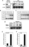
Figure 1
Figure 1. Effect of refeeding on SREBP-1 and GLUT4 mRNA levels in epidydimal WAT
(A) GLUT4 mRNA levels. Diabetes was induced by administering STZ (50 mg/kg) to Sprague–Dawley rats (approx. 200 g body weight) and the blood glucose concentration was checked to confirm the onset of diabetes. Normal rats were subjected to fasting for 48 h. Insulin (0.5 mM/kg) was administered to STZ-diabetic rats. (B) Time course measurements of GLUT4 mRNA levels. Rats were fasted for 48 h and refed a fat-free, high-carbohydrate diet ad libitum. After refeeding, the rats were sacrificed at the time periods indicated. Total RNA (20 μg) isolated from epididymal WAT was subjected to 0.9% formaldehyde–agarose gel electrophoresis. The RNA in the gel was transferred to a nylon membrane and hybridized to 32P-labeled cDNAs for GLUT4, SREBP-1 or β-actin. Representative Northern blots are shown for GLUT4, SREBP-1 and β-actin mRNA from normal (n=3) or diabetic (n=3) rats. (C) Kinetics of SCD1 and FAS mRNA induction after refeeding. (D) Time course measurement of mature SREBP-1 protein expression after refeeding. The mature form of SREBP-1 was measured by fractionating nuclear lysate from adipose tissue as described in the Experimental section. The expression level of endogenous GLUT4 gene (E) and FAS (F) induced by SREBP-1c adenovirus in 3T3-L1 adipocytes. After infecting SREBP-1c adenovirus at 100 MOI for 48 h in differentiated 3T3-L1 adipocytes, GLUT4 and FAS gene expression was confirmed by real-time PCR. Values are mean±S.E.M (n=3). P< 0.001 compared to mock group. Ad: ad libitum, Ins: insulin treated, NS: non-specific band.
Figure 2
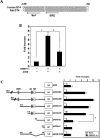
Figure 2. Localization of SRE in the human GLUT4 promoter
(A) Comparison of human and rat GLUT4 promoter sequences. (B) The effect of SREBP-1c on GLUT4 promoter activity. pHGT4-2031 promoter reporter (500 ng) was cotransfected with SREBP-1c (100 ng) or SREBP-1c plus ADDN into 3T3-L1 preadipocytes cell lines that were plated at a density of 2×10⁵ cells/35 mm dish. Transient transfection, luciferase assays and measurement of total protein concentration of the lysates were performed as described in the Experimental section. *P<0.001, untreated versus SREBP-1; #P<0.05, SREBP-1c versus SREBP-1c+ADDN. (C) The effect of deletion on the SREBP-1c-mediated GLUT4 promoter activity. Schematic diagram of serial deletion constructs of the GLUT4 promoter reporter (left) and SREBP-1c mediated GLUT4 promoter activity (right). The indicated numbers represent the number of nucleotides from the mRNA start codon. The promoter activities were measured by co-transfecting 100ng of SREBP-1c expression or empty vectors into 3T3-L1 preadipocytes. The results were normalized by the amount of total protein in the lysates, which was determined by the Bradford method and shown as the fold changes of luciferase activities compared to those of the control. Normalized luciferase activities are shown as the means±S.D. of three independent experiments in triplicate. *P<0.005, pHGT4d-119 versus pHGT4d-92.
Figure 3
Figure 3. Identification of the SRE and binding of SREBP-1 in the human GLUT4 promoter
EMSA was performed with recombinant SREBP-1 protein in 4% (w/w) non-denaturing polyacrylamide gel. Then, 50000 cpm (0.1 pmole) of 32P-labelled GLUT4 promoter fragments (−125/−79) containing putative SRE was incubated with 20–80 ng of recombinant SREBP-1 protein. The DNA–protein complexes are indicated by arrowheads. LDLR-SRE, SRE region on the promoter of the known low-density lipoprotein receptor.
Figure 4
Figure 4. Binding of Sp1 and SREBP-1 to the GLUT4 promoter
(A) Schematic diagram of wild and mutated probes used in EMSA. The preparation of the site specific mutations are described in the Experimental section. (BD) Direct and specific binding of the recombinant Sp1 zinc-finger (ZF) (80 ng) and SREBP-1 (200 ng) to each of the putative binding sites in the human GLUT4 promoter. The EMSA was performed using 32P-labelled GLUT4 promoter oligonucleotides, Sp1 and SREBP-1. SS, supershift.
Figure 5
Figure 5. The functional relationship between Sp1 and SREBP-1 in the GLUT4 promoter
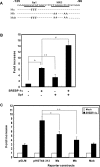
(A) The sequences of wild-type and the mutated construct of putative Sp1 and SREBP-1c binding sites. The mutated bases are represented by bold characters. (B) The effect of the SREBP-1c and Sp1 on human GLUT4 promoter activity. SREBP-1c or Sp1 expression vector was transfected to 3T3-L1 preadipocytes. The luciferase activities were represented as fold changes compared with the control group. *P<0.001, untransfected versus SREBP-1c; **P<0.05, untransfected versus Sp1; #P<0.001, SREBP-1c versus SREBP-1c+Sp1. (C) GLUT4–luciferase constructs containing mutations in the Sp1 or SREBP-1c were generated as described in the Experimental section. 3T3-L1 preadipocytes were co-transfected with each promoter fragment linked to luciferase with or without SREBP-1c expression vectors. Ma, promoter reporter of mutated Sp1 sequence. Mb, promoter reporter of mutated SRE sequence. Mab, promoter reporter of double mutated Sp1 and SRE sequences. Values are the means±S.D. of three independent experiments performed in triplicate. *P<0.05, pHGT4d-313 versus Ma, **P<0.05, pHGT4d-313 versus Mb.
Figure 6
Figure 6. SREBP-1c mediates GLUT4 gene expression in differentiated 3T3-L1 adipocytes
3T3-L1 adipocytes were treated with insulin (100 nM) and ADDN or null virus (adeno-GFP) at the indicated MOI for 16 h at 37 °C with serum-free media and replaced with media including serum. Total RNA was extracted for the measurement of GLUT4 mRNA using (A) RT-PCR or (B) real-time PCR. (C) The effect of ADDN on FAS gene expression. The mRNA level was normalized to that of α-tubulin in real-time PCR. β-Actin gene expression measured by RT-PCR was used as a negative control. M, DNA marker. Values are the means±S.D. of three independent experiments performed in triplicate. #P<0.005, GFP versus ADDN (200 MOI).
Figure 7
Figure 7. SREBP-1c binding to putative GLUT4-SRE is increased by refeeding and insulin treatment in vivo
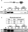
(A) Scheme of amplifying the GLUT4 promoter in the ChIP assay. The region between −280 to −55 was amplified. (B) For the ChIP assay, chromatin extracted from epididymal WAT was precipitated by SREBP-1 antibody and GLUT4-SRE was amplified by PCR. The amount of chromosomal DNA used for the precipitation reaction was normalized by input chromatin (1/100th of chromosomal DNA used for precipitation). F, fasted; R, refed. (C) The effect of insulin on the binding of SREBP-1 to the GLUT4 promoter in adipose tissue of STZ-induced rats. Insulin depleted diabetic rats were prepared by STZ. For the insulin treatment group, 0.1 mM/kg of insulin was administrated to STZ-induced diabetic rats. The ChIP assay is representative of three independent experiments. Blood glucose levels for the animal groups are shown in the Table. F: 48 h fasted, R: refeeding rats, S: STZ, I: insulin.

References

    1. Shepherd P. R., Kahn B. B. Glucose transporters and insulin action: implications for insulin resistance and diabetes mellitus. N. Engl. J. Med. 1999;341:248–257. - PubMed
    1. Furtado L. M., Somwar R., Sweeney G., Niu W., Klip A. Activation of the glucose transporter GLUT4 by insulin. Biochem. Cell Biol. 2002;80:569–578. - PubMed
    1. Porte D., Jr Banting lecture 1990. Beta-cells in type II diabetes mellitus. Diabetes. 1991;40:166–180. - PubMed
    1. Kahn C. R. Banting lecture 1994. Insulin action, diabetogenes and the cause of type II diabetes. Diabetes. 1994;43:1066–1084. - PubMed
    1. Reaven G. M. Pathophysiology of insulin resistance in human disease. Physiol. Rev. 1995;75:473–486. - PubMed

Publication types

MeSH terms

Substances