. 2006 Oct 1;399(1):37-46.
doi: 10.1042/BJ20060662.

Cloning of rat amelotin and localization of the protein to the basal lamina of maturation stage ameloblasts and junctional epithelium

Affiliations

Cloning of rat amelotin and localization of the protein to the basal lamina of maturation stage ameloblasts and junctional epithelium

Pierre Moffatt et al. Biochem J. .

Abstract

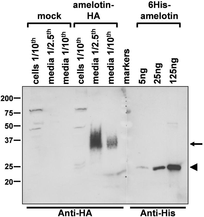
Formation of tooth enamel is a very complex process in which a specific set of proteins secreted by ameloblasts play a primordial role. As part of a screening procedure to identify novel proteins secreted by EO (enamel organ) cells of rat incisors, we isolated a partial cDNA fragment (EO-017) that is the homologue of the recently described mouse Amtn (amelotin) gene [Iwasaki, Bajenova, Somogyi-Ganss, Miller, Nguyen, Nourkeyhani, Gao, Wendel and Ganss (2005) J. Dent. Res. 84, 1127-1132]. Presented herein is the cloning of rat and pig full-length cDNAs with their deduced protein sequences. Detailed expression profiling by Northern-blot analysis and RT (reverse transcriptase)-PCR on rat and mouse tissues revealed highest expression in the mandible, more specifically in the maturation stage of the EO. Among all tissues tested, low expression was detected only in periodontal ligament, lung, thymus and gingiva. In silico analyses revealed that the Amtn gene is highly conserved in seven other mammals, but is absent from fish, birds and amphibians. The Amtn protein is enriched in proline, leucine, glutamine and threonine (52% of total) and contains a perfectly conserved protein kinase CK2 phosphorylation site. Transient transfection experiments in HEK-293 cells (human embryonic kidney cells) showed that secreted Amtn is post-translationally modified possibly through O-linked oligosaccharides on threonine residues. In concordance with its predominant expression site, immunofluorescence localization within the rat and mouse mandibles revealed Amtn localized to the basal lamina of maturation stage ameloblasts of incisors and unerupted molars. Intense Amtn protein expression was also detected in the internal basal lamina of junctional epithelium in molars. The peculiar and unique cellular localization of Amtn suggests a role in cell adhesion.


Figures

Figure 1
Figure 1. Northern-blot analyses of the expression of Amtn in rat tissues
Total RNA was extracted from tissues of adult rat unless otherwise indicated (4d: 4-days-old; e19: embryo at day 19). Either 10 μg (*) or 20 μg of RNA was separated on agarose gels and processed for hybridization. Integrity of RNA was monitored by hybridization with a Gapdh probe (A, bottom panel) or after staining the membrane with Methylene Blue (B, bottom panel). RNA isolated from EOs at the secretion and maturation stages of development was analysed separately (B). In (A), the middle panel is a longer exposure of the same blot shown in the top panel (bracketed area), and illustrates weaker Amtn expression levels present in periodontal ligament and lung. The positions of 28 S and 18 S ribosomal RNAs are indicated.
Figure 2
Figure 2. Full-length cDNA for rat Amtn and its deduced protein sequence
The normal and italicized numbers at left refer to the nucleotide and protein sequences respectively. The 5′- and 3′-untranslated regions are in lower-case, and the coding region is in upper-case. The start and stop codons are boxed and two polyadenylation signals are shown in boldface. The nucleotides corresponding to different exons are depicted by alternating white/grey shadings. The signal peptide is underlined and a downward arrowhead indicates its predicted cleavage site. A putative CK2 serine phosphorylation site (SSEE) is boxed, and the peptidic sequence used for antibody production is highlighted black. Predicted O-glycosylated serine and threonine residues are circled.
Figure 3
Figure 3. Schematic representation of the location and structure of the rat Amtn gene and its derived transcripts
The rat Amtn gene is located on chromosome 14 (A) flanked by the enamel-specific genes Ambn and Enam, and the salivary gland mucin 10 (Muc10) (B). The 600 kb locus containing Amtn is rich in genes encoding secreted proteins such as the submandibular glands androgen regulated genes (Smr) and at the other extremity casein kappa (Cask), follicular dendritic cells secreted peptide (Fdcsp) and APin (B). The Amtn gene is composed of nine exons covering roughly 12 kb (C). The three different transcripts identified are depicted with the matching exons location (D). Semi-arrowheads above and below the longest cDNA (D) indicate location of the primers used for RT–PCR (Figure 5). ORF, open reading frame; SP, signal peptide. 5′UTR, 5′ untranslated region.
Figure 4
Figure 4. Alignment of the Amtn protein sequences from various mammalian species
Numbering at top refers to the human sequence, while those at the end of each sequence indicate the total number of residues in each protein. Non-identical residues are highlighted in black, and similar residues are highlighted in grey. Sequences were obtained from GenBank® entries AY358528 (human), CN440607 (cow) and AK017352 (mouse), or adapted from Ensembl transcript ENSCAFT00000004283 (dog), ENSMODT000000015781 (opossum), ENSPTRT00000030049 (chimp) and ENSMMUT00000004282 (macaque). The signal peptide (underlined) and predicted CK2 phosphorylation site (boxed) are conserved among all species.
Figure 5
Figure 5. The rat Amtn protein is secreted into the culture media of transfected HEK-293 cells
The rat Amtn cDNA, tagged at its C-terminus with the HA epitope, was transiently transfected into HEK-293 cells. After 48 h, media and cells were collected and analysed by Western blotting with an anti-HA antibody. Proportion of samples loaded relative to total is indicated. Cells transfected with the empty vector (mock) served as control. The Amtn-specific band was detected in the conditioned media (arrow) attesting to its secretory nature. A recombinant rat His6-tagged Amtn (6His–Amtn) was produced in and purified from bacteria by Ni-affinity chromatography. The His6–Amtn was detected with an anti-His monoclonal antibody (arrowhead). Note the higher apparent molecular mass (35 kDa) of the mammalian Amtn protein relative to the recombinant form (22 kDa). Molecular masses (kDa) are given on the extreme left.
Figure 6
Figure 6. RT–PCR analyses of Amtn gene expression in rat and mouse tissues
Total RNA was extracted from adult rat (A, B) and mouse (C) tissues and used in one-step RT–PCR reactions. Unless otherwise specified, 200 ng of RNA was used per reaction. The PCR products were resolved on 2% agarose gels and stained with ethidium bromide. Amtn gene-specific primers produce a band at 379 bp. Gapdh primers, yielding a 433 bp product, were used in parallel reactions to assess the integrity and uniformity of the RNAs. PCR reactions were stopped after 35 cycles (A, C) and 24 cycles (B) for Amtn, and after 25 cycles for Gapdh. A 100 bp DNA ladder was used as markers. Molecular size (bp) is given on the extreme left.
Figure 7
Figure 7. Immunofluorescence localization of Amtn protein in rat and mouse mandibular incisors
Composite image showing Amtn immunofluorescence localization over a 3-mm-long segment of the rat lower incisor EO (A). Pictures in (BD) are magnified regions boxed in (A) (from left to right respectively). No specific signal is visible in secretory stage ameloblasts in rat (B) and mouse (F). Golgi-like staining is apparent in post-secretory transition (C, arrows), and early maturation stage ameloblasts (D, arrows). Prominent labelling is observed at the basal lamina-like structure along the interface between the maturation stage ameloblasts and the developing enamel (D, E, arrowheads). The enamel surface is immunoreactive where the cell layer splits away from the enamel surface as a result of sectioning (E). The mouse lower incisor (FH) shows a similar pattern of Amtn labelling, being detectable at basal lamina-like surface only in the post-secretory (G) to maturation regions (H). Control incubations performed with a purified anti-rabbit-IgG on a serial section like that shown in (E) remained unlabelled (I). Scale bars, 200 μm (A) and 25 μm (I). Sections were counterstained with Hoechst (blue). Green coloration is tissue autofluorescence. ES, enamel space; Am, ameloblasts; PL, papillary layer; CT, connective tissues.
Figure 8
Figure 8. Immunofluorescence localization of Amtn on rat and mouse mandibular molars
Composite image showing Amtn immunofluorescence in rat (A) and mouse (F) molars. Boxes in (A) and (F) represent areas enlarged in (BE) and (G) respectively. Amtn labelling is present at the basal lamina-like surface of the rat unerupted molar (B). Detection of Amtn at the internal basal lamina of JE in first and second molars in rat (CE) and mouse (G). Control incubation performed with a purified anti-rabbit-IgG on a serial section as that shown in (E) remained unlabelled (H). Scale bars, 500 μm (A), 250 μm (F) and 25 μm (H). Sections were counterstained with Hoechst (blue). Green coloration is tissue autofluorescence. ES, enamel space; Am, ameloblasts; De, dentin; CEJ, cemento-enamel junction.
Figure 9
Figure 9. High-resolution immunolocalization of Amtn to the basal lamina of rat maturation stage ameloblasts
Low-magnification, scanning electron micrograph of an EO cap showing the ameloblast layer and a surface view of the overlying basal lamina (A). Backscattered electron imaging of the immunogold labelling for Amtn in a region similar to the boxed area in (A) is shown in (B). The pattern of distribution of gold particles, which appear as white particles with this type of imaging, indicates that Amtn is distributed throughout the basal lamina (B). Transmission electron micrographs of EO caps labelled for Amtn (C) and incubated under control conditions (D). The basal lamina appears as an electron-dense layer at the ameloblast apex decorated at its surface with gold particles, which appear black in transmission microscopy. The control shows virtually no gold particles.
Figure 10
Figure 10. Immunogold localization of Amtn to the internal basal lamina (IBL) of the rat JE
Transmission electron micrographs showing the presence of Amtn over the inner basal lamina (IBL) (A) and the almost complete absence of gold particles under control conditions (B). HD, hemidesmosome; m, mitochondria, N, nucleus.

References

    1. Gibson C. W. Regulation of amelogenin gene expression. Crit. Rev. Eukaryot. Gene Expr. 1999;9:45–57. - PubMed
    1. Nanci A., Zalzal S., Lavoie P., Kunikata M., Chen W., Krebsbach P. H., Yamada Y., Hammarstrom L., Simmer J. P., Fincham A. G., et al. Comparative immunochemical analyses of the developmental expression and distribution of ameloblastin and amelogenin in rat incisors. J. Histochem. Cytochem. 1998;46:911–934. - PubMed
    1. Smith C. E. Cellular and chemical events during enamel maturation. Crit. Rev. Oral Biol. Med. 1998;9:128–161. - PubMed
    1. Kawasaki K., Weiss K. M. Mineralized tissue and vertebrate evolution: the secretory calcium-binding phosphoprotein gene cluster. Proc. Natl. Acad. Sci. U.S.A. 2003;100:4060–4065. - PMC - PubMed
    1. Moradian-Oldak J., Paine M. L., Lei Y. P., Fincham A. G., Snead M. L. Self-assembly properties of recombinant engineered amelogenin proteins analyzed by dynamic light scattering and atomic force microscopy. J. Struct. Biol. 2000;131:27–37. - PubMed

Publication types

MeSH terms

LinkOut - more resources