

The normal human chondro-osseous junctional region: evidence for contact of uncalcified cartilage with subchondral bone and marrow spaces
- PMID: 16787529
- PMCID: PMC1550228
- DOI: 10.1186/1471-2474-7-52
The normal human chondro-osseous junctional region: evidence for contact of uncalcified cartilage with subchondral bone and marrow spaces
Abstract
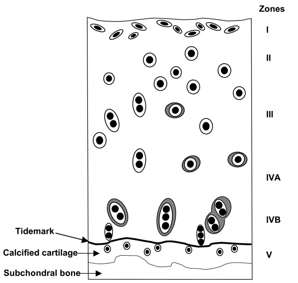
Background: The chondro-osseous junctional region of diarthrodial joints is peculiarly complex and may be considered to consist of the deepest layer of non-calcified cartilage, the tidemark, the layer of calcified cartilage, a thin cement line (between the calcified cartilage and the subchondral bone) and the subchondral bone. A detailed knowledge of the structure, function and pathophysiology of the normal chondro-osseous junction is essential for an understanding of the pathogenesis of osteoarthrosis.
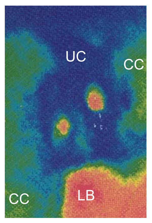
Methods: Full thickness samples from human knee joints were processed and embedded in paraffin wax. One hundred serial sections (10 mum thick) were taken from the chondro-osseous junctional region of a block from the medial tibial plateau of a normal joint. They were stained with haematoxylin and eosin and photographed. For a simple physical reconstruction images of each 10th sequential tissue section were printed and the areas of the photomicrographs containing the chondro-osseous junctional region were cut out and then overlaid so as to create a three-dimensional (3D) model of this region. A 3D reconstruction was also made using computer modelling.
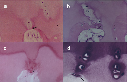
Results: Histochemical staining revealed some instances where prolongations of uncalcified cartilage, delineated by the tidemark, dipped into the calcified cartilage and, in places, abutted onto subchondral bone and marrow spaces. Small areas of uncalcified cartilage containing chondrocytes (virtual islands) were seen, in two-dimensional (2D) sections, to be apparently entombed in calcified matrix. The simple physical 3D reconstruction confirmed that these prolongations of uncalcified cartilage were continuous with the cartilage of zone IV and demonstrated that the virtual islands of uncalcified cartilage were cross-sections of these prolongations. The computer-generated 3D reconstructions clearly demonstrated that the uncalcified prolongations ran through the calcified cartilage to touch bone and marrow spaces and confirmed that the apparent entombment of chondrocytes was a 2D artefact.
Conclusion: This study demonstrates that the chondro-osseous junctional region is more complex than previously described. The tidemark is a clearly defined boundary delineating uncalcified from calcified cartilage. It is not a straight line across a joint, but a complex three-dimensional structure that follows uncalcified cartilage prolongations dipping down through the calcified cartilage to abut onto subjacent bone or marrow spaces.
Figures


References
-
- Havelka S, Horn V, Spohrova D, Valough P. The calcified-non calcified cartilage interface; the tidemark. Acta Biol Hung. 1984;35:271–279. - PubMed
-
- Green WT, Garland NM, Eanes ED, Sokoloff L. Microradiography study of the calcified layer of articular cartilage. Arch Pathol. 1970;90:151–158. - PubMed
-
- Dmitrovsky E, Lane LB, Bullough PG. The characterisation of the tidemark in human articular cartilage. Metab Bone Dis Relat Res. 1978;1:115–118. doi: 10.1016/0221-8747(78)90047-4. - DOI
MeSH terms
Substances
LinkOut - more resources
Full Text Sources
Other Literature Sources
Miscellaneous
