. 2006 Jul;188(13):4787-800.
doi: 10.1128/JB.00066-06.

Protein-protein interactions among Helicobacter pylori cag proteins

Affiliations

Protein-protein interactions among Helicobacter pylori cag proteins

Valerie J Busler et al. J Bacteriol. 2006 Jul.

Abstract

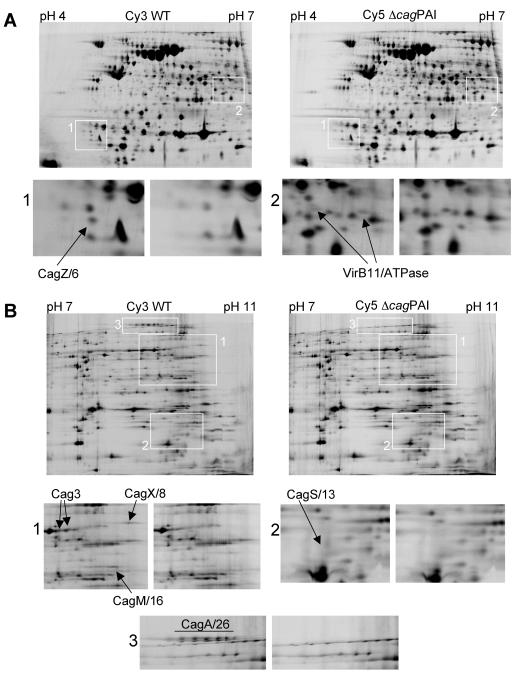
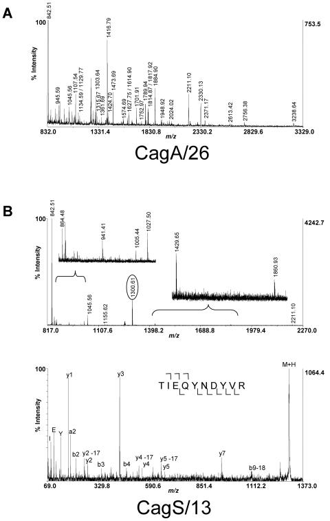
Many Helicobacter pylori isolates contain a 40-kb region of chromosomal DNA known as the cag pathogenicity island (PAI). The risk for development of gastric cancer or peptic ulcer disease is higher among humans infected with cag PAI-positive H. pylori strains than among those infected with cag PAI-negative strains. The cag PAI encodes a type IV secretion system that translocates CagA into gastric epithelial cells. To identify Cag proteins that are expressed by H. pylori during growth in vitro, we compared the proteomes of a wild-type H. pylori strain and an isogenic cag PAI deletion mutant using two-dimensional difference gel electrophoresis (2D-DIGE) in multiple pH ranges. Seven Cag proteins were identified by this approach. We then used a yeast two-hybrid system to detect potential protein-protein interactions among 14 Cag proteins. One heterotypic interaction (CagY/7 with CagX/8) and two homotypic interactions (involving H. pylori VirB11/ATPase and Cag5) were similar to interactions previously reported to occur among homologous components of the Agrobacterium tumefaciens type IV secretion system. Other interactions involved Cag proteins that do not have known homologues in other bacterial species. Biochemical analysis confirmed selected interactions involving five of the proteins that were identified by 2D-DIGE. Protein-protein interactions among Cag proteins are likely to have an important role in the assembly of the H. pylori type IV secretion apparatus.


Figures

FIG. 1.
FIG. 1.
Translocation of CagA into AGS cells. AGS gastric cells were cocultured with a wild-type H. pylori strain (H. pylori strain 26695 Sc#7) (WT), with an isogenic mutant strain that lacked the cag PAI (ΔcagPAI), or without bacteria. After 6 h, the cells were lysed, and the proteins in the lysates were analyzed by immunoblotting with an anti-phosphotyrosine (anti-PY99) antibody (top panel). The membrane was then stripped and immunoblotted with an anti-CagA antibody (bottom panel). The wild-type strain expressed a CagA protein that underwent tyrosine phosphorylation (CagAp-Tyr), and the isogenic cag PAI deletion mutant strain did not express CagA.
FIG. 2.
FIG. 2.
Expression of proteins encoded by the cag pathogenicity island. H. pylori strain 26695 Sc#7 (WT) and an isogenic cag PAI deletion mutant strain (ΔcagPAI) were lysed, and the proteomes of these strains were analyzed by 2D-DIGE/MS, as described in Materials and Methods. Seven H. pylori Cag proteins (Cag3, VirB11/ATPase, CagZ/6, CagX/8, CagS/13, CagM/16, and CagA/26) (Table 2) were expressed by the wild-type strain but not by the mutant strain. (A) Representative data from a 2D-DIGE experiment using a gel with pH range of 4 to 7. CagZ/6 and VirB11/ATPase are indicated. VirB11/ATPase was detected in two isoforms (panel 2). (B) Representative data from a 2D-DIGE experiment using a gel with a pH range of 7 to 11. Cag3, CagX/8, CagM/16, CagS/13, and CagA/26 are indicated. Cag3 was detected in two isoforms (panel 1), and CagA/26 was detected in multiple isoforms (panel 3).
FIG. 3.
FIG. 3.
Identification of Cag proteins by mass spectrometry. Examples of MALDI-TOF and tandem TOF/TOF mass spectra. (A) Peptide mass map spectrum for CagA/26 displaying intact peptide ion masses (M + H) to within a 20-ppm mass accuracy. (B) Similar peptide mass map spectrum for CagS/13, with accompanying MS/MS fragmentation spectrum for an ion at m/z 1,300.61 (circled). Fragment ions containing the carboxyl terminus (y ions) are denoted below the peptide sequence (numbering follows the peptide right to left), and fragment ions containing the amino terminus (b ions) are denoted above the peptide sequence (numbering follows the peptide left to right). CagS/13 was the faintest of the seven Cag proteins identified by 2D-DIGE analysis.
FIG. 4.
FIG. 4.
Protein-protein interactions among H. pylori Cag proteins. Yeast cells were cotransformed with plasmids encoding the indicated fusion proteins. Cotransformed yeast cells (normalized based on optical density) were 10-fold serially diluted. Identical inocula were plated onto two types of media (SD medium lacking tryptophan and leucine and SD medium lacking tryptophan, leucine, and histidine). All of the cotransformed yeast cells grew on SD plates containing 2% glucose and lacking tryptophan and leucine. Growth of the cotransformed yeast cells on SD medium lacking tryptophan, leucine, and histidine provided evidence for the occurrence of protein-protein interactions. Panel A depicts three of the four homotypic interactions identified in the current study. Cag3(A) is a full-length protein, and Cag3(B) is a gene fragment lacking the predicted signal sequence. Panel B shows representative heterotypic interactions. GAL4AD-WT plus GAL4BD-WT are a positive control, and GAL4AD-WT plus GAL4BD-pLC are a negative control.
FIG. 5.
FIG. 5.
Interaction of H. pylori CagY/7 (VirB10 homologue) with H. pylori CagX/8 (VirB9 homologue). (A) Alignment of the full-length H. pylori CagY protein with A. tumefaciens VirB10. Three CagY/7 fragments [CagY/7(A), CagY/7(B), and CagY/7(C)] were expressed in the yeast two-hybrid system, as shown. The C-terminal region of CagY/7 [CagY/7(C)] is homologous to VirB10 from A. tumefaciens. (B) Yeast cells were cotransformed with the plasmids encoding the indicated fusion proteins and plated as described in the Fig. 4 legend. Yeast cotransformed with plasmids encoding GAL4BD-CagY/7(C) (the C terminus of CagY/7, which is homologous to VirB10) and GAL4AD-CagX/8 (a VirB9 homologue) grew on SD medium lacking tryptophan, leucine, and histidine, indicating an interaction between the two proteins encoded by these genes.
FIG. 6.
FIG. 6.
Analysis of β-galactosidase activity. Yeast cells cotransformed with the indicated plasmid pairs (AD indicates the GAL4 activation domain, and BD indicates the GAL4 binding domain) were tested in triplicate for β-galactosidase activity as described in Materials and Methods. β-Galactosidase activity provides evidence for protein-protein interactions. The activity produced by each putative interacting pair was classified into one of three categories as shown in Table 4. High activity (+++) is comparable to that of the positive control, medium activity (++) is comparable to that of the mutant positive control, and low activity (+) is lower than that of the mutant positive control but higher than that of the negative control. Representative data from each of the three categories are shown.
FIG. 7.
FIG. 7.
Detection of interactions among Cag fusion proteins. (A) The indicated proteins were expressed in E. coli, and E. coli lysates were then immunoblotted with anti-GST or anti-MBP antibodies. (B) E. coli extracts containing Cag fusion proteins, a GST control protein without a fused Cag protein, or an MBP control protein without a fused Cag protein were mixed as indicated. In each case, a GST-Cag fusion protein or GST control was mixed with an MBP-Cag fusion protein or MBP control. GST-Cag fusion proteins or GST without a fused Cag protein and any interacting MBP fusion proteins were affinity purified using glutathione-Sepharose beads. Affinity-purified proteins were analyzed by SDS-PAGE and immunoblotting. Each membrane was first analyzed by anti-MBP immunoblotting and then stripped and analyzed by anti-GST immunoblotting, as shown. MBP-CagV/10 and MBP-CagM/16 copurified with GST-Cag3 but not with GST alone. MBP-CagV/10, MBP-CagM/16, MBP-CagX/8, MBP-CagS/13, and MBP-CagI/19 copurified with GST-CagZ/6 but not with GST alone.

References

    1. Agatep, R., R. D. Kirkpatrick, D. L. Parchaliuk, R. A. Woods, and R. D. Gietz. 1998. Transformation of Saccharomyces cerevisiae by the lithium acetate/single-stranded carrier DNA/polyethylene glycol (LiAc/ss-DNA/PEG) protocol. Technical Tips Online. [Online.] http://tto.trends.com.
    1. Akopyants, N. S., S. W. Clifton, D. Kersulyte, J. E. Crabtree, B. E. Youree, C. A. Reece, N. O. Bukanov, E. S. Drazek, B. A. Roe, and D. E. Berg. 1998. Analyses of the cag pathogenicity island of Helicobacter pylori. Mol. Microbiol. 28:37-53. - PubMed
    1. Alban, A., S. O. David, L. Bjorkesten, C. Andersson, E. Sloge, S. Lewis, and I. Currie. 2003. A novel experimental design for comparative two-dimensional gel analysis: two-dimensional difference gel electrophoresis incorporating a pooled internal standard. Proteomics 3:36-44. - PubMed
    1. Amieva, M. R., R. Vogelmann, A. Covacci, L. S. Tompkins, W. J. Nelson, and S. Falkow. 2003. Disruption of the epithelial apical-junctional complex by Helicobacter pylori CagA. Science 300:1430-1434. - PMC - PubMed
    1. Anderson, L. B., A. V. Hertzel, and A. Das. 1996. Agrobacterium tumefaciens VirB7 and VirB9 form a disulfide-linked protein complex. Proc. Natl. Acad. Sci. USA 93:8889-88894. - PMC - PubMed

Publication types

MeSH terms

LinkOut - more resources