. 2006 Jul;2(7):e116.
doi: 10.1371/journal.pgen.0020116. Epub 2006 Jun 12.

Lysine 63-polyubiquitination guards against translesion synthesis-induced mutations

Affiliations

Lysine 63-polyubiquitination guards against translesion synthesis-induced mutations

Roland K Chiu et al. PLoS Genet. 2006 Jul.

Abstract

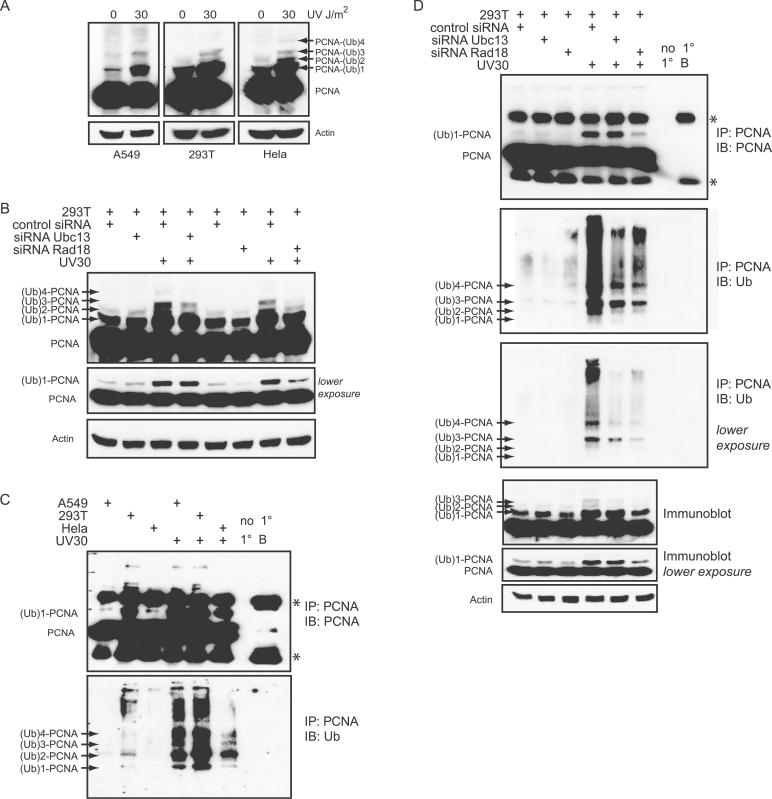
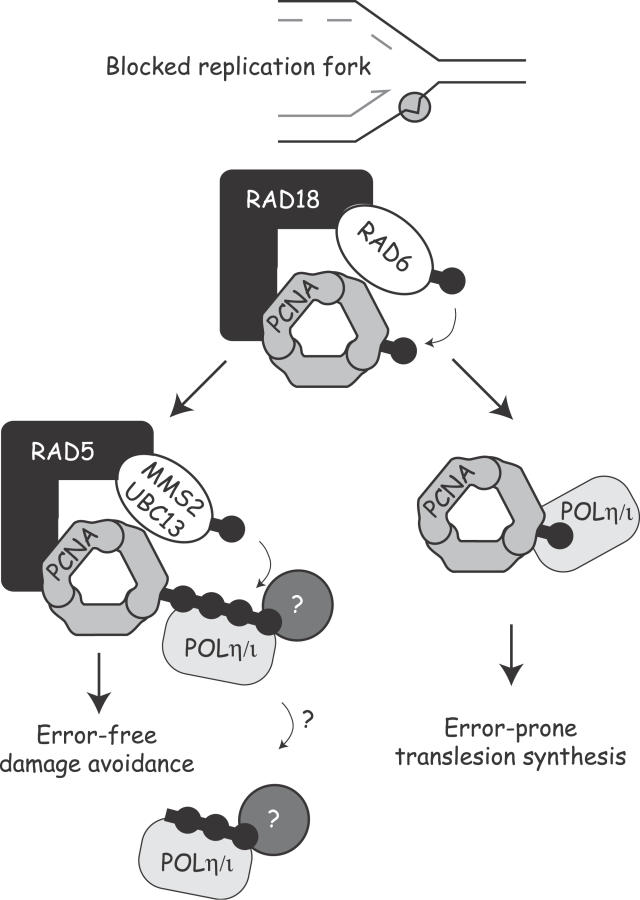
Eukaryotic cells possess several mechanisms to protect the integrity of their DNA against damage. These include cell-cycle checkpoints, DNA-repair pathways, and also a distinct DNA damage-tolerance system that allows recovery of replication forks blocked at sites of DNA damage. In both humans and yeast, lesion bypass and restart of DNA synthesis can occur through an error-prone pathway activated following mono-ubiquitination of proliferating cell nuclear antigen (PCNA), a protein found at sites of replication, and recruitment of specialized translesion synthesis polymerases. In yeast, there is evidence for a second, error-free, pathway that requires modification of PCNA with non-proteolytic lysine 63-linked polyubiquitin (K63-polyUb) chains. Here we demonstrate that formation of K63-polyUb chains protects human cells against translesion synthesis-induced mutations by promoting recovery of blocked replication forks through an alternative error-free mechanism. Furthermore, we show that polyubiquitination of PCNA occurs in UV-irradiated human cells. Our findings indicate that K63-polyubiquitination guards against environmental carcinogenesis and contributes to genomic stability.


Conflict of interest statement

Competing interests. The authors have declared that no competing interests exist.

Figures

Figure 1
Figure 1. Disruption of K63-PolyUb Chain Assembly
(A) Cartoon depicting dominant negative K63R-Ub-GFP construct. The expressed fusion protein is processed by endogenous Ub proteases generating free GFP used for detection on a flow cytometer and mono-K63R-Ub. Incorporation of this mutant will terminate K63-polyUb chains while not affecting canonical K48-polyUb chain assembly. (B) Whole-cell lysates were isolated from untransfected cells, and from cells stably expressing either WT-Ub or K63R-Ub, followed by immunoblot analysis with antibodies directed against Ub, His, and GFP. (C) The growth of untransfected, WT-Ub, or K63R-Ub cells was followed by cell counting over the course of 7 d.
Figure 2
Figure 2. Cells Deficient in K63-Ub Chain Formation Are Sensitized to Cisplatin Treatment while UV Sensitivity Is Revealed only upon POLη Knockdown
(A and B) Clonogenic survival assays were used to determine sensitivity to 1 h acute treatment with cisplatin in untransfected A549 cells or in A549 cells stably expressing WT-Ub or K63R-Ub. The mean values of three independent experiments are shown with standard error of the mean (error bars). Cells expressing K33R-Ub or cells that lost K63R-Ub expression revert to WT-Ub cisplatin sensitivity. (C) Cells were treated for 24 h with 100 μM cisplatin followed by Hoechst staining to detect apoptosis. The mean values of three independent experiments are shown with standard deviation. (D) Clonogenic survival assays were used to determine sensitivity to UV irradiation in untransfected A549 cells or in A549 cells stably expressing WT-Ub or K63R-Ub. (E) Clonogenic survival of A549 cells stably expressing WT-Ub or K63R-Ub with or without POLη RNAi following 10 J/m2 UV treatment.
Figure 3
Figure 3. Cells Deficient in K63-Ub Chain Formation Are Mutagenic in Response to UV Treatment
(A and B) Cells were treated with cisplatin for 1 h or UV irradiation and subcultured for 7 d. Cells were then plated and grown in 6-TG to select for HPRT mutants. The mean values of three independent experiments are shown with standard deviation. (C) Normal fibroblasts stably expressing WT-Ub or K63R-Ub were UV-irradiated (10 J/m2) and cultured for 5 d. Cells were then plated and grown in 6-TG to select for HPRT mutants. (D) The number of HPRT mutants was quantitated for A549 cells stably expressing WT-Ub or K63R-Ub with or without POLη RNAi. Cells were treated as described in Figure 3C. (E) Cells were UV-irradiated and plated in the absence or presence of 0.4 mM caffeine. The mean values of three independent experiments are shown with standard error of the mean (error bars).
Figure 4
Figure 4. Disrupting K63-PolyUb Chain Formation Increases Reliance of Cells on the Error-Prone TLS Pathway
(A) HeLa cells stably expressing WT-Ub-puro or K63R-Ub-puro were transiently transfected with a plasmid expressing a POLη-GFP fusion. Twenty-four hours post-transfection, cells were UV-irradiated (10 J/m2). POLη (green) and PCNA (red) were detected using antibodies. Shown are representative confocal photographs of cells 6 h post-UV treatment. (B) Kinetics of POLη foci formation in WT-Ub– and K63R-Ub–expressing HeLa cell lines. (C) HPRT mutation spectra. RNA was isolated from 6-TG resistant 10 J/m2 UV-treated clones followed by RT-PCR and sequence analysis of the HPRT locus. The UV-induced mutations are shown in the upper table. Most of the point mutations were G→A or C→T transitions indicated as G/C→A/T. The lower table in (C) shows the same mutants in sequence context. (D) Foci were quantitated 6 h post-UV treatment using a live-cell imaging fluorescent microscope. (E) The number of HPRT mutants was quantitated for A549 cells stably expressing K63R-Ub with or without RAD18 RNAi. Cells were treated as described in Figure 4B.
Figure 5
Figure 5. Modification of PCNA by Polyubiquitin in Human Cells after DNA Damage
(A) A549, 293T, and Hela cells were irradiated with 0 or 30 J/m2 UV and lysed 6 h posttreatment followed by immunoblotting for PCNA. (B) 293T cells were transfected with 100 nM of either control siRNA, siRNA Ubc13, or siRNA RAD18. Seventy-two hours post-transfection, cells were treated as described in Figure 1A. A darker and lighter exposure of the PCNA immunoblot is shown. (C) A549, 293T, and Hela cells were irradiated with 30 J/m2 UV and lysed in boiling SDS, diluted in lysis buffer and subjected to immunoprecipitation with a PCNA antibody and detected with PCNA or Ub antibodies. The controls in the immunoprecipitations were “no 1”, in which lysates were incubated with beads but no PCNA antibody, and “1 B” in which PCNA antibody was incubated with beads alone. (D) 293T cells were transfected as described in Figure 5B. Seventy-two hours post-transfection, cells were irradiated with 30 J/m2 of UV and lysed 6 h later in boiling SDS, diluted in lysis buffer, and subjected to immunoprecipitation with a PCNA antibody and immunoblotted for PCNA (upper panel) and Ub (lower panel). A lighter exposure of the PCNA IP immunoblotted for Ub is also shown. A PCNA immunoblot with darker and lighter exposure performed on protein lysates from the same samples used in the immunoprecipitations is also shown. Asterisks denote immunoglobulin heavy and light chains as detected on the immunoprecipitations.
Figure 6
Figure 6. Model of the DDT Pathway in Mammalian Cells
Recovery from a stalled replication fork at sites of DNA damage can occur by one of two alternative pathways. Previous work has shown that PCNA mono-ubiquitination by the RAD6/RAD18 complex stimulates lesion bypass through recruitment of the error-prone TLS polymerases. Here we show that an alternative error-free pathway requires formation of K63-polyUb chains. Blockade of this error-free pathway results in increased use of the TLS polymerases after DNA damage and a corresponding increase in mutations. As the TLS polymerases POLη and POLι both bind directly and avidly to polyUb chains [20], it is hypothesized that the interaction with K63-polyUb causes a disengagement of the polymerase from the DNA, allowing other proteins to migrate to the site of damage to perform error-free repair. This model predicts that K63-polyubiquitination acts to suppress environmental carcinogenesis by preventing genomic instability that would otherwise be introduced by the TLS polymerases.

References

    1. Spivak G, Hanawalt PC. Translesion DNA synthesis in the dihydrofolate reductase domain of UV-irradiated CHO cells. Biochemistry. 1992;31:6794–6800. - PubMed
    1. Wu HI, Brown JA, Dorie MJ, Lazzeroni L, Brown JM. Genome-wide identification of genes conferring resistance to the anticancer agents cisplatin, oxaliplatin, and mitomycin C. Cancer Res. 2004;64:3940–3948. - PubMed
    1. Simon JA, Szankasi P, Nguyen DK, Ludlow C, Dunstan HM, et al. Differential toxicities of anticancer agents among DNA repair and checkpoint mutants of Saccharomyces cerevisiae . Cancer Res. 2000;60:328–333. - PubMed
    1. Bailly V, Lauder S, Prakash S, Prakash L. Yeast DNA repair proteins Rad6 and Rad18 form a heterodimer that has ubiquitin conjugating, DNA binding, and ATP hydrolytic activities. J Biol Chem. 1997;272:23360–23365. - PubMed
    1. Hoege C, Pfander B, Moldovan GL, Pyrowolakis G, Jentsch S. RAD6-dependent DNA repair is linked to modification of PCNA by ubiquitin and SUMO. Nature. 2002;419:135–141. - PubMed

Publication types