. 2006 Jul;2(7):e120.
doi: 10.1371/journal.pgen.0020120. Epub 2006 Jun 15.

Who ate whom? Adaptive Helicobacter genomic changes that accompanied a host jump from early humans to large felines

Affiliations

Who ate whom? Adaptive Helicobacter genomic changes that accompanied a host jump from early humans to large felines

Mark Eppinger et al. PLoS Genet. 2006 Jul.

Abstract

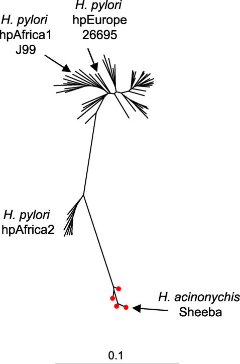
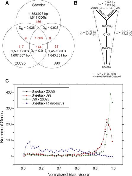
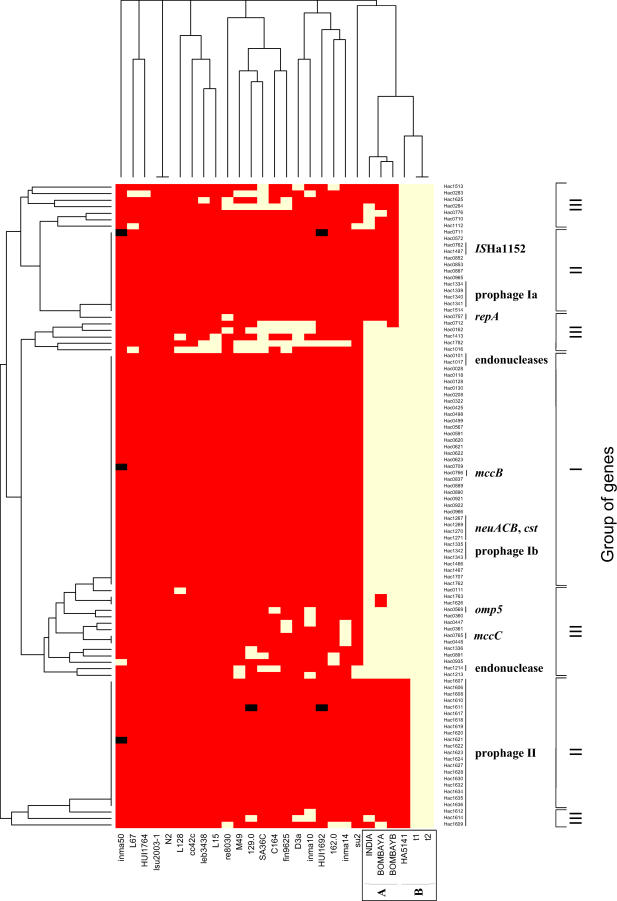
Helicobacter pylori infection of humans is so old that its population genetic structure reflects that of ancient human migrations. A closely related species, Helicobacter acinonychis, is specific for large felines, including cheetahs, lions, and tigers, whereas hosts more closely related to humans harbor more distantly related Helicobacter species. This observation suggests a jump between host species. But who ate whom and when did it happen? In order to resolve this question, we determined the genomic sequence of H. acinonychis strain Sheeba and compared it to genomes from H. pylori. The conserved core genes between the genomes are so similar that the host jump probably occurred within the last 200,000 (range 50,000-400,000) years. However, the Sheeba genome also possesses unique features that indicate the direction of the host jump, namely from early humans to cats. Sheeba possesses an unusually large number of highly fragmented genes, many encoding outer membrane proteins, which may have been destroyed in order to bypass deleterious responses from the feline host immune system. In addition, the few Sheeba-specific genes that were found include a cluster of genes encoding sialylation of the bacterial cell surface carbohydrates, which were imported by horizontal genetic exchange and might also help to evade host immune defenses. These results provide a genomic basis for elucidating molecular events that allow bacteria to adapt to novel animal hosts.


Conflict of interest statement

Competing interests. The authors have declared that no competing interests exist.

Figures

Figure 1
Figure 1. Neighbor-Joining Tree Based on the GTR+G+I Evolutionary Model for 3,406 bp Sequences from 58 Strains of H. pylori and Four Strains of H. acinonychis
The tree shows the phylogenetic relationships between H. acinonychis and populations within H. pylori, and arrows indicate the three strains, J99, 26695, and Sheeba, from which genome sequences are currently available. This phylogenetic tree indicates that H. pylori (lines) and H. acinonychis (lines plus red dots) are closely related but cannot resolve the direction of ancestor-descendent relationships. Genetic distance scale bar at bottom.
Figure 2
Figure 2. Similarities between the Genomes of H. acinonychis Sheeba and H. pylori 26695 and J99
(A) Venn diagram of genomic properties. Numbers in red within each arc represent numbers of genes, each of which may contain multiple CDSs if the corresponding gene is fragmented. (B) Age calculations since a common ancestor (LCA) based on synonymous pair-wise distances for 612 conserved genes according to the methods of Li et al., 1985 and the modified Nei-Gojobori method. (C) Frequencies of normalized blast scores in pair-wise comparisons between Sheeba and three other genomes.
Figure 3
Figure 3. Gene Fragmentation, Duplication, and Import
(A) Fragmentation of the vacA (vacuolating cytotoxin) gene (red) into 13 fragments and import of neuACB, cst, cst (blue-green) within Sheeba. neuACB encode acylneuraminate cytidyltransferases and cst encodes a sialyltransferase. (B) Translocation of a duplicate of the fragmented vacA gene to a different genomic location. The duplicated vacA gene (red) contains the same fragmentation pattern and differs by only one sequence polymorphism in 3,815 bp from that in part A, indicating that the duplication is recent and occurred after the fragmentation event. Next to the duplicated vacA gene are located three genes (light blue) that are unique to H. acinonychis. (C) Homologies of the neuACB, cst, cst gene cluster from part A with syntenic clusters in C. jejuni NCTC11168 and the B. cereus virulence plasmid pBC218.
Figure 4
Figure 4. Fragmentation Patterns in Ten Genes among Three H. acinonychis Strains
Ten genes that are intact in 26695 but are fragmented in the Sheeba genome (subgroup B) were re-sequenced from strains t1 and HA5141 of subgroup B and BombayA of subgroup A. Black lines indicate sequenced fragments and thick blue arrows indicate CDSs of ≥140 bp. Designations at the top indicate CDS designations in 26695 whereas designations above the Sheeba sequences indicate both the protein name and the CDS designations in Sheeba (Hac0035, Hac0036, etc.).
Figure 5
Figure 5. Hybridization of a DNA Microarray Chip Containing 99 PCR Products against Representative Strains of H. acinonychis and H. pylori
Six strains of H. acinonychis from subgroups A and B and 21 strains that represent the genetic diversity of H. pylori (Table S5) were tested for hybridization (yellow) or lack of hybridization (red) with 99 PCR products (Table S6) from genes that are present in Sheeba and lacking in 26695 and J99. The results were clustered according to genes (left) and strains (top). Genes that hybridize exclusively with all H. acinonychis strains are summarized as group I (right), genes hybridizing only with some H. acinonychis are in group II and genes hybridizing with some H. pylori are in group III. Where gene functions were attributed, they are indicated at the right and other genes encode hypothetical proteins. black, missing data.
Figure 6
Figure 6. Genomic Rearrangements in Pair-Wise Genomic Comparisons between Sheeba and other Helicobacteraceae [–18,85] or Campylobacteraceae [86] (A–B, D–F) or between 26695 and J99 (C)
Stretches consisting of two or more syntenic orthologs are indicated in green for orthologs in the same orientation relative to the origin and in red for orthologs in inverted orientation. Single orthologous CDSs are indicated in yellow. Ortholog matches were plotted using CGViz (http://www-ab.informatik.uni-tuebingen.de/software/cgviz).

References

    1. Suerbaum S, Michetti P. Helicobacter pylori infection. New England J Med. 2002;347:1175–1186. - PubMed
    1. Marshall BJ, Warren JR. Unidentified curved bacilli in the stomach of patients with gastritis and peptic ulceration. Lancet. 1984;1:1311–1315. - PubMed
    1. Solnick JV, Schauer DB. Emergence of diverse Helicobacter species in the pathogenesis of gastric and enterohepatic diseases. Clin Microbiol Rev. 2001;14:59–97. - PMC - PubMed
    1. Kersulyte D, Mukhopadhyay AK, Velapatino B, Su W, Pan Z, et al. Differences in genotypes of Helicobacter pylori from different human populations. J Bacteriol. 2000;182:3210–3218. - PMC - PubMed
    1. Dailidiene D, Dailide G, Ogura K, Zhang M, Mukhopadhyay AK, et al. Helicobacter acinonychis: Genetic and rodent infection studies of a Helicobacter pylori-like gastric pathogen of cheetahs and other big cats. J Bacteriol. 2004;186:356–365. - PMC - PubMed

Publication types

Substances