. 2006 Jun;2(6):e49.
doi: 10.1371/journal.ppat.0020049. Epub 2006 Jun 9.

CCR5 is essential for NK cell trafficking and host survival following Toxoplasma gondii infection

Affiliations

CCR5 is essential for NK cell trafficking and host survival following Toxoplasma gondii infection

Imtiaz A Khan et al. PLoS Pathog. 2006 Jun.

Abstract

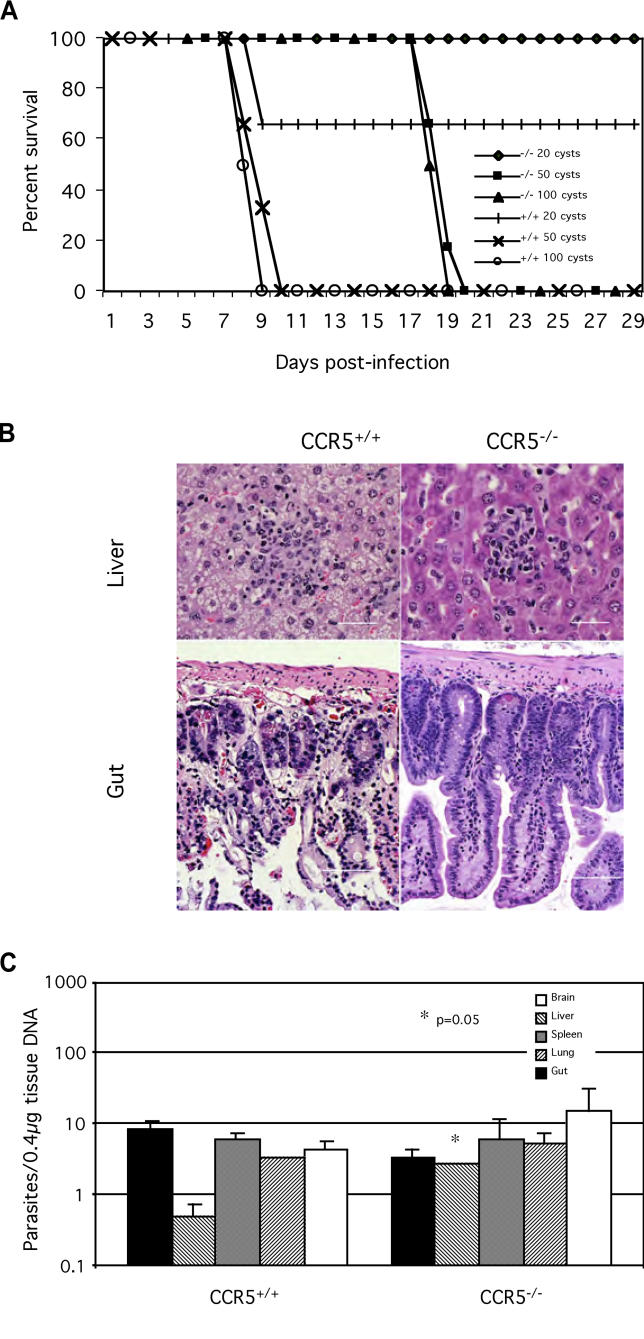
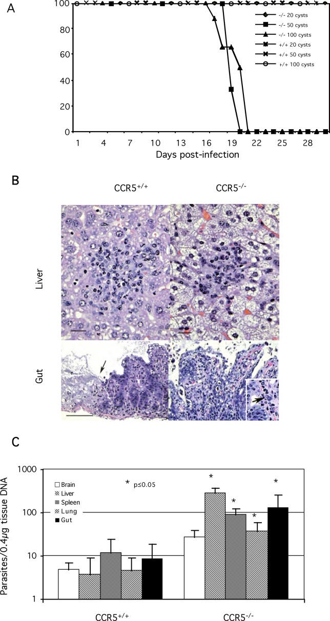
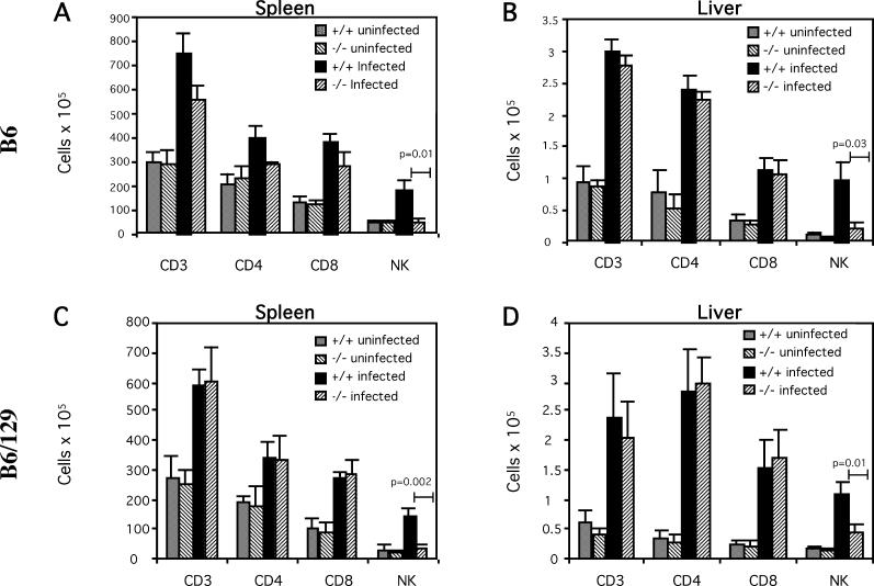
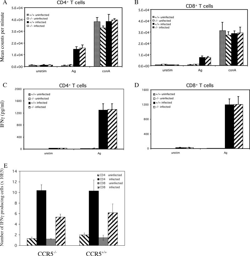
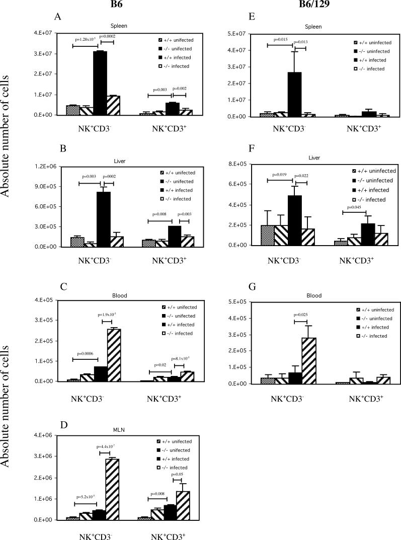
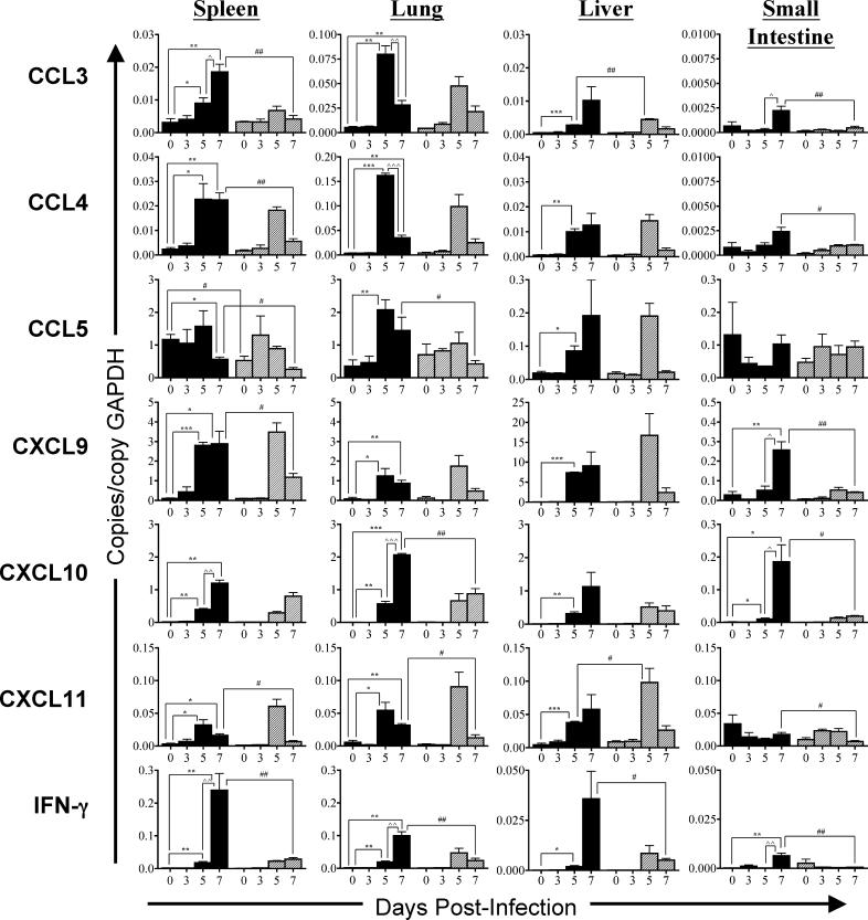
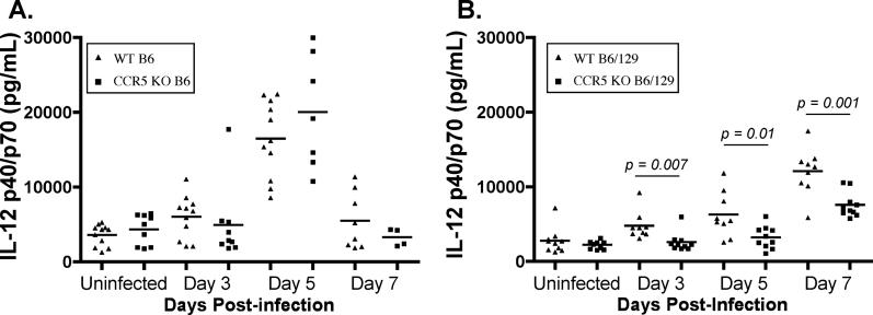
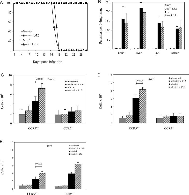
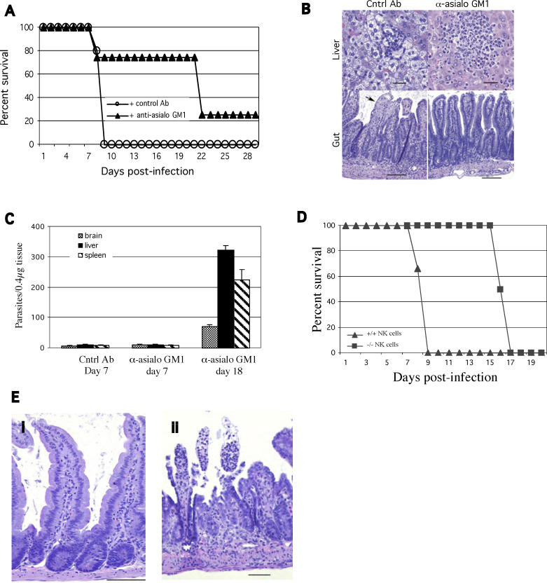
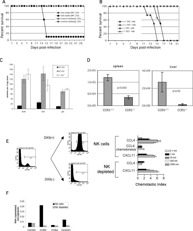
The host response to intracellular pathogens requires the coordinated action of both the innate and acquired immune systems. Chemokines play a critical role in the trafficking of immune cells and transitioning an innate immune response into an acquired response. We analyzed the host response of mice deficient in the chemokine receptor CCR5 following infection with the intracellular protozoan parasite Toxoplasma gondii. We found that CCR5 controls recruitment of natural killer (NK) cells into infected tissues. Without this influx of NK cells, tissues from CCR5-deficient (CCR5-/-) mice were less able to generate an inflammatory response, had decreased chemokine and interferon gamma production, and had higher parasite burden. As a result, CCR5-/- mice were more susceptible to infection with T. gondii but were less susceptible to the immune-mediated tissue injury seen in certain inbred strains. Adoptive transfer of CCR5+/+ NK cells into CCR5-/- mice restored their ability to survive lethal T. gondii infection and demonstrated that CCR5 is required for NK cell homing into infected liver and spleen. This study establishes CCR5 as a critical receptor guiding NK cell trafficking in host defense.


Conflict of interest statement

Competing interests. The authors have declared that no competing interests exist.

Figures

Figure 1
Figure 1. Survival, Histopathology, and Parasite Load of CCR5−/− and CCR5+/+ Mice in a C57BL/6 Background following Infection with Different Doses of T. gondii
(A) Survival. Female CCR5−/− mice (5–7 wk old) and age-matched CCR5+/+ wild-type controls in the C57BL/6 background were challenged perorally with the cysts of the 76K strain of T. gondii. The animals were monitored for survival on a daily basis. There were six animals per group and the experiment was performed three times with similar results. (B) Histopathology. Photomicrographs of hematoxylin and eosin-stained liver and ileum of small bowel isolated from infected CCR5−/− and CCR5+/+ mice. Liver of infected CCR5+/+ mouse reveals hepatocytes with extensive fatty degeneration. A nodule of inflammatory cells is seen in the center of the field, composed of lymphocytes and granulocytes. Hepatocyte morphology is preserved in liver of an infected CCR5−/− mouse. A small nodule of inflammatory cells, largely composed of lymphocytes, is seen in the center of the field. Ileum of an infected CCR5+/+ mouse contains extensive epithelial necrosis and ulceration. Dilated blood vessels with hemorrhage are evident. The epithelial architecture is preserved in ileum of infected CCR5−/− mouse. Bars: ileum, 100 μm; liver, 50 μm. (C) Level of parasite DNA. Groups of CCR5−/− and CCR5+/+ mice on the C57BL/6 background were infected perorally with 20 cysts of T. gondii. At day 7 PI, small intestine, liver, spleen, lung, and brain from both CCR5−/− and wild-type mice (three mice per group) were collected and the level of parasite load in the tissues was determined by competitive DNA PCR. The experiment was performed twice with similar results.
Figure 2
Figure 2. Survival, Histopathology, and Parasite Load of CCR5−/− and CCR5+/+ Mice in a C57BL/6x129 Background following Infection with Different Doses of T. gondii
(A) Survival. Female CCR5−/− mice (5–8 wk old) and age-matched CCR5−/− wild-type littermate controls, both in a C57BL/6x129 background, were challenged perorally with the cysts of the 76K strain of T. gondii. The animals were monitored for survival on daily basis. There were six animals per group and the experiment was performed three times with similar results. All CCR5+/+ mice survived at the three doses of cysts tested. (B) Histopathology. Photomicrographs of hematoxylin and eosin-stained liver and ileum of small intestine isolated from infected CCR5+/+ mice and CCR5−/− mice at day 14 PI. Wild-type liver: Moderate fatty change is seen in hepatocytes, with small foci of mixed polymorphonuclear and mononuclear infiltration. Bar, 10 μm. CCR5−/− liver: Severe fatty changes are seen in hepatocytes with mononuclear and polymorphonuclear inflammatory nodules. Bar, 10 μm. Wild-type small intestine: The superficial mucosa is missing in some places (arrow) and no parasite multiplication is evident. Bar, 100 μm. CCR5−/− small intestine. The superficial mucosa is largely missing and a mixed inflammatory infiltrate including polymorphonuclear cells is evident within the lamina propria and superficial mucosa. Bar, 100 μm. Inset: Higher magnification with arrow pointing to tachyzoites in lamina propria of CCR5−/− mice. Bar, 75 μm. (C) Level of parasite DNA. Groups of CCR5−/− mice and wild-type littermate controls both on a C57BL/6x129 background were infected perorally with 20 cysts of T. gondii. At day 14 PI, small intestine, liver, spleen, lung, and brain from both CCR5−/− and wild-type mice (three mice per group) were collected and the parasite load in the tissues was determined by competitive PCR. The experiment was performed twice with similar results.
Figure 3
Figure 3. T Cell and NK Cell Numbers in the Spleen and Liver of T. gondii-Infected CCR5−/− and CCR5+/+ Mice in the C57BL/6 and C57BL/6x129 Backgrounds
Cell numbers. T cell and NK cell numbers in spleen (A and C) and liver (B and D) of mice infected with 15 cysts and harvested 7 d PI. Single-cell suspensions of spleen and hepatic lymphocytes were prepared and phenotyped for the expression of CD3, CD4, CD8, and NK1.1 by FACS analysis. The experiment was performed twice with similar results (three or four mice per group).
Figure 4
Figure 4. CD4+ and CD8+ T Cell T. gondii-Induced Proliferation and IFNγ Production
(A and B) Proliferation. Magnetically purified positively selected CD4+ (A) and CD8+ (B) T cells (>95% pure) isolated from the spleens of C57BL/6x129 on day 7 PI. Cells were stimulated either with ConA (2.5 μg/ml) or Toxoplasma lysate antigen (15 μg/ml). After 72 h incubation, proliferation was measured by 3H thymidine incorporation. Data are represented as mean cpm ± standard deviation and are representative of two experiments. (C and D) IFNγ secretion. 106 purified CD4+ (C) and CD8+ (D) T cells from 7 d infected C57BL/6x129 mice were cultured in presence of 15 μg/ml of Toxoplasma lysate antigen and irradiated feeder cells (5 × 10⁵ cells/well) in 24-well plates. After 72 h of incubation the supernatants were collected, centrifuged, and assayed for IFNγ production by ELISA. (E) Intracellular IFNγ production. Female CCR5−/− (5–8 wk old) and wild-type mice were infected perorally with T. gondii cysts and splenocytes were harvested at day 7 PI, pooled (three mice per group), and cultured in vitro with phorbol 12-myristate 13-acetate, ionomycin, and monensin for 4 h. The cultured cells were stained for CD4 or CD8 before intracellular staining for IFNγ. Data are presented as percentage (mean ± standard deviation) of CD4+ or CD8+ T cells positive for IFNγ and are pooled from two experiments.
Figure 5
Figure 5. NK and NK T Cell Numbers in T. gondii-Infected CCR5−/− and CCR5+/+ Mice in the C57BL/6 and C57BL/6x129 Backgrounds
CCR5+/+ and CCR5−/− mice in C57BL/6 and C57BL/6 × 129 backgrounds (three mice per group) were infected with 15 T. gondii cysts. At day 7 PI, lymphocytes from spleen (A and E), liver (B and F), blood (C and G), and mesenteric lymph nodes (D) were isolated and phenotyped for CD3+ and NK1.1+ by FACS analysis. NK1.1+CD3 and NK1.1+CD3+ cell numbers are graphed. The experiment was performed twice with similar results.
Figure 6
Figure 6. Expression of CCR5 and CXCR3 Chemokine Ligands and IFNγ in T. gondii-Infected Mice
Quantitative PCR was performed on total RNA isolated from spleen, lung, liver, and small intestine of wild-type control and CCR5−/− C57BL/6 mice (female, 5–6 wk old, three or four mice per group) infected orally with 15–50 cysts of 76K strain of T. gondii. Tissues were harvested from uninfected and at days 3, 5, and 7 PI. Data are displayed as copies of cytokine or chemokine per copies of GAPDH, with wild type in black bars and CCR5−/− in hatched bars ± standard error of the mean. Statistics were performed using Student's t-test. Sets of symbols: * indicates comparisons with uninfected wild type, ^ indicates comparisons between days 5 and 7 PI; and # indicates comparisons between wild type and CCR5−/−. * p < 0.05, ** p < 0.01, *** p < 0.001, ^ p < 0.05, ^ ^ p < 0.01, ^ ^ ^ p < 0.001, # p < 0.05, ## p < 0.01, ### p < 0.001.
Figure 7
Figure 7. Serum IL-12 Levels for Wild-Type and CCR5−/− Mice
Wild-type (WT) and CCR5−/− (CCR5 KO) mice in the C57BL/6 background (A) and C57BL/6x129 background (B) are represented. Serum IL-12 levels were measured by ELISA before infection or at days 3, 5, and 9 PI with 50 cysts of strain 76K T. gondii. Each data point was measured in duplicate and indicates IL-12 levels for an individual mouse. The bar represents the average at each time point. Statistics were performed using Student's t-test.
Figure 8
Figure 8. Survival and NK Cell Response following Exogenous IL-12 Treatment of CCR5+/+ and CCR5−/− Mice
(A and B) Effect of IL-12 administration on survival in the C57BL/6 x129 background. CCR5−/− and CCR5+/+ mice (three mice per group) were treated with recombinant murine IL-12 (0.33 μg/mouse) daily intraperitoneally starting one day prior to infection with T. gondii. Uninfected controls treated with either IL-12 or saline were included in the study (six mice per group). Mice were challenged orally with 50 cysts and survival was monitored daily. Parasite load (B) was determined for mice described in (A). Mice were sacrificed on day 14 PI (three mice per group), and tissues (brain, liver, small intestine, and spleen) analyzed for parasite load by competitive DNA PCR. (C–E) Effect of exogenous IL-12 treatment on the NK cell response. CCR5−/− and CCR5+/+ mice were infected orally with the 76K strain of T. gondii (three mice per group) and were treated with recombinant murine IL-12 (0.33 μg/mouse) via intraperitoneal route starting one day prior to infection. Uninfected controls treated with either IL-12 or saline were included in the study (three mice per group). At day 7 PI, single-cell suspensions from the spleens (C), and lymphocytes from liver (D) and blood (E) were prepared and cells were analyzed for the expression of the NK1.1 marker by FACS (experiment was performed twice with similar results).
Figure 9
Figure 9. Effect of NK Cells on Survival following T. gondii Infection of C57BL/6 Mice
(A–B) NK cell depletion of wild type. C57BL/6 mice were infected orally with 50 cysts. One day before and every day after infection mice were injected intraperitoneally with anti-asialo GM1 antibody. The treatment was continued for the duration of the experiment. Eight animals were treated per group, and data are representative of two separate experiments. Survival (A) and histopathology (B) of NK cell depleted and nondepleted T. gondii-infected CCR5+/+ C57BL6 mice. Photomicrographs of hematoxylin and eosin-stained liver and ileum of small intestine isolated from infected anti-asialo GM1 and control antibody-treated mice. NK cell-depleted mice had essentially normal small bowel morphology, and the livers showed an acute inflammatory response compatible with acute T. gondii infection. In nondepleted wild-type mice, the small bowel showed extensive, scattered, superficial necrosis, with acute inflammation, including polymorphonuclear cells in the lamina propria. The livers show marked, confluent fatty degeneration of hepatocytes, with small inflammatory nodules in the parenchyma consistent with a hyperimmune inflammatory response. Size bars: liver, 20 μm; small bowel 100 μm. The arrow indicates an area of superficial necrosis of epithelial cells. (C) Parasite load. Wild-type C57BL/6 mice were infected with 50 cysts orally and NK cell depletion performed as described above. At day 7 and 18 PI mice (three per group) were sacrificed and tissues (brain, liver, and spleen) analyzed for parasite load by competitive DNA PCR. (D–E) NK cell adoptive transfer. NK cells from CCR5−/− and wild-type C57BL/6 mice were isolated and 107 purified NK cells were injected to naïve CCR5−/− mice. Recipients were subsequently challenged with 50 cysts of T. gondii. Six animals were treated per group, and data represent two separate experiments. Survival (D) and histopathology (E) of T. gondii-infected CCR5−/− C57BL/6 treated with either CCR5−/− or CCR5+/+ NK cells. Mice described above (two mice each) were sacrificed on day 9 and ileum isolated and stained with hematoxylin and eosin. In (E), small bowel of CCR5−/− C57BL/6 recipients treated with CCR5−/− NK cells (i) demonstrates preservation of the mucosa, as seen in large areas of the bowel wall. Bar, 100 μm. In contrast, in CCR5−/− C57BL/6 recipients treated with wild-type NK cells (ii), the small bowel mucosa is characterized with superficial necrosis and with an inflammatory cell infiltrate of in the lamina propria, typical of hyperinflammatory response. Bar, 100 μm.
Figure 10
Figure 10. Effect of NK Cells on Survival following T. gondii Infection of C57BL/6x129 Mice and CCR5-Dependent Homing of NK cells
(A) NK cell depletion of wild-type C57BL/6x129 mice. Mice were infected orally with 20 or 50 cysts of T. gondii. One day prior to infection and then three times a week for 2 wk, mice were injected intraperitoneally with anti-asialo GM1 antibody or a control rabbit IgG. Data represent two separate experiments (six mice per group). (B) Adoptive transfer of NK cells protects CCR5−/− mice in the C57BL/6 x129 background against T. gondii infection. DX5+ cells from spleens of uninfected C57BL/6x129 wild-type and CCR5−/− mice were isolated by magnetic separation. 1 × 107 DX5+ NK cells and the same number of DX5 splenocytes or an equal volume of saline were injected intravenously into naïve syngeneic C57BL/6x129 CCR5−/− animals. At 48 h after transfer, animals were challenged orally with 50 cysts of T. gondii and survival monitored daily. The experiment was performed twice with similar results, and representative data are shown. (C) Parasite load. Mice described in (B) (three mice per group) were sacrificed on day 12 and tissues (brain, liver, and spleen) analyzed for parasite load by competitive DNA PCR. (D) CCR5-dependent homing of adoptively transferred NK cells. DX5+ CD45.2+ cells from uninfected CCR5−/− and wild-type mice were isolated by affinity purification. 2 × 10⁶ purified (CD45.2) DX5+ cells were transferred to naïve congenic CD45.1 mice. One group (three mice per group) received DX5+ CD45.2+ cells from wild-type mice, while the other group was injected with DX5+ CD45.2+ cells isolated from CCR5−/− mice. Recipients were subsequently challenged with 50 cysts orally and 3 d later animals were sacrificed. Splenocytes and hepatocytes isolated from the recipients were stained with FITC-labeled anti-CD45.2 antibody and number of transferred CD45.2+ cells determined by flow cytometry. (E) CCR5-dependent NK cell chemotaxis. NK cells respond to the CCR5 specific agonist CCL4 (MIP-1β). Purified DX5+ splenocytes (NK cells) and DX5-depleted splenocytes (NK cell-depleted) were analyzed in transwell chemotaxis assays. Chemotaxis and chemokinesis (control) of NK cells and NK-depleted splenocytes were tested at the indicated CCL4 (MIP-1β CCR5-specific agonist) and CXCL11 (I-TAC) (CXCR3-specific agonist) concentrations. Each data point is the mean ± standard deviation of triplicate measurements and represents three independent experiments. (F) DX5+ NK cells express CCR5 mRNA. DX5+ NK cells and DX5 C57BL/6 splenocytes were analyzed for chemokine receptor expression profile by quantitative real-time PCR. These data represent three experiments.

References

    1. Sher A, Denkers E, Gazzinelli R. Induction and regulation of host cell-mediated immunity by Toxoplasma gondii . Ciba Found Symp. 1995;195:95–104. - PubMed
    1. Bliss SK, Butcher BA, Denkers EY. Rapid recruitment of neutrophils containing prestored IL-12 during microbial infection. J Immunol. 2000;165:4515–4521. - PubMed
    1. Khan IA, Murphy PM, Casciotti L, Schwartzman JD, Collins J, et al. Mice lacking the chemokine receptor CCR1 show increased susceptibility to Toxoplasma gondii infection. J Immunol. 2001;166:1930–1937. - PubMed
    1. Suzuki Y, Orellana M, Schreiber R, Remington J. Interferon-gammon: The major mediator of resistance against Toxoplasma gondii . Science. 1988;22:516–518. - PubMed
    1. Suzuki Y, Remington J. Dual regulation of resistance against Toxoplasma gondii infection by Lyt-2+ and Lyt-1+, L3T4+ T cells in mice. J Immunol. 1988;140:3943–3946. - PubMed

Publication types

MeSH terms