The tip of the iceberg: a giant pelvic atypical lipoma presenting as a sciatic hernia
- PMID: 16790047
- PMCID: PMC1526433
- DOI: 10.1186/1477-7819-4-33
The tip of the iceberg: a giant pelvic atypical lipoma presenting as a sciatic hernia
Abstract
Background: This case report highlights two unusual surgical phenomena: lipoma-like well-differentiated liposarcomas and sciatic hernias. It illustrates the need to be aware that hernias may not always simply contain intra-abdominal viscera.
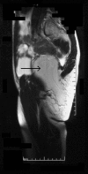
Case presentation: A 36 year old woman presented with an expanding, yet reducible, right gluteal mass, indicative of a sciatic hernia. However, magnetic resonance imaging demonstrated a large intra- and extra-pelvic fatty mass traversing the greater sciatic foramen. The tumour was surgically removed through an abdomino-perineal approach. Subsequent pathological examination revealed an atypical lipomatous tumour (synonym: lipoma-like well-differentiated liposarcoma). The patient remains free from recurrence two years following her surgery.
Conclusion: The presence of a gluteal mass should always suggest the possibility of a sciatic hernia. However, in this case, the hernia consisted of an atypical lipoma spanning the greater sciatic foramen. Although lipoma-like well-differentiated liposarcomas have only a low potential for recurrence, the variable nature of fatty tumours demands that patients require regular clinical and radiological review.
Figures


References
-
- Sadek HM, Kiss DR, Vasconselos E. Sciatic hernia caused by a neurofibroma. Surgical repair with a stainless wire mesh. Int Surg. 1970;54:135–141. - PubMed
-
- Rommel FM, Boline GB, Huffnagle HW. Ureterosciatic hernia: an anatomical radiographic correlation. J Urol. 1993;150:1232–1234. - PubMed
-
- Spring DB, Vandeman F, Watson RA. Computed tomographic demonstration of ureterosciatic hernia. AJR. 1983;141:579–580. - PubMed
-
- Curry N. Hernias of the urinary tract. In: Pollack HM, editor. Clinical urography: an atlas and textbook of urological imaging. Philadelphia: Saunders; 1990. pp. 2570–2578.
LinkOut - more resources
Full Text Sources

