. 2006 Jul;74(7):3773-82.
doi: 10.1128/IAI.01902-05.

Invasion of epithelial cells and proteolysis of cellular focal adhesion components by distinct types of Porphyromonas gingivalis fimbriae

Affiliations

Invasion of epithelial cells and proteolysis of cellular focal adhesion components by distinct types of Porphyromonas gingivalis fimbriae

Ichiro Nakagawa et al. Infect Immun. 2006 Jul.

Abstract

Porphyromonas gingivalis fimbriae are classified into six types (types I to V and Ib) based on the fimA genes encoding FimA (a subunit of fimbriae), and they play a critical role in bacterial interactions with host tissues. In this study, we compared the efficiencies of P. gingivalis strains with distinct types of fimbriae for invasion of epithelial cells and for degradation of cellular focal adhesion components, paxillin, and focal adhesion kinase (FAK). Six representative strains with the different types of fimbriae were tested, and P. gingivalis with type II fimbriae (type II P. gingivalis) adhered to and invaded epithelial cells at significantly greater levels than the other strains. There were negligible differences in gingipain activities among the six strains; however, type II P. gingivalis apparently degraded intracellular paxillin in association with a loss of phosphorylation 30 min after infection. Degradation was blocked with cytochalasin D or in mutants with fimA disrupted. Paxillin was degraded by the mutant with Lys-gingipain disrupted, and this degradation was prevented by inhibition of Arg-gingipain activity by Nalpha-p-tosyl-l-lysine chloromethyl ketone. FAK was also degraded by type II P. gingivalis. Cellular focal adhesions with green fluorescent protein-paxillin macroaggregates were clearly destroyed, and this was associated with cellular morphological changes and microtubule disassembly. In an in vitro wound closure assay, type II P. gingivalis significantly inhibited cellular migration and proliferation compared to the cellular migration and proliferation observed with the other types. These results suggest that type II P. gingivalis efficiently invades epithelial cells and degrades focal adhesion components with Arg-gingipain, which results in cellular impairment during wound healing and periodontal tissue regeneration.


Figures
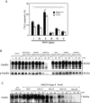
FIG. 1.
FIG. 1.
Degradation of paxillin associated with adhesion to and invasion of epithelial cells by P. gingivalis. (A) Adhesion to and invasion of HeLa cells by P. gingivalis strains with distinct types of fimbriae (types I to V and Ib). HeLa cells (1 × 10⁵ cells in a 24-well plate) were infected with [3H]thymidine-labeled P. gingivalis cells at an MOI of 200 for 90 min. The numbers of adhering and/or invading bacteria were determined as described in Materials and Methods. An asterisk indicates that the P value is <0.05. Multiple comparisons of the numbers of type II P. gingivalis and the numbers of other strains were performed. (B) Degradation of paxillin in P. gingivalis-infected epithelial cells. HeLa cells (4 × 10⁵ cells in a 60-mm dish) were infected with P. gingivalis with distinct types of fimbriae at an MOI of 200 for 5, 30, and 60 min. Degradation was assayed by immunoblotting the cellular lysates with specific antibodies. (C) Degradation of cellular paxillin with different numbers of type II P. gingivalis (strain OMZ314) cells. HeLa cells (4 × 10⁵ cells in a 60-mm dish) were infected with P. gingivalis strain OMZ314 at various MOIs for 5, 30, and 60 min. Degradation was assayed by immunoblotting the cellular lysates with specific antibodies. p-Paxillin, phospo-paxillin.
FIG. 2.
FIG. 2.
Paxillin degradation is dependent on P. gingivalis invasion of epithelial cells. (A) Adhesion to and invasion of HeLa cells by P. gingivalis in the presence of cytochalasin D (cytoD). HeLa cells (1 × 10⁵ cells in a 24-well plate) were infected with type I (ATCC 33277) and type II (OMZ314) P. gingivalis strains at an MOI of 200 for 90 min, similar to the method used for the experiment whose results are shown in Fig. 1. Cytochalasin D in dimethyl sulfoxide (DMSO) (final concentration, 10 μg/ml) was added to the cell culture 1 h prior to infection. Dimethyl sulfoxide (1/1,000, vol/vol) was used as a negative control. (B) Effect of cytochalasin D on paxillin degradation by P. gingivalis. Degradation was assayed by immunoblotting cellular lysates with specific antibodies. (C) Adhesion to and invasion of HeLa cells by P. gingivalis OMZ314 (type II wild type) and a mutant with fimA disrupted [OMZ314ΔfimA(II)] at an MOI of 200. An asterisk indicates that the P value is <0.05. (D) Paxillin degradation by P. gingivalis with fimA disrupted. HeLa cells (1 × 10⁵ cells in a 24-well plate) were infected with P. gingivalis strain OMZ314 or OMZ314ΔfimA(II) at an MOI of 200 for 5, 30, and 60 min. p-Paxillin, phospo-paxillin.
FIG. 3.
FIG. 3.
Involvement of Rgp in paxillin degradation in P. gingivalis-infected epithelial cells. (A) HeLa cells (1 × 10⁵ cells in a 24-well plate) were infected with P. gingivalis ATCC 33277 (type I fimbriae), its isogenic mutant with kgp disrupted (KDP129), a mutant with both rgpA and rgpB disrupted (KDP133), a mutant with rgpA, rgpB, and kgp disrupted (KDP136), and a mutant with fimA disrupted (KDP150) for 5, 30, and 60 min at an MOI of 1,000. Cellular lysates from the P. gingivalis-infected cells were analyzed by Western blotting using antipaxillin antibodies. (B) Effect of TLCK on invasion of HeLa cells by P. gingivalis. HeLa cells (1 × 10⁵ cells in a 24-well plate) were infected with [3H]thymidine-labeled P. gingivalis cells at an MOI of 200 for 90 min. TLCK (10 mM) in dimethyl sulfoxide (DMSO) (final concentration, 0.1%) or 0.1% dimethyl sulfoxide (negative control) was added to the culture 30 min prior to infection. The numbers of adherent and/or invading bacteria were determined as described in Materials and Methods. Statistical analyses were performed by multiple comparisons. (C) HeLa cells (1 × 10⁵ cells in a 24-well plate) were infected with P. gingivalis strain ATCC 33277 (type I) or OMZ314 (type II) at an MOI of 200 for 5, 30, and 60 min with or without TLCK (10 mM). Paxillin degradation was analyzed by Western blotting.
FIG. 4.
FIG. 4.
Degradation of FAK by P. gingivalis strains with distinct types of fimbriae. HeLa cells were infected with the P. gingivalis strains at an MOI of 200 for 5, 30, and 60 min. Cellular lysates of P. gingivalis-infected cells were analyzed by Western blotting using anti-FAK antibodies or anti-phosphorylated FAK (p-FAK) antibodies.
FIG. 5.
FIG. 5.
Effect of P. gingivalis with type II fimbriae on formation of focal adhesions by epithelial cells. An enhanced green fluorescent protein EGFP-paxillin expression vector (Paxillin-EGFP) was transfected into HeLa cells, and then the cells were infected with type II P. gingivalis (OMZ314) and OMZ314ΔfimA(II) for 1 h. The cells were fixed with 4% paraformaldehyde-PBS and stained with Alexa Fluor 594-conjugated phalloidin. Fluorescent images were obtained with a laser scanning confocal microscope at a magnification of ×630. Red, actin; green, paxillin. Bar = 10 μm.
FIG. 6.
FIG. 6.
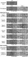
Microscopic views of wound closure by HeLa cells infected with P. gingivalis strains with distinct types of fimbriae. Confluent HeLa cell layers were scratched with a plastic tip. The cells were infected with P. gingivalis with distinct types of fimbriae at an MOI of 100, after which the cellular migration and proliferation to the scratched areas were analyzed at 37°C for 24 and 48 h. The images show the scratched wound regions at zero time and 24 and 48 h, and the rates of wound closure, indicated under the images, were determined by assays performed in triplicate on three separate occasions (n = 9), as described in Materials and Methods.

Similar articles

Cited by

References

    1. Amano, A., I. Nakagawa, N. Okahashi, and N. Hamada. 2004. Variations of Porphyromonas gingivalis fimbriae in relation to microbial pathogenesis. J. Periodontal Res. 39:136-142. - PubMed
    1. Amano, A., M. Kuboniwa, I. Nakagawa, S. Akiyama, S., I. Morisaki, and S. Hamada. 2000. Prevalence of specific genotypes of Porphyromonas gingivalis fimA and periodontal health status. J. Dent. Res. 79:1664-1668. - PubMed
    1. Amano, A., I. Nakagawa, K. Kataoka, I. Morisaki, and S. Hamada. 1999. Distribution of Porphyromonas gingivalis strains with fimA genotypes in periodontitis patients. J. Clin. Microbiol. 37:1426-1430. - PMC - PubMed
    1. Belton, C. M., K. T. Izutsu, P. C. Goodwin, Y. Park, and R. J. Lamont. 1999. Fluorescence image analysis of the association between Porphyromonas gingivalis and gingival epithelial cells. Cell. Microbiol. 1:215-223. - PubMed
    1. Chen, Z., C. A. Casiano, and H. M. Fletcher. 2001. Protease-active extracellular protein preparations from Porphyromonas gingivalis W83 induce N-cadherin proteolysis, loss of cell adhesion, and apoptosis in human epithelial cells. J. Periodontol. 72:641-650. - PubMed

Publication types

MeSH terms

LinkOut - more resources