Comparative Study
. 2006 Jul;74(7):3946-57.
doi: 10.1128/IAI.00112-06.

Comparison of different live vaccine strategies in vivo for delivery of protein antigen or antigen-encoding DNA and mRNA by virulence-attenuated Listeria monocytogenes

Affiliations
Comparative Study

Comparison of different live vaccine strategies in vivo for delivery of protein antigen or antigen-encoding DNA and mRNA by virulence-attenuated Listeria monocytogenes

Daniela I M Loeffler et al. Infect Immun. 2006 Jul.

Abstract

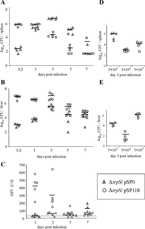
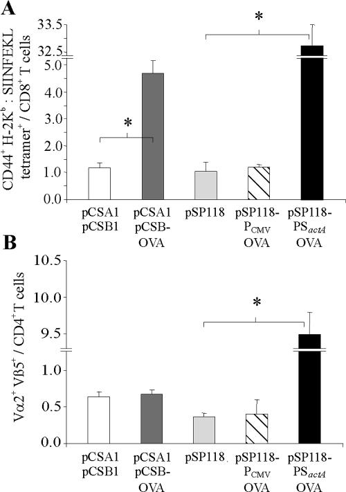
Listeria monocytogenes can be used to deliver protein antigens or DNA and mRNA encoding such antigens directly into the cytosol of host cells because of its intracellular lifestyle. In this study, we compare the in vivo efficiencies of activation of antigen-specific CD8 and CD4 T cells when the antigen is secreted by L. monocytogenes or when antigen-encoding plasmid DNA or mRNA is released by self-destructing strains of L. monocytogenes. Infection of mice with self-destructing L. monocytogenes carriers delivering mRNA that encodes a nonsecreted form of ovalbumin (OVA) resulted in a significant OVA-specific CD8 T-cell response. In contrast, infection with L. monocytogenes delivering OVA-encoding DNA failed to generate specific T cells. Secretion of OVA by the carrier bacteria yielded the strongest immune response involving OVA-specific CD8 and CD4 T cells. In addition, we investigated the antigen delivery capacity of a self-destructing, virulence-attenuated L. monocytogenes aroA/B mutant. In contrast to the wild-type strain, this mutant exhibited only marginal liver toxicity when high doses (5 x 10(7) CFU per animal administered intravenously) were used, and it was also able to deliver sufficient amounts of secreted OVA into mice. Therefore, the results presented here could lay the groundwork for a rational combination of L. monocytogenes as an attenuated carrier for the delivery of protein and nucleic acid vaccines in novel vaccination strategies.


Figures

FIG. 1.
FIG. 1.
Schematic maps of different antigen expression plasmids for delivery by L. monocytogenes. (A) Shuttle plasmid for expression and secretion of OVA by L. monocytogenes. (B) Plasmid for expression of OVA under control of the CMV promoter (PCMV). (C) Two-plasmid system for transcription of ova mRNA by L. monocytogenes under control of a T7 promoter (PT7) and for translation of OVA protein by host cells via an internal ribosomal entry site (IRES). In contrast to plasmid pSP0-PSactAOVA, plasmid pSP118-PSactAOVA contains the phage lysin 118 gene (ply118) under control of the promoter of the L. monocytogenes actA gene (PactA). (Panel i) Plasmid pCSA1 encodes the expression cassettes for T7 polymerase (T7 pol) and phage lysin 118 (ply118), both under control of PactA. (Panel ii) Plasmid pCSB-OVA encodes the ova mRNA expression cassette under control of the T7 promoter. trpS, gene encoding L. monocytogenes tryptophanyl tRNA synthetase; REm, erythromycin resistance marker; RTc, tetracycline resistance marker; ori Lm, origin of replication for L. monocytogenes; oriE1, origin of replication for E. coli; PSactA, promoter and secretion signal of actA; ova, partial ovalbumin gene; poly A, polyadenylation site.
FIG. 2.
FIG. 2.
Expression of truncated OVA protein (∼27 kDa) determined by Western blot analysis using a purified rabbit anti-ovalbumin antiserum. (A) Expression of OVA by L. monocytogenes ΔtrpS containing the expression plasmids indicated, grown to the late logarithmic phase in BHI medium. Lanes 1 to 5, proteins from culture supernatants; lanes 6 to 10, total cellular proteins. (B) Expression of OVA by COS-1 cells 3 days after transfection with the expression plasmids indicated. (C) Expression of OVA by Caco-2 cells 24 h after infection with 50 CFU/cell of L. monocytogenes ΔtrpS containing different expression plasmids. Proteins were obtained by immunoprecipitation prior to Western blot analysis. The arrowheads indicate bands representing truncated OVA protein. Lm, L. monocytogenes.
FIG. 3.
FIG. 3.
Replication of the L. monocytogenes ΔtrpS carrier strains containing the expression plasmids indicated. (A) Growth in BHI medium. mRNA delivery strains, L. monocytogenes ΔtrpS/pCSA1/pCSB1 (control strain), and L. monocytogenes ΔtrpS/pCSA1/pCSB-OVA have increased generation times. The DNA delivery strain was L. monocytogenes ΔtrpS/pSP118-PCMVOVA, the protein delivery strain was L. monocytogenes ΔtrpS/pSP118-PSactAOVA, and the control strain was L. monocytogenes ΔtrpS/pSP118. The optical densities at 600 nm of cultures were determined, and the results are expressed as means ± standard deviations. The results of one of three experiments are shown. (B) Intracellular growth of the L. monocytogenes delivery strains in macrophage cell line P388.D1. The mRNA delivery strains L. monocytogenes ΔtrpS/pCSA1/pCSB1 and L. monocytogenes ΔtrpS/pCSA1/pCSB-OVA are significantly less invasive than the other delivery strains but are able to replicate within the host cell cytosol. The OVA-secreting strain L. monocytogenes ΔtrpS/pSP118-PSactAOVA is more invasive than the mRNA delivery strains but is not able to replicate intracellularly. The results of one of three experiments are shown.
FIG. 4.
FIG. 4.
Viable bacterial counts in spleens and livers of C57BL/6 mice infected intravenously with 5 × 10³ CFU (0.5 LD50) of L. monocytogenes ΔtrpS/pSP0 or 5 × 10⁷ CFU (0.25 to 0.5 LD50) of the self-destructing carrier strain L. monocytogenes ΔtrpS/pSP118. (A) Bacterial loads in spleens of infected animals. (B) Bacterial loads in livers of infected animals. (C) GPT levels of infected mice at different times after infection. The dotted line indicates the mean GPT level for mice that were inoculated with a saline solution as a control. (D) Bacterial loads in spleens of animals infected with equal doses (5 × 10³ CFU each) of L. monocytogenes ΔtrpS/pSP0 and L. monocytogenes ΔtrpS/pSP118. In this experiment, a third group of mice was also infected with 5 × 10⁷ CFU of L. monocytogenes ΔtrpS/pSP118. Each symbol represents a single animal. The lines indicate the means for the experimental groups (n = 5). The results of one of two experiments are shown.
FIG. 5.
FIG. 5.
Clonal expansion of OVA-specific CD8 and CD4 T cells following infection with 5 × 10⁷ CFU of L. monocytogenes ΔtrpS containing the plasmids indicated. (A) One day prior to infection, 107 spleen cells derived from OT-I mice were transferred into C57BL/6 mice. The frequencies of CD44+, H-2Kb-SIINFEKL tetramer-positive cells in all the CD8+ T cells in spleens were determined 3 days after infection, which indicated presentation of OVA epitopes via MHC class I. (B) To investigate presentation via MHC class II, 107 spleen cells derived from OT-II mice were transferred into C57BL/6 mice 1 day prior to infection. Three days later, the frequencies of Vα2+ Vβ5+ cells in all the CD4+ T cells in spleens of infected animals were determined. L. monocytogenes ΔtrpS/pCSA1/pCSB1 and L. monocytogenes ΔtrpS/pSP118 were the control strains. The results are expressed as means ± standard deviations (n = 3) and are representative of the results of at least two experiments. An asterisk indicates that there is a significant difference (P < 0.05, as determined two-tailed Student's t test) between experimental groups.
FIG. 6.
FIG. 6.
Delivery properties and level of virulence attenuation for a self-destructing L. monocytogenes carrier strain with additional deletions of the aroA and aroB genes. (A) Viable bacterial counts in livers and spleens of C57BL/6 mice that were infected intravenously with 5 × 10⁷ CFU of the self-destructing carrier strains L. monocytogenes ΔtrpS/pSP118 and L. monocytogenes Δ(trpS/aroA/B)/pSP118. (B) GPT levels of mice infected with 5 × 10⁷ CFU of the self-destructing carrier strains L. monocytogenes ΔtrpS/pSP118 and L. monocytogenes Δ(trpS/aroA/B)/pSP118, determined at different times after infection. (C) Clonal expansion of OVA-specific CD8+ cells following infection with 1 × 10⁸ CFU of L. monocytogenes Δ(trpS/aroA/B) containing the plasmids indicated. One day prior to infection, 107 OT-I spleen cells were transferred into C57BL/6 recipient mice. The frequencies of Vα2+ Vβ5+ cells in the total CD8+ T cells in spleens were determined 3 days after infection. Each symbol represents a single animal. The lines indicate the means for the experimental groups (n = 5). The results of one of two experiments are shown.
FIG. 7.
FIG. 7.
OVA-specific T-cell responses of mice infected three times at 3-week intervals with 5 × 10⁷ CFU of the self-destructing L. monocytogenes carrier strain ΔtrpS containing the expression plasmids indicated. Spleen cells of infected mice were harvested at day 49 postinfection. Control mice received injections of a saline solution (0.9% NaCl). (A) CD8+ T-cell responses. For ELISPOT analysis, 105 lymphocytes purified from spleen cells were treated in triplicate for 24 h with OVA peptide 257-264 (SIINFEKL) and were assayed for IFN-γ-producing cells. (B) CD4+ T-cell responses. Frequencies of IFN-γ producing cells were determined following stimulation of total spleen cells (4 × 10⁵ cells per well) for 24 h in triplicate with purified ovalbumin. Unstimulated cells (medium controls) had low levels of cytokine responses, and the values for these cells were not subtracted. The results are the results of one of two experiments and are expressed as means ± standard deviations for groups of five mice. An asterisk indicates that there is a significant difference (P < 0.05, as determined by two-tailed Student's t test) between experimental groups.

Similar articles

Cited by

References

    1. Angelakopoulos, H., K. Loock, D. M. Sisul, E. R. Jensen, J. F. Miller, and E. L. Hohmann. 2002. Safety and shedding of an attenuated strain of Listeria monocytogenes with a deletion of actA/plcB in adult volunteers: a dose escalation study of oral inoculation. Infect. Immun. 70:3592-3601. - PMC - PubMed
    1. Badovinac, V. P., B. B. Porter, and J. T. Harty. 2002. Programmed contraction of CD8(+) T cells after infection. Nat. Immunol. 3:619-626. - PubMed
    1. Brockstedt, D. G., M. A. Giedlin, M. L. Leong, K. S. Bahjat, Y. Gao, W. Luckett, W. Liu, D. N. Cook, D. A. Portnoy, and T. W. Dubensky, Jr. 2004. Listeria-based cancer vaccines that segregate immunogenicity from toxicity. Proc. Natl. Acad. Sci. USA 101:13832-13837. - PMC - PubMed
    1. Corbin, G. A., and J. T. Harty. 2004. Duration of infection and antigen display have minimal influence on the kinetics of the CD4+ T cell response to Listeria monocytogenes infection. J. Immunol. 173:5679-5687. - PubMed
    1. DeGrendele, H. C., M. Kosfiszer, P. Estess, and M. H. Siegelman. 1997. CD44 activation and associated primary adhesion is inducible via T cell receptor stimulation. J. Immunol. 159:2549-2553. - PubMed

Publication types

MeSH terms