

Circulating gamma delta T cells in response to Salmonella enterica serovar enteritidis exposure in chickens
- PMID: 16790770
- PMCID: PMC1489728
- DOI: 10.1128/IAI.01128-05
Circulating gamma delta T cells in response to Salmonella enterica serovar enteritidis exposure in chickens
Abstract
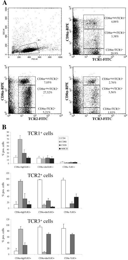
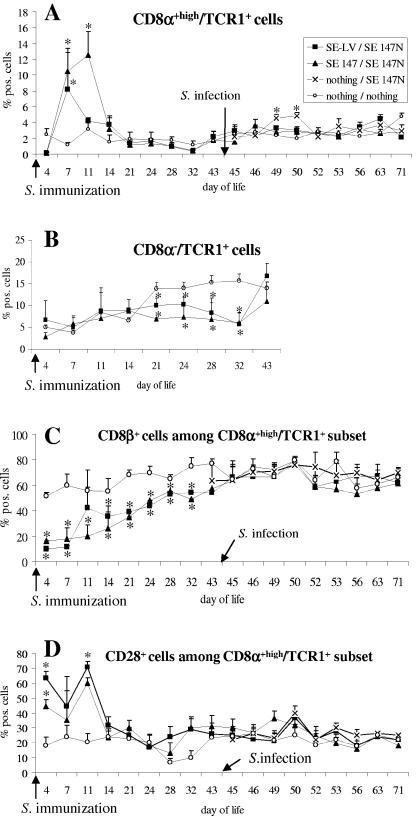
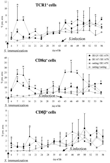
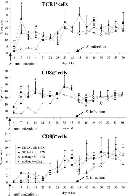
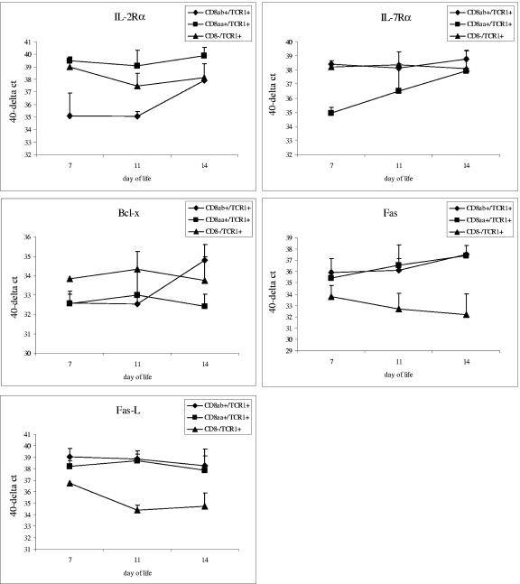
gammadelta T cells are considered crucial to the outcome of various infectious diseases. The present study was undertaken to characterize gammadelta (T-cell receptor 1(+) [TCR1(+)]) T cells phenotypically and functionally in avian immune response. Day-old chicks were orally immunized with Salmonella enterica serovar Enteritidis live vaccine or S. enterica serovar Enteritidis wild-type strain and infected using the S. enterica serovar Enteritidis wild-type strain on day 44 of life. Between days 3 and 71, peripheral blood was examined flow cytometrically for the occurrence of gammadelta T-cell subpopulations differentiated by the expression of T-cell antigens. Three different TCR1(+) cell populations were found to display considerable variation regarding CD8alpha antigen expression: (i) CD8alpha(+high) TCR1(+) cells, (ii) CD8alpha(+dim) TCR1(+) cells, and (iii) CD8alpha(-) TCR1(+) cells. While most of the CD8alpha(+high) TCR1(+) cells expressed the CD8alphabeta heterodimeric antigen, the majority of the CD8alpha(+dim) TCR1(+) cells were found to express the CD8alphaalpha homodimeric form. After immunization, a significant increase of CD8alphaalpha(+high) gammadelta T cells was observed within the CD8alpha(+high) TCR1(+) cell population. Quantitative reverse transcription-PCR revealed reduced interleukin-7 receptor alpha (IL-7Ralpha) and Bcl-x expression and elevated IL-2Ralpha mRNA expression of the CD8alphaalpha(+high) gammadelta T cells. Immunohistochemical analysis demonstrated a significant increase of CD8alpha(+) and TCR1(+) cells in the cecum and spleen and a decreased percentage of CD8beta(+) T cells in the spleen after Salmonella immunization. After infection of immunized animals, immune reactions were restricted to intestinal tissue. The study showed that Salmonella immunization of very young chicks is accompanied by an increase of CD8alphaalpha(+high) gammadelta T cells in peripheral blood, which are probably activated, and thus represent an important factor for the development of a protective immune response to Salmonella organisms in chickens.
Figures
Similar articles
-
Characterization of avian γδ T-cell subsets after Salmonella enterica serovar Typhimurium infection of chicks.Infect Immun. 2011 Feb;79(2):822-9. doi: 10.1128/IAI.00788-10. Epub 2010 Nov 15. Infect Immun. 2011. PMID: 21078853 Free PMC article.
-
Compensatory mechanisms in γδ T cell-deficient chickens following Salmonella infection.Front Immunol. 2025 May 14;16:1576766. doi: 10.3389/fimmu.2025.1576766. eCollection 2025. Front Immunol. 2025. PMID: 40438105 Free PMC article.
-
Avian CD25(+) gamma/delta (γδ) T cells after Salmonella exposure.Vet Immunol Immunopathol. 2015 Nov 15;168(1-2):14-8. doi: 10.1016/j.vetimm.2015.09.010. Epub 2015 Sep 28. Vet Immunol Immunopathol. 2015. PMID: 26553561
-
AMPK and mTOR: sensors and regulators of immunometabolic changes during Salmonella infection in the chicken.Poult Sci. 2016 Feb;95(2):345-53. doi: 10.3382/ps/pev349. Epub 2015 Dec 25. Poult Sci. 2016. PMID: 26706353 Review.
-
Chicken CD4, CD8alphabeta, and CD8alphaalpha T cell co-receptor molecules.Poult Sci. 1998 Dec;77(12):1858-73. doi: 10.1093/ps/77.12.1858. Poult Sci. 1998. PMID: 9872590 Review.
Cited by
-
Chicken γδ T cells proliferate upon IL-2 and IL-12 treatment and show a restricted receptor repertoire in cell culture.Front Immunol. 2024 Feb 13;15:1325024. doi: 10.3389/fimmu.2024.1325024. eCollection 2024. Front Immunol. 2024. PMID: 38420118 Free PMC article.
-
Chicken cecum immune response to Salmonella enterica serovars of different levels of invasiveness.Infect Immun. 2007 Dec;75(12):5993-6007. doi: 10.1128/IAI.00695-07. Epub 2007 Aug 20. Infect Immun. 2007. PMID: 17709416 Free PMC article.
-
Temporal Changes in Splenic Immune Cell Populations following Infection with a Very Virulent plus MDV in Commercial Meat-Type Chickens.Viruses. 2024 Jul 6;16(7):1092. doi: 10.3390/v16071092. Viruses. 2024. PMID: 39066253 Free PMC article.
-
A Novel Approach against Salmonella: A Review of Polymeric Nanoparticle Vaccines for Broilers and Layers.Vaccines (Basel). 2021 Sep 18;9(9):1041. doi: 10.3390/vaccines9091041. Vaccines (Basel). 2021. PMID: 34579278 Free PMC article. Review.
-
Effects of Selected Prebiotics or Synbiotics Administered in ovo on Lymphocyte Subsets in Bursa of the Fabricius, Thymus, and Spleen in Non-Immunized and Immunized Chicken Broilers.Animals (Basel). 2021 Feb 11;11(2):476. doi: 10.3390/ani11020476. Animals (Basel). 2021. PMID: 33670391 Free PMC article.
References
-
- Allison, J. P. 1993. γδ T cell development. Curr. Opin. Immunol. 5:241-246. - PubMed
-
- Arstila, T. P., and O. Lassila. 1993. Androgen-induced expansion of the peripheral blood γδ T cell population in the chicken. J. Immunol. 151:6627-6633. - PubMed
-
- Babu, U., M. Scott, M. J. Myers, M. Okamura, D. Gaines, H. F. Yancy, H. Lillehoj, R. A. Heckert, and R. B. Raybourne. 2003. Effects of live attenuated and killed Salmonella vaccine on T-lymphocyte mediated immunity in laying hens. Vet. Immunol. Immunopathol. 91:39-44. - PubMed
-
- Bank, I., R. A. DePinho, B. Brenner, J. Cassimeris, F. W. Alt, and L. A. Chess. 1986. Functional T3 molecule associated with a novel heterodimer on the surface of immature human thymocytes. Nature 322:179-181. - PubMed
-
- Barrow, P. A., J. M. Simpson, and M. A. Lovell. 1988. Intestinal colonization in the chicken of food-poisoning Salmonella serotypes; microbial characteristics associated with faecal excretion. Avian Pathol. 17:571-588. - PubMed
Publication types
MeSH terms
Substances
LinkOut - more resources
Full Text Sources
Other Literature Sources
Research Materials
