

Identification, recombinant expression, immunolocalization in macrophages, and T-cell responsiveness of the major extracellular proteins of Francisella tularensis
- PMID: 16790773
- PMCID: PMC1489726
- DOI: 10.1128/IAI.00257-06
Identification, recombinant expression, immunolocalization in macrophages, and T-cell responsiveness of the major extracellular proteins of Francisella tularensis
Abstract
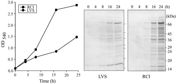
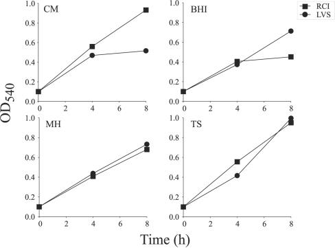
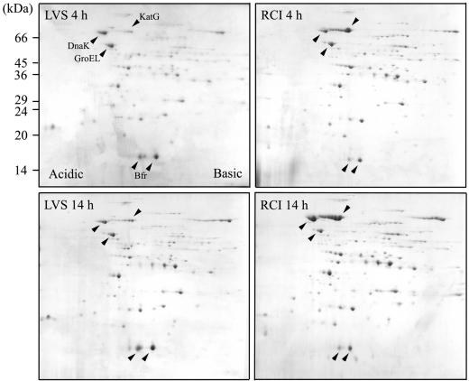
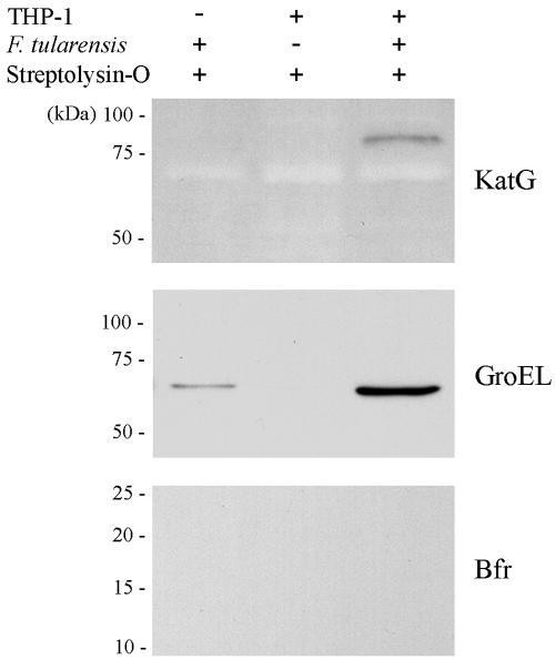
A safer and more effective vaccine than the previously developed live attenuated vaccine is needed for combating Francisella tularensis, a highly infectious bacterial pathogen. To search for potential candidates for inclusion in a new vaccine, we characterized the proteins present in the culture filtrates of a virulent recent clinical isolate and the attenuated live vaccine strain of F. tularensis using a proteomic approach. We identified a total of 12 proteins; among these, catalase-peroxidase was much more abundant in the culture filtrate of the virulent clinical isolate, whereas bacterioferritin was more abundant in the culture filtrate of the live vaccine strain. Streptolysin O treatment of infected human macrophages indicated that catalase-peroxidase and the heat shock protein GroEL are released intracellularly by actively growing F. tularensis. Mice immunized with F. tularensis developed significant cell-mediated immune responses to catalase-peroxidase, the heat shock protein GroEL, and bacterioferritin as measured by splenic lymphocyte proliferation and gamma interferon production. Finally, we expressed the major culture filtrate proteins that are promising vaccine candidates in Escherichia coli at high levels in soluble form to facilitate study of their immunobiology and potential role in vaccines.
Figures

Similar articles
-
Francisella tularensis Live Vaccine Strain deficient in capB and overexpressing the fusion protein of IglA, IglB, and IglC from the bfr promoter induces improved protection against F. tularensis respiratory challenge.Vaccine. 2016 Sep 22;34(41):4969-4978. doi: 10.1016/j.vaccine.2016.08.041. Epub 2016 Aug 28. Vaccine. 2016. PMID: 27577555 Free PMC article.
-
Construction and characterization of an attenuated purine auxotroph in a Francisella tularensis live vaccine strain.Infect Immun. 2006 Aug;74(8):4452-61. doi: 10.1128/IAI.00666-06. Infect Immun. 2006. PMID: 16861631 Free PMC article.
-
Deletion of IglH in virulent Francisella tularensis subsp. holarctica FSC200 strain results in attenuation and provides protection against the challenge with the parental strain.Microbes Infect. 2012 Feb;14(2):177-87. doi: 10.1016/j.micinf.2011.08.017. Epub 2011 Sep 8. Microbes Infect. 2012. PMID: 21930232
-
Tularemia vaccines: recent developments and remaining hurdles.Future Microbiol. 2011 Apr;6(4):391-405. doi: 10.2217/fmb.11.22. Future Microbiol. 2011. PMID: 21526941 Review.
-
Vaccination strategies for Francisella tularensis.Adv Drug Deliv Rev. 2005 Jun 17;57(9):1403-14. doi: 10.1016/j.addr.2005.01.030. Adv Drug Deliv Rev. 2005. PMID: 15919131 Review.
Cited by
-
The francisella tularensis proteome and its recognition by antibodies.Front Microbiol. 2011 Jan 7;1:143. doi: 10.3389/fmicb.2010.00143. eCollection 2010. Front Microbiol. 2011. PMID: 21687770 Free PMC article.
-
Identification of Francisella tularensis Himar1-based transposon mutants defective for replication in macrophages.Infect Immun. 2007 Nov;75(11):5376-89. doi: 10.1128/IAI.00238-07. Epub 2007 Aug 6. Infect Immun. 2007. PMID: 17682043 Free PMC article.
-
Adaptation of Francisella tularensis to the mammalian environment is governed by cues which can be mimicked in vitro.Infect Immun. 2008 Oct;76(10):4479-88. doi: 10.1128/IAI.00610-08. Epub 2008 Jul 21. Infect Immun. 2008. PMID: 18644878 Free PMC article.
-
Generation and characterization of hybridoma antibodies for immunotherapy of tularemia.Immunol Lett. 2007 Oct 15;112(2):92-103. doi: 10.1016/j.imlet.2007.07.006. Epub 2007 Aug 8. Immunol Lett. 2007. PMID: 17764754 Free PMC article.
-
A mucosal subunit vaccine protects against lethal respiratory infection with Francisella tularensis LVS.PLoS One. 2012;7(11):e50460. doi: 10.1371/journal.pone.0050460. Epub 2012 Nov 28. PLoS One. 2012. PMID: 23209745 Free PMC article.
References
-
- Amemura-Maekawa, J., S. Mishima-Abe, F. Kura, T. Takahashi, and H. Watanabe. 1999. Identification of a novel periplasmic catalase-peroxidase KatA of Legionella pneumophila. FEMS Microbiol. Immunol. 176:339-344. - PubMed
Publication types
MeSH terms
Substances
Grants and funding
LinkOut - more resources
Full Text Sources
Other Literature Sources
Research Materials
