. 2006 Jul;74(7):4114-23.
doi: 10.1128/IAI.00328-06.

Blastocystis ratti induces contact-independent apoptosis, F-actin rearrangement, and barrier function disruption in IEC-6 cells

Affiliations

Blastocystis ratti induces contact-independent apoptosis, F-actin rearrangement, and barrier function disruption in IEC-6 cells

Manoj K Puthia et al. Infect Immun. 2006 Jul.

Abstract

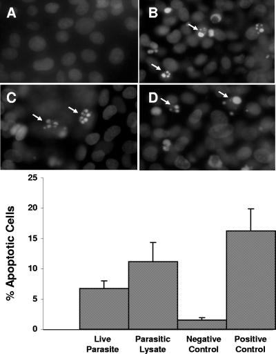
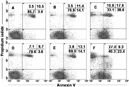
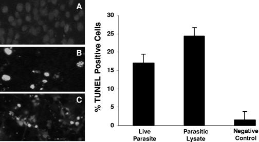
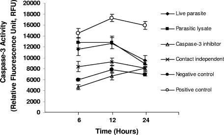
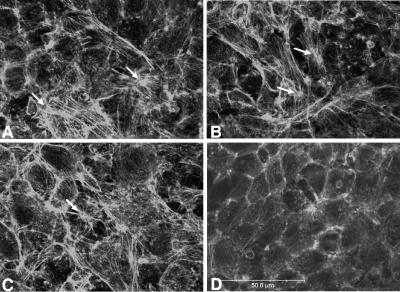
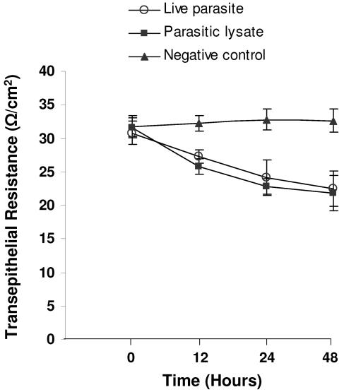
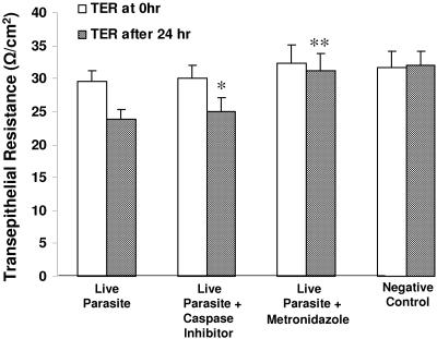
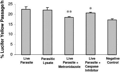
Blastocystis is an enteric protozoan purportedly associated with numerous clinical cases of diarrhea, flatulence, vomiting, and other gastrointestinal symptoms. Despite new knowledge of Blastocystis cell biology, genetic diversity, and epidemiology, its pathogenic potential remains controversial. Numerous clinical and epidemiological studies either implicate or exonerate the parasite as a cause of intestinal disease. Therefore, the aim of this study was to investigate the pathogenic potential of Blastocystis by studying the interactions of Blastocystis ratti WR1, an isolate of zoonotic potential, with a nontransformed rat intestinal epithelial cell line, IEC-6. Here, we report that B. ratti WR1 induces apoptosis in IEC-6 cells in a contact-independent manner. Furthermore, we found that B. ratti WR1 rearranges F-actin distribution, decreases transepithelial resistance, and increases epithelial permeability in IEC-6 cell monolayers. In addition, we found that the effects of B. ratti on transepithelial electrical resistance and epithelial permeability were significantly abrogated by treatment with metronidazole, an antiprotozoal drug. Our results suggest for the first time that Blastocystis-induced apoptosis in host cells and altered epithelial barrier function might play an important role in the pathogenesis of Blastocystis infections and that metronidazole has therapeutic potential in alleviating symptoms associated with Blastocystis.


Figures

FIG. 1.
FIG. 1.
Fluorescence photomicrographs and histograms obtained from DAPI fluorescence assay for apoptosis of nontransformed rat intestinal epithelial IEC-6 cells. Cell monolayers were grown on glass coverslips and incubated for 24 h with either growth medium (A), B. ratti WR1 live parasites (B), parasitic lysate (C), and 0.25 μM staurosporin as a positive control (D). Cells coincubated with live parasites, parasitic lysate, and staurosporin show nuclear condensation and fragmentation (B, C, and D). From the histogram, a significant increase in the percentage of apoptotic cells in monolayers coincubated with live parasites and lysate can be noticed in comparison to the negative control. For each sample, 1,000 cells were counted under a magnification of ×40. Values are means ± standard deviations (error bars) (three monolayers in each group). The values were significantly different (P < 0.05) from the value for the negative control.
FIG. 2.
FIG. 2.
Annexin V-FITC staining of IEC-6 cells by flow cytometry. Representative dot plots of cells subjected to different treatments are shown. Untreated IEC-6 cells (A) are shown as a control. IEC-6 cells were incubated for 5 h with B. ratti WR1 live parasites (B), parasitic lysate (C), live parasites after pretreatment with caspase inhibitor Z-VAD-fmk (D), live parasites on Millicell-HA filter for contact-independent assay (E), and 0.25 μM staurosporin as a positive control (F). Panels B, C, E, and F show significant increases in annexin V-positive apoptotic cell population (lower right quadrants). A total of 2 × 10⁴ cells from each sample were analyzed. The upper right quadrant represents apoptotic or necrotic cells positive for both annexin V and PI. The lower left quadrant represents healthy cells negative for annexin V and PI staining, and the upper left quadrant represents necrotic cells positive for only PI. Results are representative from two experiments, and the values were significantly different (P < 0.05) from the value for the negative control for all samples.
FIG. 3.
FIG. 3.
TUNEL for in situ DNA fragmentation of IEC-6 cells. Fluorescence micrographs of cells grown on glass coverslips and coincubated for 12 h with growth medium (A), B. ratti WR1 live parasites (B), and parasitic lysate (C). A significant number of TUNEL-positive cells (green fluorescence) can be seen in panels B and C. (Right) Histogram of TUNEL-positive cell population determined by flow cytometry shows a significant increase in TUNEL-positive IEC-6 cells after coincubation with live parasites and parasitic lysates. Values are means ± standard deviations (error bars) (two sets of cells grown on coverslips per group). The values were significantly different (P < 0.05) from the value for the negative control.
FIG. 4.
FIG. 4.
Effect of B. ratti WR1 on caspase-3 activity of IEC-6 cells. Caspase-3 activity was measured at the indicated time points as described in Materials and Methods. A significant increase in caspase-3 activity was observed in cells treated with live parasites, parasitic lysates, and positive control (0.25 μM staurosporin) after 6 and 12 h. Caspase-3 activity was gradually reduced at 24 h. Pretreatment of cells with caspase-3 inhibitor Z-DEVD-fmk reduced caspase-3 activity significantly. Values are means ± standard deviations (error bars) (n = 3 per group). The values were significantly different (P < 0.05) from the value for the negative control.
FIG. 5.
FIG. 5.
Effect of B. ratti WR1 exposure on actin cytoskeleton. IEC-6 cells were stained with fluorescein-phalloidin and analyzed by confocal microscopy. Prominent formation of stress fibers (white arrows) can be noticed in monolayers coincubated with B. ratti WR1 live parasites (A), parasitic lysate (B), and 5 μg/ml cholera toxin (C) as a positive control. Negative control (D) showing normal distribution of F-actin in the cortex zone of cells. Bar, 50 μm.
FIG. 6.
FIG. 6.
Effect of B. ratti WR1 on the transepithelial resistance of IEC-6 cell monolayers. Confluent monolayers of IEC-6 cells were grown on Millicell-HA filters and coincubated for the indicated times with live parasites, parasitic lysate, or growth medium (negative control). Thereafter, transepithelial resistance was measured as described in Materials and Methods. Live-parasite- and lysate-treated monolayers showed a significant drop in TER after 12, 24, and 48 h. Values are means ± standard deviations (error bars) (three monolayers per group). The values were significantly different (P < 0.05) from the value for the negative control.
FIG. 7.
FIG. 7.
Effects of caspase inhibition and metronidazole on B. ratti WR1-induced decrease in transepithelial resistance of IEC-6 monolayers. Confluent monolayers of IEC-6 cells were grown on Millicell-HA filters and incubated for 24 h with live parasites, live parasites after pretreatment of cells with broad-spectrum caspase inhibitor Z-VAD-fmk, or live parasites along with the antiprotozoal drug metronidazole. Monolayers incubated with growth medium served as a negative control. Thereafter, transepithelial resistance was measured as described in Materials and Methods. Pretreatment of IEC-6 cells with caspase inhibitors does not considerably rescue these cells from Blastocystis-induced effect, but exposure of Blastocystis to metronidazole abolished the effect significantly. Values are means ± standard deviations (error bars) (three monolayers per group). Values that were significantly different from the values for live-parasite-treated samples are indicated (*, P = 0.07; **, P < 0.05).
FIG. 8.
FIG. 8.
Flux measurement with Lucifer yellow. Confluent monolayers of IEC-6 cells were grown on Millicell-HA filters and incubated for 24 h with live parasites, live parasites after pretreatment of cells with the broad-spectrum caspase inhibitor Z-VAD-fmk, or live parasites along with the antiprotozoal drug metronidazole. Monolayers incubated with only growth medium served as a negative control. Permeability was determined by measurement of Lucifer yellow fluxes across the monolayer as described in Materials and Methods. A significant increase in the epithelial permeability can be noticed after incubation with live parasites and parasitic lysate in comparison to the control monolayer. Pretreatment of IEC-6 cells with caspase inhibitors does not considerably rescue these cells from Blastocystis-induced effect on permeability, but exposure of Blastocystis to metronidazole abolished the effect significantly. Values are means ± standard deviations (error bars) (three monolayers per group). Values that were significantly different from the values for live-parasite-treated samples are indicated (*, P = 0.2; **, P < 0.05).

Similar articles

Cited by

References

    1. Alexeieff, A. 1911. Sur la nature des formations dites kystes de Trichomonas intestinalis. C. R. Soc. Biol. 71:296-298.
    1. Ashford, R. W., and E. A. Atkinson. 1992. Epidemiology of Blastocystis hominis infection in Papua New Guinea: age-prevalence and associations with other parasites. Ann. Trop. Med. Parasitol. 86:129-136. - PubMed
    1. Berkes, J., V. K. Viswanathan, S. D. Savkovik, and G. Hecht. 2003. Intestinal epithelial responses to enteric pathogens: effects on the tight junction barrier, ion transport, and inflammation. Gut 52:439-451. - PMC - PubMed
    1. Bjarnason, I., A. MacPherson, and D. Hollander. 1995. Intestinal permeability: an overview. Gastroenterology 108:1566-1581. - PubMed
    1. Bojarski, C., K. Bendfeldt, A. H. Gitter, J. Mankertz, M. Fromm, S. Wagner, E. O. Riecken, and J. D. Schulzke. 2000. Apoptosis and intestinal barrier function. Ann. N. Y. Acad. Sci. 915:270-274. - PubMed

Publication types

LinkOut - more resources