. 2006 Jul;74(7):4200-13.
doi: 10.1128/IAI.00493-06.

Identification and characterization of an antigen I/II family protein produced by group A Streptococcus

Affiliations

Identification and characterization of an antigen I/II family protein produced by group A Streptococcus

Shizhen Zhang et al. Infect Immun. 2006 Jul.

Abstract

Group A Streptococcus (GAS) is a gram-positive human bacterial pathogen that causes infections ranging in severity from pharyngitis to life-threatening invasive disease, such as necrotizing fasciitis. Serotype M28 strains are consistently isolated from invasive infections, particularly puerperal sepsis, a severe infection that occurs during or after childbirth. We recently sequenced the genome of a serotype M28 GAS strain and discovered a novel 37.4-kb foreign genetic element designated region of difference 2 (RD2). RD2 is similar in gene content and organization to genomic islands found in group B streptococci (GBS), the major cause of neonatal infections. RD2 encodes seven proteins with conventional gram-positive secretion signal sequences, six of which have not been characterized. Herein, we report that one of these six proteins (M28_Spy1325; Spy1325) is a member of the antigen I/II family of cell surface-anchored molecules produced by oral streptococci. PCR and DNA sequence analysis found that Spy1325 is very well conserved in GAS strains of distinct M protein serotypes. As assessed by real-time TaqMan quantitative PCR, the Spy1325 gene was expressed in vitro, and Spy1325 protein was present in culture supernatants and on the GAS cell surface. Western immunoblotting and enzyme-linked immunosorbent assays indicated that Spy1325 was produced by GAS in infected mice and humans. Importantly, the immunization of mice with recombinant Spy1325 fragments conferred protection against GAS-mediated mortality. Similar to other antigen I/II proteins, recombinant Spy1325 bound purified human salivary agglutinin glycoprotein. Spy1325 may represent a shared virulence factor among GAS, GBS, and oral streptococci.


Figures

FIG. 1.
FIG. 1.
Spy1325 is a member of the antigen I/II protein family. (A) Phylogenetic relationships of Spy1325 and orthologous proteins. Spy1325 is most closely related to a group of five inferred and uncharacterized cell surface proteins in GBS (S. agalactiae) (group I). The second closest-related group of proteins is composed of antigen I/II protein family members (group II). The tree was generated with MacVector, version 8.0. Numbers indicate the genetic distance between Spy1325 and various orthologous groups. Species and strain designations are given in parentheses. (B) The molecular architecture of Spy1325 is conserved among antigen I/II family proteins. Comparison of Spy1325 with SspB of Streptococcus gordonii showing the signal peptide (S), N-terminal region (N), alanine-rich repeats (AR), divergent region, proline-rich repeats (PR), conserved region, and the cell wall anchor motif (CWA). Spy1325 and SspB contain an LDV (Leu-Asp-Val) integrin-binding motif in the conserved region. SspB is representative of antigen I/II family proteins. The percent amino acid identity between the domains is indicated.
FIG. 2.
FIG. 2.
Allelic variation in Spy1325 in natural populations of GAS. (A) Agarose gel electrophoresis showing three amplicon size variants corresponding to sequence variation in the proline-rich region. Size (in base pairs) is indicated on the left, and arrowheads denote an amplicon size corresponding to two, three, or four proline-rich repeats. All strains are serotype M28 except where indicated. L, 100-bp ladder, lane 1, MGAS8423; lane 2, MGAS8448; lane 3, MGAS11097; lane 4, MGAS11116; lane 5, MGAS9664; lane 6, MGAS9707; lane 7, MGAS10890; lane 8, MGAS10270 (serotype M2); lane 9, MGAS6180. (B) PCR and DNA sequence analysis identified seven distinct Spy1325 alleles among GAS strains of diverse M protein serotypes. Ninety-two percent of strains contained three proline-rich repeats (Spy1325.1), whereas four and three strains had two or four repeats (Spy1325.2 and Spy1325.3), respectively. Four nucleotide substitutions, each resulting in an amino acid replacement, also were identified (Spy1325.4 to Spy1325.7). nt, nucleotide.
FIG. 3.
FIG. 3.
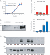
Characterization of Spy1325 in vitro expression. (A) Growth curve of strain MGAS6180 (serotype M28) and strain MGAS10270 (serotype M2) in THY broth. (B) The Spy1325 gene is transcribed in vitro. cDNA prepared from strains MGAS6180 and MGAS10270 harvested at early (EE), mid-exponential (ME), late exponential (LE), and stationary (S) phase was analyzed by real-time TaqMan quantitative PCR for Spy1325 transcript level. Values are expressed as the increase (n-fold) in transcript standardized to proS. The data represent values obtained with two independently isolated RNA samples analyzed in quadruplicate. (C) Spy1325 is present on the cell surface of GAS. MGAS5005, which does not contain the Spy1325 gene, and strain MGAS10270 (serotype M2) were harvested at stationary phase, and cell-wall proteins were extracted with (+) or without (−) mutanolysin. Proteins were separated by SDS-PAGE and analyzed by Western immunoblotting with Spy1325-specific antisera. (D) Spy1325 is present in the culture supernatant of various M protein serotype strains. Precipitated proteins prepared from culture supernatants from mid-exponential-phase (upper panel) or stationary-phase (lower panel) cultures were separated by SDS-PAGE and analyzed for the presence of immunoreactive Spy1325 with mouse antisera raised against purified rSpy1325. Strains tested: MGAS5005 (M1), MGAS6180 (M28), MGAS9455 (M28), MGAS9538 (M28), MGAS10208 (M28), MGAS10270 (M2), MGAS10318 (M2), MGAS10422 (M2), MGAS9482 (M77), MGAS11932 (M124), MGAS12431 (M124).
FIG. 4.
FIG. 4.
Spy1325 has trypsin-resistant and trypsin-sensitive fragments. (A) Schematic of recombinant Spy1325 fragments used in analysis. r1325 (aa 40 to 1319), mature Spy1325 without cell wall anchor motif; rAR (aa 201 to 434), alanine-rich repeats; rD (aa 435 to 646), divergent region; rNAD (aa 40 to 646), N-terminal, alanine-rich, and divergent regions; rPC (aa 647 to 1319), proline-rich repeats and conserved region; rC (aa 976 to 1319), conserved region. (B and C) Purified rSpy1325 (r1325) and fragments thereof were incubated in digestion buffer in the absence (B, −) or presence (C, +) of trypsin (10 ng/ml) for 17 h. Proteins were separated by SDS-PAGE and stained with Coomassie brilliant blue. The arrowhead in panel C indicates trypsin.
FIG. 5.
FIG. 5.
The alanine- and proline-rich regions of Spy1325 interact. (A) Plates (96 well) were coated with purified recombinant fragments of Spy1325, including NAD (aa 40 to 646), AR (aa 201 to 434, alanine-rich repeats), or D (aa 435 to 646, divergent region). The C terminus with (PC, aa 647 to 1319) or without (C, aa 767 to 1319) the proline-rich repeats was added as probe, and polyclonal antiserum raised against the C-terminal half of the protein was used to detect bound protein. (B) The converse experiment, in which C-terminal fragments were bound to the plate and probed with N-terminal fragments, was performed. Polyclonal antiserum directed against the N-terminal region of Spy1325 was used to detect bound protein.
FIG. 6.
FIG. 6.
Spy1325 is expressed in vivo in mice infected with serotype M28 strain MGAS6180. Purified rSpy1325 (r1325) and various purified recombinant protein fragments thereof were separated by SDS-PAGE and analyzed by Western immunoblotting. Sera obtained from mice infected with strain MGAS6180 (day 30, II) recognized rSpy1325 and various domain fragments, whereas preimmune sera (I) did not. (A) Purified rSpy1325; (B) rAR, alanine-rich repeat region (aa 201 to 434); (C) rPC, proline-rich, and conserved regions (aa 647 to 1319); (D) rNAD, N-terminal, alanine-rich, and divergent regions (aa 40 to 646).
FIG. 7.
FIG. 7.
Immunization of mice with recombinant fragments of Spy1325 confers protection against lethal GAS challenge. CD-1 Swiss outbred mice were immunized subcutaneously with 50 μg rPC (aa 647 to 1319), 50 μg rNAD (aa 40 to 646), or a mixture of 25 μg each of rNAD and rPC or were mock immunized with PBS. Mice were challenged intraperitoneally with a lethal dose of serotype M28 strain MGAS6180 (7.6 × 10⁸ CFU/mouse) and monitored for mortality or near mortality.
FIG. 8.
FIG. 8.
Spy1325 binds salivary agglutinin glycoprotein (SAG) complex. (A) The high-molecular-weight species eluted from the surface of GAS is SAG. Human saliva was adsorbed with serotype M28 strain MGAS6180, and eluted components were purified by size exclusion chromatography. Fractions were separated by SDS-PAGE (left panel) and analyzed by Western immunoblotting (right panel). The high-molecular-weight species reacted with anti-gp340 monoclonal antibody. Molecular masses and fraction numbers are indicated along the left and bottom, respectively. (B) Spy1325 contains two distinct SAG binding sites. Mature rSpy1325 and purified protein fragments (represented schematically in left panel) were separated by SDS-PAGE and analyzed by Coomassie brilliant blue staining (middle panel). Proteins were transferred to nitrocellulose, and the membrane was incubated with purified SAG and analyzed by Western immunoblotting with anti-gp340 monoclonal antibody (Far Western, right panel). SAG bound to mature Spy1325 (r1325), rNAD, and rPC fragments.

Similar articles

Cited by

References

    1. Anonymous. 2005. Active bacterial core surveillance (ABCs) report: emerging infections program network group A Streptococcus, 2004—provisional. [Online.] http://www.cdc.gov/ncidod/dbmd/abcs/survreports/gas04prelim.htm.
    1. Banks, D. J., S. B. Beres, and J. M. Musser. 2005. The contribution of phages to group A Streptococcus genetic diversity and pathogenesis, p. 319-334. In M. K. Waldor, D. I. Friedman, and S. L. Adhya (ed.), Phages: their role in bacterial pathogenesis and biotechnology. ASM Press, Washington, D.C.
    1. Banks, D. J., S. F. Porcella, K. D. Barbian, S. B. Beres, L. E. Philips, J. M. Voyich, F. R. DeLeo, J. M. Martin, G. A. Somerville, and J. M. Musser. 2004. Progress toward characterization of the group A Streptococcus metagenome: complete genome sequence of a macrolide-resistant serotype M6 strain. J. Infect. Dis. 190:727-738. - PubMed
    1. Beres, S. B., E. W. Richter, M. J. Nagiec, P. Sumby, S. F. Porcella, F. R. DeLeo, and J. M. Musser. 2006. Molecular genetic anatomy of inter- and intraserotypic variation in the human bacterial pathogen group A Streptococcus. Proc. Natl. Acad. Sci. USA 103:7059-7064. - PMC - PubMed
    1. Beres, S. B., G. L. Sylva, K. D. Barbian, B. Lei, J. S. Hoff, N. D. Mammarella, M. Y. Liu, J. C. Smoot, S. F. Porcella, L. D. Parkins, D. S. Campbell, T. M. Smith, J. K. McCormick, D. Y. Leung, P. M. Schlievert, and J. M. Musser. 2002. Genome sequence of a serotype M3 strain of group A Streptococcus: phage-encoded toxins, the high-virulence phenotype, and clone emergence. Proc. Natl. Acad. Sci. USA 99:10078-10083. - PMC - PubMed

MeSH terms