. 2006 Jul;74(7):4246-53.
doi: 10.1128/IAI.01620-05.

Exploitation of the endocytic pathway by Orientia tsutsugamushi in nonprofessional phagocytes

Affiliations

Exploitation of the endocytic pathway by Orientia tsutsugamushi in nonprofessional phagocytes

Hyuk Chu et al. Infect Immun. 2006 Jul.

Erratum in

  • Infect Immun. 2006 Sep;74(9):5422

Abstract

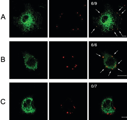
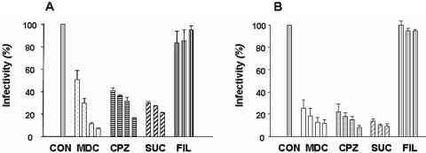
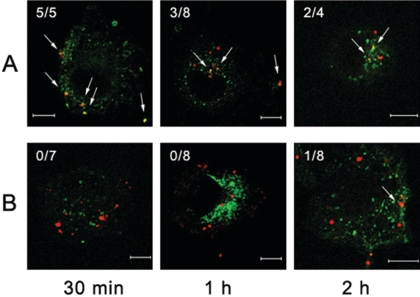
Orientia tsutsugamushi, a causative agent of scrub typhus, is an obligate intracellular bacterium that requires the exploitation of the endocytic pathway in the host cell. We observed the localization of O. tsutsugamushi with clathrin or adaptor protein 2 within 30 min after the infection of nonprofessional phagocytes. We have further confirmed that the infectivity of O. tsutsugamushi is significantly reduced by drugs that block clathrin-mediated endocytosis but not by filipin III, an inhibitor that blocks caveola-mediated endocytosis. In the present study, with a confocal microscope, O. tsutsugamushi was sequentially colocalized with the early and late endosomal markers EEA1 and LAMP2, respectively, within 1 h after infection. The colocalization of O. tsutsugamushi organisms with EEA1 and LAMP2 gradually disappeared until 2 h postinfection, and then free O. tsutsugamushi organisms were found in the cytoplasm. When the acidification of endocytic vesicles was blocked by treating the cells with NH(4)Cl or bafilomycin A, the escape of O. tsutsugamushi organisms from the endocytic pathway was severely impaired, and the infectivity of O. tsutsugamushi was drastically reduced. To our knowledge, this is the first report that the invasion of O. tsutsugamushi is dependent on the clathrin-dependent endocytic pathway and the acidification process of the endocytic vesicles in nonprofessional phagocytes.


Figures

FIG. 1.
FIG. 1.
Colocalization of O. tsutsugamushi with clathrin, α-adaptin, or caveolin 1. ECV304 cells were infected with O. tsutsugamushi for 30 min. The cells were subjected to anti-clathrin (A), anti-α-adaptin (B), or anti-caveolin 1 (C) antibodies with anti-O. tsutsugamushi antibody. Colocalization of each protein (green) with O. tsutsugamushi (red) was examined with a confocal microscope. Colocalization of each protein and O. tsutsugamushi produced yellow fluorescence. The ratios are the number of O. tsutsugamushi organisms that colocalized with clathrin (A), α-adaptin (B), or caveolin 1 (C) to the total number of infecting O. tsutsugamushi organisms. The colocalizations of O. tsutsugamushi with clathrin, α-adaptin, or caveolin 1 are indicated by arrows. Bars, 10 μm.
FIG. 2.
FIG. 2.
Inhibitory effects of endocytosis-disrupting agents on O. tsutsugamushi invasion of nonprofessional phagocytes. ECV304 (A) and L929 (B) cells were infected at 2 × 10⁵ ICU (about 15 O. tsutsugamushi bacteria were found per cell) for 2 h in the presence or absence of the inhibitors. Cells were labeled with KI-37 (monoclonal antibody against the Boryong strain's 56-kDa outer membrane protein) and goat anti-mouse IgG-FITC. The number of infecting bacteria in every 100 cells was counted by observation with a fluorescence microscope. The concentrations of inhibitors were maintained throughout the study. Each experiment was performed a minimum of three times. Inhibition assays were performed by preincubation of inhibitors for 30 min followed by O. tsutsugamushi infection for 2 h. The concentrations of inhibitors were as follows: MDC, 0.1, 0.2, 0.3, and 0.4 mM; CPZ, 1, 5, 10, and 25 μM; SUC, 0.1, 0.2, and 0.3 M; and filipin III (FIL), 0.375, 0.75, and 1.5 mM. CON, control.
FIG. 3.
FIG. 3.
Colocalization of O. tsutsugamushi with EEA1 (A) and LAMP2 (B). Representative immunofluorescence with the confocal microscopic images of ECV304 cells infected with O. tsutsugamushi for the time periods indicated below. Cells were fixed, permeabilized, and double labeled with human serum against O. tsutsugamushi (goat anti-human rhodamine conjugate) and anti-EEA1 (Alexa Fluor 488) or LAMP2 (Alexa Fluor 488). (A) Merged images of EEA1 (green) and O. tsutsugamushi (red) at 30 min (left panels), 1 h (middle panels), and 2 h (right panels). (B) Merged images of LAMP2 (green) and O. tsutsugamushi (red) at 30 min, 1 h, and 2 h. The ratios are the number of O. tsutsugamushi organisms that colocalized with EEA1 (A) or LAMP2 (B) to the total number of infecting O. tsutsugamushi organisms. The colocalization of O. tsutsugamushi with EEA1 or LAMP2 is indicated by arrows. Bars, 10 μm.
FIG. 4.
FIG. 4.
Colocalization of O. tsutsugamushi and early endosome antigen 1 or LAMP2 in the presence of NH4Cl (20 mM). Representative immunofluorescence as seen in confocal microscopic images of ECV304 cells infected with O. tsutsugamushi for the indicated time periods. Infected cells were washed, fixed, permeabilized, and double labeled with human serum against O. tsutsugamushi (goat anti-human rhodamine conjugate) together with anti-clathrin, anti-EEA1, or anti-LAMP2 (Alexa Fluor 488). (A) Colocalization of EEA1 (green) with O. tsutsugamushi (red) at 30 min, 1 h, and 2 h. (B) Colocalization of LAMP2 (green) with O. tsutsugamushi (red) at 30 min, 1 h, and 2 h. Ratios are the number of O. tsutsugamushi organisms that colocalized with EEA1 (A) or LAMP2 (B) to the total number of infecting O. tsutsugamushi organisms. The colocalization of O. tsutsugamushi with EEA1 or LAMP2 is indicated by arrows. Bars, 10 μm.
FIG. 5.
FIG. 5.
Effects of bafilomycin A and NH4Cl on O. tsutsugamushi infection. Cells were infected at 2 × 10⁵ ICU for the indicated time periods in the presence or absence of bafilomycin A (20 nM) or NH4Cl (20 nM). ECV304 cells were fixed and labeled with KI-37 and goat anti-mouse IgG-FITC conjugate. Infective bacteria in every 100 cells were counted with a fluorescence microscope. Values represent the means from three independent experiments. C, control; BC, bafilomycin A-supplemented medium replaced with regular medium (without bafilomycin A); B, bafilomycin A-treated cells; NC, NH4Cl-supplemented medium replaced with regular medium (without NH4Cl); N, NH4Cl-treated cells.

Similar articles

Cited by

References

    1. Alvarez-Dominguez, C., R. Roberts, and P. D. Stahl. 1997. Internalized Listeria monocytogenes modulates intracellular trafficking and delays maturation of the phagosome. J. Cell Sci. 110:731-743. - PubMed
    1. Anderson, H. A., Y. Chen, and L. C. Norkin. 1996. Bound simian virus 40 translocates to caveolin-enriched membrane domains, and its entry is inhibited by drugs that selectively disrupt caveolae. Mol. Biol. Cell 7:1825-1834. - PMC - PubMed
    1. Beron, W., C. Alvarez-Dominguez, L. Mayorga, and P. D. Stahl. 1995. Membrane trafficking along the phagocytic pathway. Trends Cell Biol. 5:100-104. - PubMed
    1. Caruana, S. 1974. Rickettsial diseases. Trop. Dis. Bull. 71:781-786. - PubMed
    1. Chang, W.-H., J.-S. Kang, W.-K. Lee, M.-S. Choi, and J.-H. Lee. 1990. Serological classification by monoclonal antibodies of Rickettsia tsutsugamushi isolated in Korea. J. Clin. Microbiol. 28:685-688. - PMC - PubMed

Publication types

LinkOut - more resources