. 2006 Jul;74(7):4322-9.
doi: 10.1128/IAI.00260-06.

The Yersinia enterocolitica invasin protein promotes major histocompatibility complex class I- and class II-restricted T-cell responses

Affiliations

The Yersinia enterocolitica invasin protein promotes major histocompatibility complex class I- and class II-restricted T-cell responses

O T Bühler et al. Infect Immun. 2006 Jul.

Abstract

Yersinia enterocolitica invasin (Inv) protein confers internalization into and expression of proinflammatory cytokines by host cells. Both events require binding of Inv to beta1 integrins, which initiates signaling cascades including activation of focal adhesion complexes, Rac1, mitogen-activated protein kinase, and NF-kappaB. Here we tested whether Inv might be suitable as a delivery molecule and adjuvant if used as a component of a vaccine. For this purpose, hybrid proteins composed of Inv and ovalbumin (OVA) were prepared, applied as a coating to microparticles, and used for vaccination. Fusion of OVA to Inv did not significantly disturb the ability of Inv to promote host cell binding, internalization, and interleukin-8 (IL-8) secretion when applied as a coating to microparticles. The microparticles were used for vaccination of mice adoptively transferred with OVA-specific T cells from OT-1 or DO11.10 mice. Administration of OVA-Inv-coated microparticles induced OVA-specific T-cell responses. OVA-specific CD4 T cells produced both gamma interferon (IFN-gamma) and IL-4 as determined by enzyme-linked immunosorbent assay. Likewise, pronounced OVA-specific CD8 T-cell responses associated with IFN-gamma production were observed. Together, these results suggest that Inv might be an attractive tool in vaccination as it confers both host cell uptake and adjuvant activity by engagement of beta1 integrins of host cells, which leads to CD4 as well as CD8 T-cell responses.


Figures

FIG. 1.
FIG. 1.
Internalization of protein-coated microparticles into HeLa cells. HeLa cells were incubated with microparticles at a ratio of 100 microparticles/cell for 1 h at 37°C. Internalization was visualized by immunofluorescence staining. The cytoskeleton was stained with phalloidin (red). Extracellularly localized microparticles are green (arrowheads); intracellularly localized microparticles are blue (arrows). The results are representative of two independent experiments.
FIG. 2.
FIG. 2.
IL-8 production by HeLa cells induced by GST fusion protein-coated microparticles. HeLa cells were incubated with microparticles coated with fusions of the indicated proteins to GST at a ratio of 4,000 microparticles/cell or with medium alone. After 8 h, culture supernatants were collected and the levels of IL-8 were determined by ELISA. Values represent the means ± standard deviations of triplicate samples. The results are representative of three independent experiments.
FIG. 3.
FIG. 3.
Frequency of antigen-specific CD4+ T cells in vivo. Three mice received transgenic T cells 3 days prior to immunization with protein-coated microparticles or PBS. The percentage of T cells positive for specific TCR and CD4 out of all CD4+ T cells was determined by fluorescence-activated cell sorting analysis. (A) Double staining with anti-CD4-PE and anti-DO11.10 clonotypic TCR-FITC antibody. Numbers in quadrants indicate the percentages of gated viable cells. (B) CD4+ OVA-specific TCR+ T cells per number of total splenic CD4+ T cells after intraperitoneal immunization. Means ± standard deviations of three animals per group are shown. The results are representative of three independent experiments.
FIG. 4.
FIG. 4.
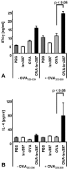
Cytokine production by OVA-specific CD4+ T cells. Five days after immunization, three spleens per group were pooled. Spleen cells were prepared and restimulated with OVA323-339. (A) Supernatants of cultures 3 days after restimulation of 3 × 10⁶ T cells with 10 μg/ml of OVA peptide were tested for IFN-γ by ELISA. (B) Supernatants of cultures 3 days after stimulation of 1.5 × 10⁶ T cells with 1 μg/ml of OVA peptide were tested for IL-4 by ELISA using triplicates. Means ± standard deviations of triplicate samples per group are shown. The results are representative of three independent experiments.
FIG. 5.
FIG. 5.
Frequency of antigen-specific CD8+ T cells in vivo. Three mice received transgenic T cells 3 days prior to immunization with protein-coated microparticles or PBS. Percentages of T cells positive for specific TCR and CD8 out of all CD8+ T cells were determined by fluorescence-activated cell sorting analysis. (A) Double staining with anti-CD8-PE and anti-Vα2-FITC antibody. Numbers in quadrants indicate the percentages of gated viable cells. (B) Ratio of CD8+ OVA-specific TCR+ T cells/number of splenic CD8+ T cells after intraperitoneal immunization. Means ± standard deviations of three animals per group are shown. The results are representative of three independent experiments.
FIG. 6.
FIG. 6.
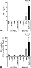
IFN-γ production by OVA-specific CD8+ T cells. Five days after immunization, three spleens per group were pooled. Spleen cells were prepared and stimulated with OVA257-264 (SIINFEKL) peptide (1 μg/ml). (A) Supernatants of cultures 3 days after restimulation were tested for the presence of IFN-γ by ELISA. (B) Flow cytometry analysis for intracellular IFN-γ was performed using anti-CD8-PE, anti-Vα2-FITC, and anti-IFN-γ-allophycocyanin antibody. Percentages of T cells positive for IFN-γ, OVA-specific TCR, and CD8 out of all CD8+ T cells are shown. Means ± standard deviations of triplicates per group are shown. The results are representative of three independent experiments.

Similar articles

Cited by

References

    1. Ackerman, A. L., and P. Cresswell. 2004. Cellular mechanisms governing cross-presentation of exogenous antigens. Nat. Immunol. 5:678-684. - PubMed
    1. Alrutz, M. A., and R. R. Isberg. 1998. Involvement of focal adhesion kinase in invasin-mediated uptake. Proc. Natl. Acad. Sci. USA 95:13658-13663. - PMC - PubMed
    1. Alrutz, M. A., A. Srivastava, K. W. Wong, C. D'Souza-Schorey, M. Tang, L. E. Ch'Ng, S. B. Snapper, and R. R. Isberg. 2001. Efficient uptake of Yersinia pseudotuberculosis via integrin receptors involves a Rac1-Arp 2/3 pathway that bypasses N-WASP function. Mol. Microbiol. 42:689-703. - PubMed
    1. Autenrieth, I. B., M. Beer, E. Bohn, S. H. E. Kaufmann, and J. Heesemann. 1994. Immune responses to Yersinia enterocolitica in susceptible BALB/c and resistant C57BL/6 mice: an essential role for gamma interferon. Infect. Immun. 62:2590-2599. - PMC - PubMed
    1. Autenrieth, I. B., V. Kempf, T. Sprinz, S. Preger, and A. Schnell. 1996. Defence mechanisms in Peyer's patches and mesenteric lymph nodes against Yersinia enterocolitica involve integrins and cytokines. Infect. Immun. 64:1357-1368. - PMC - PubMed

Publication types

Substances