

Three-dimensional virtual model of the human temporal bone: a stand-alone, downloadable teaching tool
- PMID: 16791035
- PMCID: PMC1805780
- DOI: 10.1097/01.mao.0000188353.97795.c5
Three-dimensional virtual model of the human temporal bone: a stand-alone, downloadable teaching tool
Abstract
Objective: To develop a three-dimensional virtual model of a human temporal bone based on serial histologic sections.
Background: The three-dimensional anatomy of the human temporal bone is complex, and learning it is a challenge for students in basic science and in clinical medicine.
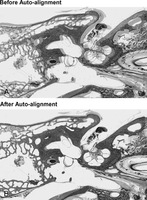
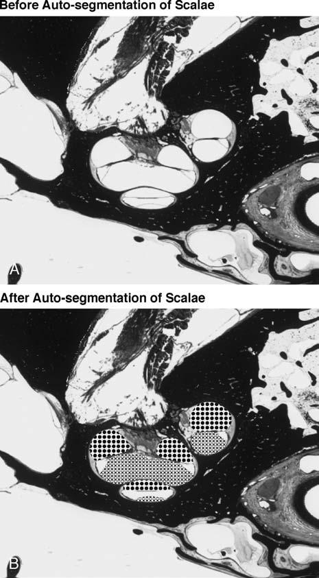
Methods: Every fifth histologic section from a normal 14-year-old male was digitized and imported into a general purpose three-dimensional rendering and analysis software package called Amira (version 3.1). The sections were aligned, and anatomic structures of interest were segmented.
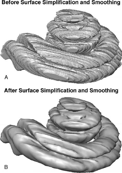
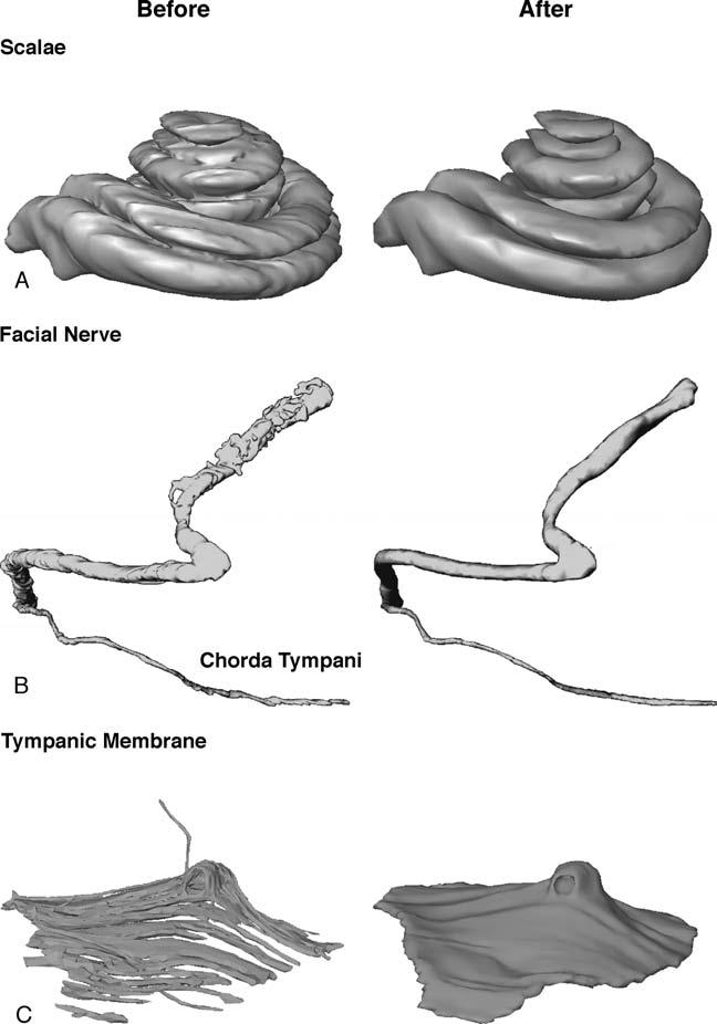
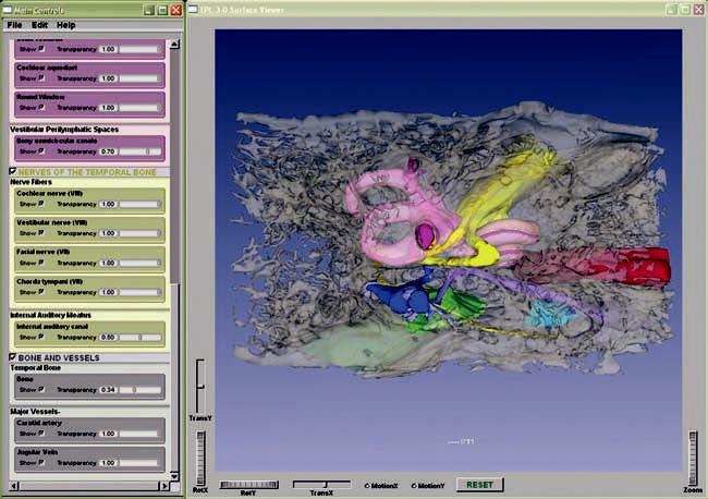
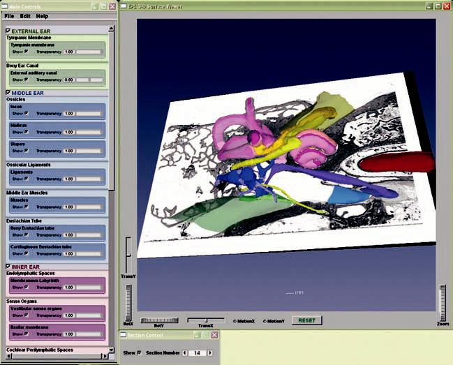
Results: The three-dimensional model is a surface rendering of these structures of interest, which currently includes the bone and air spaces of the temporal bone; the perilymph and endolymph spaces; the sensory epithelia of the cochlear and vestibular labyrinths; the ossicles and tympanic membrane; the middle ear muscles; the carotid artery; and the cochlear, vestibular, and facial nerves. For each structure, the surface transparency can be individually controlled, thereby revealing the three-dimensional relations between surface landmarks and underlying structures. The three-dimensional surface model can also be "sliced open" at any section and the appropriate raw histologic image superimposed on the cleavage plane. The image stack can also be resectioned in any arbitrary plane.
Conclusion: This model is a powerful teaching tool for learning the complex anatomy of the human temporal bone and for relating the two-dimensional morphology seen in a histologic section to the three-dimensional anatomy. The model can be downloaded from the Eaton-Peabody Laboratory web site, packaged within a cross-platform freeware three-dimensional viewer, which allows full rotation and transparency control.
Figures
References
-
- Takagi A, Sando I. Computer-aided three-dimensional reconstruction and measurement of the vestibular end-organs. Otolaryngol Head Neck Surg. 1988;98:195–202. - PubMed
-
- Takagi A, Sando I, Takahashi H. Computer-aided three-dimensional reconstruction and measurement of semicircular canals and their cristae in man. Acta Otolaryngol. 1989;107:362–5. - PubMed
-
- Nakashima S, Sando I, Tkahashi H, et al. Computer-aided 3-D reconstruction and measurement of the facial canal and facial nerve: I. cross-sectional area and diameter: preliminary report. Laryngoscope. 1993;103:1150–6. - PubMed
-
- Takahashi H, Sando I. Stereophotography of computer-aided three-dimensional reconstructions of the temporal bone structures. Otolaryngol Head Neck Surg. 1992;106:110–3. - PubMed
-
- Harada T, Ishii S, Tayama N. Three-dimensional reconstruction of the temporal bone from histologic sections. Arch Otolaryngol Head Neck Surg. 1988;114:1139–42. - PubMed
Publication types
MeSH terms
Grants and funding
LinkOut - more resources
Full Text Sources
