

Near-infrared fluorescence imaging of tumor integrin alpha v beta 3 expression with Cy7-labeled RGD multimers
- PMID: 16791749
- PMCID: PMC1643841
- DOI: 10.1007/s11307-006-0041-8
Near-infrared fluorescence imaging of tumor integrin alpha v beta 3 expression with Cy7-labeled RGD multimers
Abstract
Purpose: Cell adhesion molecule integrin alpha v beta 3 is an excellent target for tumor interventions because of its unique expression on the surface of several types of solid tumor cells and on almost all sprouting tumor vasculatures. Here, we describe the development of near-infrared (NIR) fluorochrome Cy7-labeled RGD peptides for tumor integrin targeting.
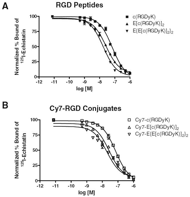
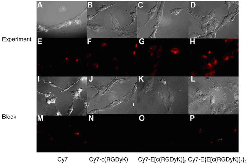
Procedures: Mono-, di-, and tetrameric RGD peptides were synthesized and conjugated with Cy7. The integrin specificity of these fluorescent probes was tested in vitro for receptor binding assay and fluorescence microscopy and in vivo for subcutaneous U87MG tumor targeting.
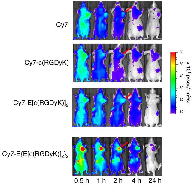
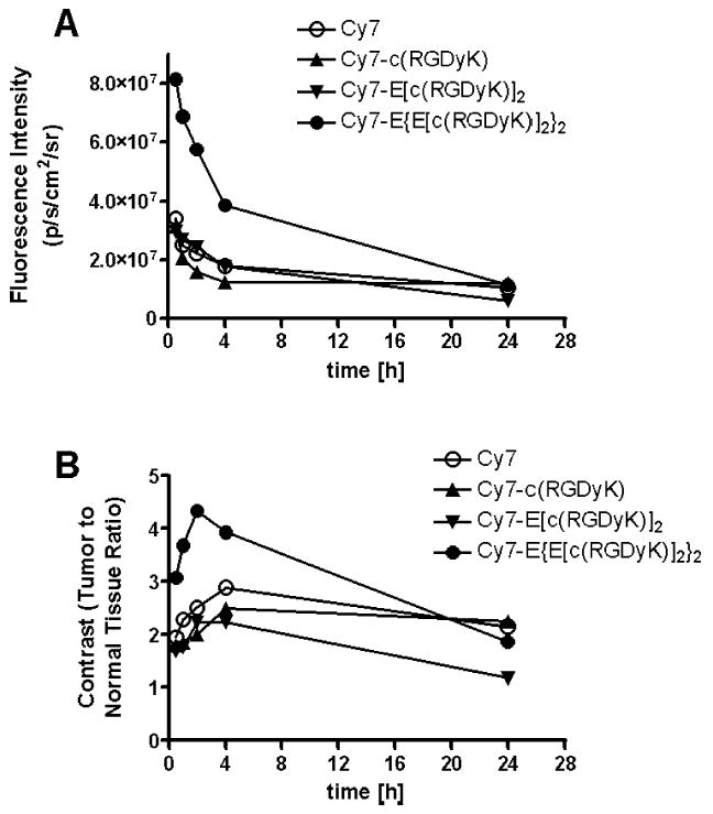
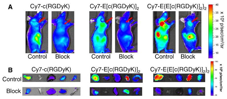
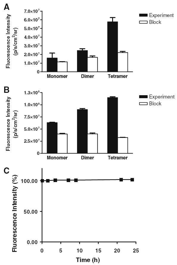
Results: The tetrameric RGD peptide probe with the highest integrin affinity showed the highest tumor activity accumulation and strongest tumor-to-normal tissue contrast. This uptake is integrin-specific as the signal accumulated in the tumor can be effectively blocked by unconjugated RGD peptide antagonist of integrin alpha v beta 3.
Conclusions: Noninvasive NIR fluorescence imaging is able to detect and semiquantify tumor integrin expression based upon the highly potent tetrameric RGD peptide probe.
Figures

Similar articles
-
Fast clearing RGD-based near-infrared fluorescent probes for in vivo tumor diagnosis.Contrast Media Mol Imaging. 2012 Jul-Aug;7(4):390-402. doi: 10.1002/cmmi.1464. Contrast Media Mol Imaging. 2012. PMID: 22649045
-
Near-infrared fluorescent RGD peptides for optical imaging of integrin alphavbeta3 expression in living mice.Bioconjug Chem. 2005 Nov-Dec;16(6):1433-41. doi: 10.1021/bc0501698. Bioconjug Chem. 2005. PMID: 16287239 Free PMC article.
-
(64)Cu-labeled tetrameric and octameric RGD peptides for small-animal PET of tumor alpha(v)beta(3) integrin expression.J Nucl Med. 2007 Jul;48(7):1162-71. doi: 10.2967/jnumed.107.039859. Epub 2007 Jun 15. J Nucl Med. 2007. PMID: 17574975
-
Cy7-Tetrameric arginine-glycine-aspartic acid peptide.2007 Mar 9 [updated 2008 May 13]. In: Molecular Imaging and Contrast Agent Database (MICAD) [Internet]. Bethesda (MD): National Center for Biotechnology Information (US); 2004–2013. 2007 Mar 9 [updated 2008 May 13]. In: Molecular Imaging and Contrast Agent Database (MICAD) [Internet]. Bethesda (MD): National Center for Biotechnology Information (US); 2004–2013. PMID: 20641381 Free Books & Documents. Review.
-
Integrin (αvβ3) Targeted RGD Peptide Based Probe for Cancer Optical Imaging.Curr Protein Pept Sci. 2016;17(6):570-81. doi: 10.2174/1389203717666160101124015. Curr Protein Pept Sci. 2016. PMID: 26721402 Review.
Cited by
-
Click chemistry for (18)F-labeling of RGD peptides and microPET imaging of tumor integrin alphavbeta3 expression.Bioconjug Chem. 2007 Nov-Dec;18(6):1987-94. doi: 10.1021/bc700226v. Epub 2007 Nov 21. Bioconjug Chem. 2007. PMID: 18030991 Free PMC article.
-
Glutathione-responsive biodegradable polyurethane nanoparticles for lung cancer treatment.J Control Release. 2020 May 10;321:363-371. doi: 10.1016/j.jconrel.2020.02.021. Epub 2020 Feb 12. J Control Release. 2020. PMID: 32061622 Free PMC article.
-
Peptide-based probes for targeted molecular imaging.Biochemistry. 2010 Feb 23;49(7):1364-76. doi: 10.1021/bi901135x. Biochemistry. 2010. PMID: 20102226 Free PMC article. Review.
-
In vivo near-infrared fluorescence imaging of CD105 expression during tumor angiogenesis.Eur J Nucl Med Mol Imaging. 2011 Nov;38(11):2066-76. doi: 10.1007/s00259-011-1886-x. Epub 2011 Aug 4. Eur J Nucl Med Mol Imaging. 2011. PMID: 21814852 Free PMC article.
-
Dual in vivo quantification of integrin-targeted and protease-activated agents in cancer using fluorescence molecular tomography (FMT).Mol Imaging Biol. 2010 Oct;12(5):488-99. doi: 10.1007/s11307-009-0279-z. Epub 2009 Dec 4. Mol Imaging Biol. 2010. PMID: 19960268
References
-
- Ruoslahti E. RGD and other recognition sequences for integrins. Annu Rev Cell Dev Biol. 1996;12:697–715. - PubMed
-
- Xiong JP, Stehle T, Zhang R, et al. Crystal structure of the extracellular segment of integrin αvβ3 in complex with an Arg–Gly–Asp ligand. Science. 2002;296:151–155. - PubMed
-
- Brooks PC, Clark RA, Cheresh DA. Requirement of vascular integrin αvβ3 for angiogenesis. Science. 1994;264:569–571. - PubMed
-
- Kumar CC. Integrin αvβ3 as a therapeutic target for blocking tumor-induced angiogenesis. Curr Drug Targets. 2003;4:123–131. - PubMed
-
- Hood JD, Cheresh DA. Role of integrins in cell invasion and migration. Nat Rev Cancer. 2002;2:91–100. - PubMed
Publication types
MeSH terms
Substances
Grants and funding
LinkOut - more resources
Full Text Sources
Other Literature Sources
Miscellaneous
