

Increased neutrophil membrane expression and plasma level of proteinase 3 in systemic vasculitis are not a consequence of the - 564 A/G promotor polymorphism
- PMID: 16792675
- PMCID: PMC1941990
- DOI: 10.1111/j.1365-2249.2006.03119.x
Increased neutrophil membrane expression and plasma level of proteinase 3 in systemic vasculitis are not a consequence of the - 564 A/G promotor polymorphism
Abstract
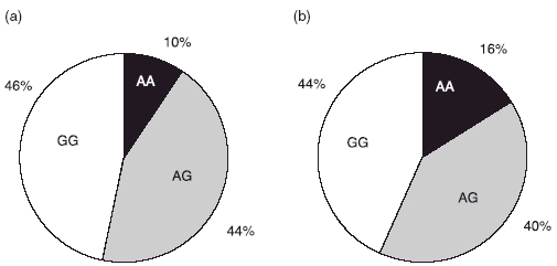
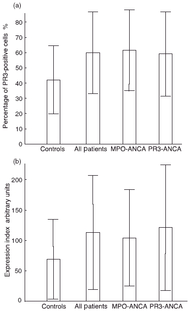
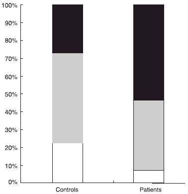
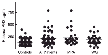
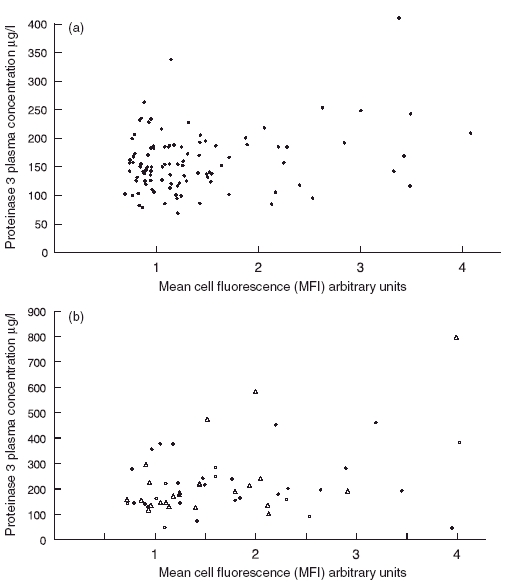
Several findings link proteinase 3 (PR3) to small vessel vasculitis. Besides being a major target of anti-neutrophil cytoplasm antibodies (ANCA), previous findings have shown increased circulating levels of PR3 in vasculitis patients, increased levels of neutrophil membrane-PR3 (mPR3) expression and a skewed distribution of the - 564 A/G polymorphism in the promotor region of the PR3 gene. In this study we elucidate how these three findings relate to each other. The plasma concentration of PR3 was measured by enzyme-linked immunosorbent assay (ELISA), mPR3 expression by fluorescence activated cell sorter (FACS) and the gene polymorphism by real-time polymerase chain reaction (PCR). We compared results from 63 patients with ANCA-associated systemic vasculitis (AASV) with 107 healthy blood donors. In accordance with previous reports, AASV patients had increased plasma concentrations of PR3 compared to healthy controls (mean 224 microg/l versus 155 microg/l, P < 0.0001). They also showed an increased number of mPR3-positive neutrophils (60%versus 42%, P < 0.001). However, contrary to a previous report, we found no skewed distribution of the polymorphism in PR3 gene. There was a weak correlation between mPR3 mean fluorescence intensity (MFI) and plasma PR3 among healthy controls and myeloperoxidase-ANCA (MPO-ANCA)-positive patients (r = 0.24, P = 0.015 and r = 0.52, P = 0.011, respectively). In conclusion, increased plasma PR3 and high expression of mPR3 are associated with small vessel vasculitis, but neither of them is a consequence of the - 564 A/G polymorphism of the PR3 gene promotor.
Figures
Similar articles
-
Coexpression of CD177 and membrane proteinase 3 on neutrophils in antineutrophil cytoplasmic autoantibody-associated systemic vasculitis: anti-proteinase 3-mediated neutrophil activation is independent of the role of CD177-expressing neutrophils.Arthritis Rheum. 2009 May;60(5):1548-57. doi: 10.1002/art.24442. Arthritis Rheum. 2009. PMID: 19404956
-
Increased monocyte transcription of the proteinase 3 gene in small vessel vasculitis.Clin Exp Immunol. 2005 Jul;141(1):174-82. doi: 10.1111/j.1365-2249.2005.02819.x. Clin Exp Immunol. 2005. PMID: 15958084 Free PMC article.
-
Increased membrane expression of proteinase 3 during neutrophil adhesion in the presence of anti proteinase 3 antibodies.J Am Soc Nephrol. 2007 Aug;18(8):2330-9. doi: 10.1681/ASN.2006121309. Epub 2007 Jul 18. J Am Soc Nephrol. 2007. PMID: 17634439
-
Membrane proteinase 3 expression on resting neutrophils as a pathogenic factor in PR3-ANCA-associated vasculitis.Clin Exp Rheumatol. 2003 Nov-Dec;21(6 Suppl 32):S64-8. Clin Exp Rheumatol. 2003. PMID: 14740429 Review.
-
Pathogenesis of PR3-ANCA associated vasculitis.J Autoimmun. 2008 Feb-Mar;30(1-2):29-36. doi: 10.1016/j.jaut.2007.11.005. Epub 2007 Dec 26. J Autoimmun. 2008. PMID: 18162369 Review.
Cited by
-
Genetics of ANCA-associated Vasculitides.Curr Rheumatol Rep. 2014 Jul;16(7):428. doi: 10.1007/s11926-014-0428-5. Curr Rheumatol Rep. 2014. PMID: 24828480 Review.
-
Gene-Specific DNA Methylation Changes Predict Remission in Patients with ANCA-Associated Vasculitis.J Am Soc Nephrol. 2017 Apr;28(4):1175-1187. doi: 10.1681/ASN.2016050548. Epub 2016 Nov 7. J Am Soc Nephrol. 2017. PMID: 27821628 Free PMC article.
-
Elevated neutrophil membrane expression of proteinase 3 is dependent upon CD177 expression.Clin Exp Immunol. 2010 Jul 1;161(1):89-97. doi: 10.1111/j.1365-2249.2010.04154.x. Epub 2010 May 10. Clin Exp Immunol. 2010. PMID: 20491791 Free PMC article.
-
Neutrophil surface presentation of the anti-neutrophil cytoplasmic antibody-antigen proteinase 3 depends on N-terminal processing.Clin Exp Immunol. 2008 Jun;152(3):508-16. doi: 10.1111/j.1365-2249.2008.03663.x. Clin Exp Immunol. 2008. PMID: 18462208 Free PMC article.
-
PRTN3 variant correlates with increased autoantigen levels and relapse risk in PR3-ANCA versus MPO-ANCA disease.JCI Insight. 2023 Feb 22;8(4):e166107. doi: 10.1172/jci.insight.166107. JCI Insight. 2023. PMID: 36626226 Free PMC article.
References
-
- Kallenberg CG, Brouwer E, Weening JJ, Tervaert JW. Anti-neutrophil cytoplasmic antibodies: current diagnostic and pathophysiological potential. Kidney Int. 1994;46:1–15. - PubMed
-
- Niles JL, McCluskey RT, Ahmad MF, Arnaout MA. Wegener’s granulomatosis autoantigen is a novel neutrophil serine proteinase. Blood. 1989;74:1888–93. - PubMed
-
- Rao NV, Wehner NG, Marshall BC, Gray WR, Gray BH, Hoidal JR. Characterization of proteinase-3 (PR-3), a neutrophil serine proteinase. Structural and functional properties. J Biol Chem. 1991;266:9540–8. - PubMed
Publication types
MeSH terms
Substances
LinkOut - more resources
Full Text Sources
Medical
Research Materials
Miscellaneous
