. 2006 Jun 22:7:51.
doi: 10.1186/1471-2202-7-51.

Insulin-like growth factor-1 (IGF-1) induces the activation/phosphorylation of Akt kinase and cAMP response element-binding protein (CREB) by activating different signaling pathways in PC12 cells

Affiliations

Insulin-like growth factor-1 (IGF-1) induces the activation/phosphorylation of Akt kinase and cAMP response element-binding protein (CREB) by activating different signaling pathways in PC12 cells

Wen-Hua Zheng et al. BMC Neurosci. .

Abstract

Background: Insulin-like growth factor-1 (IGF-1) is a polypeptide growth factor with a variety of functions in both neuronal and non-neuronal cells. IGF-1 plays anti-apoptotic and other functions by activating multiple signaling pathways including Akt kinase, a serine/threonine kinase essential for cell survival. The nuclear transcription factor cAMP response element-binding protein (CREB) may also be involved although relationships between these two proteins in IGF-1 receptor signaling and protection is not clear, especially in neuronal cells.

Results: IGF-1, in a concentration- and time-dependent manner, induces the activation/phosphorylation of Akt and CREB in PC12 cells by activating different signaling pathways. IGF-1 induced a sustained phosphorylation of Akt while only a transient one was seen for CREB. The phosphorylation of Akt is mediated by the PI3 kinase pathway while that of CREB is dependent on the activation of both MAPK kinase and p38 MAPK. Moreover, the stimulation of PKC attenuated the phosphorylation of Akt induced by IGF-1 while enhancing that of CREB. Survival assays with various kinase inhibitors suggested that the activation/phosphorylation of both Akt and CREB contributes to IGF-1 mediated cell survival in PC12 cells.

Conclusion: These data suggest that IGF-1 induced the activation of Akt and CREB using distinct pathways in PC12 cells.


Figures

Figure 1
Figure 1
IGF-1 induced time- and concentration-dependent phosphorylation of Akt and CREB in PC12 cells. PC12 cells were treated with 10 nM IGF-1 for various times and then the phosphorylation of Akt and CREB were determined as described in Methods. A). Time-dependence and (B). Concentration-dependence. Blots represent prototypical examples of experiments replicated at least 3 times.
Figure 2
Figure 2
IGF-1 induced the phosphorylation of Akt in PC12 cells via the PI3 kinase pathway while MAPK and p38 MAPK pathways mediated the activation of CREB induced by IGF-1. Following treatment with different kinase inhibitors for 40 min, PC12 cells were exposed to 10 nM IGF-1 and the phosphorylation of Akt and CREB was determined by Western blots using anti-phospho- Akt/CREB antibodies. The PI3 kinase inhibitor LY294002 inhibited IGF-1-induced phosphorylation of Akt in PC12 cells while a MAPK pathway inhibitor PD98059, a p38 MAPK inhibitor PD169316 and a p70S6 pathway inhibitor rapamycin, did not have any significant effect. In contrast, the phosphorylation of CREB induced by IGF-1 was partially blocked by inhibitors of both MEK (PD98059) and p38 MAP kinase (PD169316). Blots represent prototypical example of experiments replicated at least 3 times.
Figure 3
Figure 3
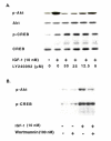
PI3 kinase inhibitors blocked the activation of Akt by IGF-1 while has no effect on the phosphorylation of CREB. PC12 cells pretreated with different concentrations of LY294002 or 100 nM wortmannin were stimulated with 10 nM IGF-1 and the phosphorylation of Akt and CREB was determined. (A) LY294002 blocked IGF-1-induced phosphorylation of Akt in a concentration-dependent manner while slightly enhancing the activation of CREB in PC12 cells. (B) 100 nM wortmannin significantly blocked the activation of Akt while having no effect on CREB phosphorylation. Blots represent prototypical example of experiments replicated at least 3 times.
Figure 4
Figure 4
The MEK kinase inhibitor PD98059 attenuated IGF-1-induced activation of MAPK kinase and CREB in PC12 cells. PC12 cells pretreated with various concentrations of the MAPK pathway inhibitor PD98059 were incubated with 10 nM IGF-1 for 10 min. Cells were collected and the activation of MAPK and CREB were determined as described in Methods. PD98059 concentration-dependently blocked the activation of MAPK stimulated by IGF-1. This inhibitor also attenuated the phosphorylation of CREB in a similar manner. Blots represent prototypical example of experiments replicated at least 3 times.
Figure 5
Figure 5
The p38 MAP kinase is involved in the phosphorylation of CREB induced by IGF-1 in PC12 cells. PC12 cells were treated with the p38 MAPK inhibitor PD169316 (2.5–20 μM) and 10 nM IGF-1, and the phosphorylation of CREB and p38 MAPK were measured by Western blot with anti-phospho-CREB and phospho-p38MARK antibodies. PD 169316 significantly blocked the phosphorylation of p38 MAPK and CREB induced by IGF-1. Blots represent prototypical example of experiments replicated at least 3 times.
Figure 6
Figure 6
PMA attenuated the phosphorylation of IGF-1-induced activation of Akt while enhancing the phosphorylation of CREB. PC12 cells were treated with 400 nM PMA before incubation with 10 nM IGF-1 and the phosphorylation of Akt and CREB determined. PMA treatment decreased the phosphorylation of Akt while increasing the activation of CREB. Blots represent prototypical example of experiments replicated at least 3 times.
Figure 7
Figure 7
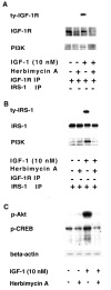
The tyrosine kinase inhibitor herbimycin A blocked IGF-1 signalling, including Akt and CREB activation, in PC12 cells. PC12 cells pretreated with or without herbimycin A were stimulated with 10 nM IGF-1 and the tyrosine phosphorylation of IGF-1 R (A) and IRS-1 (B), their association with PI3K, as well as the phosphorylation of Akt and CREB (C) were determined. IGF-1 stimulated the tyrosine phosphorylation of these markers. Pretreatment of PC12 cells with herbimycin A blocked these events. Blots represent prototypical example of experiments replicated at least 3 times.
Figure 8
Figure 8
Multiple signaling pathways are involved in the survival effect of IGF-1 in PC12 cells. Following treatment with different kinase inhibitors for 40 min, PC12 cells were exposed to 10 nM IGF-1 for 24 hours and cell viability was determined by MTT as described in Methods. Cell survival of PC12 cells induced by IGF-1 is partially inhibited by various blockers, the PI3K inhibitor being the most effective. Data represent assays from at least three independent experiments.
Figure 9
Figure 9
EGF, FGF, FBS, PMA and the calcium ionophore-A23187 stimulated CREB phosphorylation in PC12 cells. PC12 cells were treated with 10 nM IGF-1, 4 nM EGF, 4 nM FGF, 10% FBS, 200 nM PMA and 10 μM A23187 and the phosphorylation of CREB was determined as described in Methods. All these agents stimulated CREB phosphorylation in PC12 cells. Blots represent prototypical examples of experiments replicated at least 3 times.

Similar articles

Cited by

References

    1. Zheng WH, Kar S, Quirion R. Insulin-like growth factor-1-induced phosphorylation of transcription factor FKHRL1 is mediated by phosphatidylinositol 3-kinase/Akt kinase and role of this pathway in insulin-like growth factor-1-induced survival of cultured hippocampal neurons. Mol Pharmacol. 2002;62:225–233. doi: 10.1124/mol.62.2.225. - DOI - PubMed
    1. Butler AA, Yakar S, Gewolb IH, Karas M, Okubo Y, LeRoith D. Insulin-like growth factor-I receptor signal transduction: at the interface between physiology and cell biology. Comp Biochem Physiol B. Biochem Mol Biol. 1998;121:19–26. doi: 10.1016/S0305-0491(98)10106-2. - DOI - PubMed
    1. LeRoith D, Werner H, Beitner-Johnson D, Roberts CT., Jr Molecular and cellular aspects of the insulin-like growth factor I receptor. Endocr Rev. 1995;16:143–163. doi: 10.1210/er.16.2.143. - DOI - PubMed
    1. Myers MG, Jr, Sun XJ, Cheatham B, Jachna BR, Glasheen EM, Backer JM, White MF. IRS-1 is a common element in insulin and insulin-like growth factor-I signaling to the phosphatidylinositol 3'-kinase. Endocrinology. 1993;132:1421–1430. doi: 10.1210/en.132.4.1421. - DOI - PubMed
    1. Sasaoka T, Rose DW, Jhun BH, Saltiel AR, Draznin B, Olefsky JM. Evidence for a functional role of Shc proteins in mitogenic signaling induced by insulin, insulin-like growth factor-1, and epidermal growth factor. J Biol Chem. 1994;269:13689–13694. - PubMed

Publication types

MeSH terms

Substances

LinkOut - more resources