

Histopathological evaluation of ocular microsporidiosis by different stains
- PMID: 16792818
- PMCID: PMC1513581
- DOI: 10.1186/1472-6890-6-6
Histopathological evaluation of ocular microsporidiosis by different stains
Abstract
Background: There is limited data on comparing stains in the detection of microsporidia in corneal biopsies. Hence we wanted to evaluate various stains for their ability to detect microsporidia in corneal tissue sections.
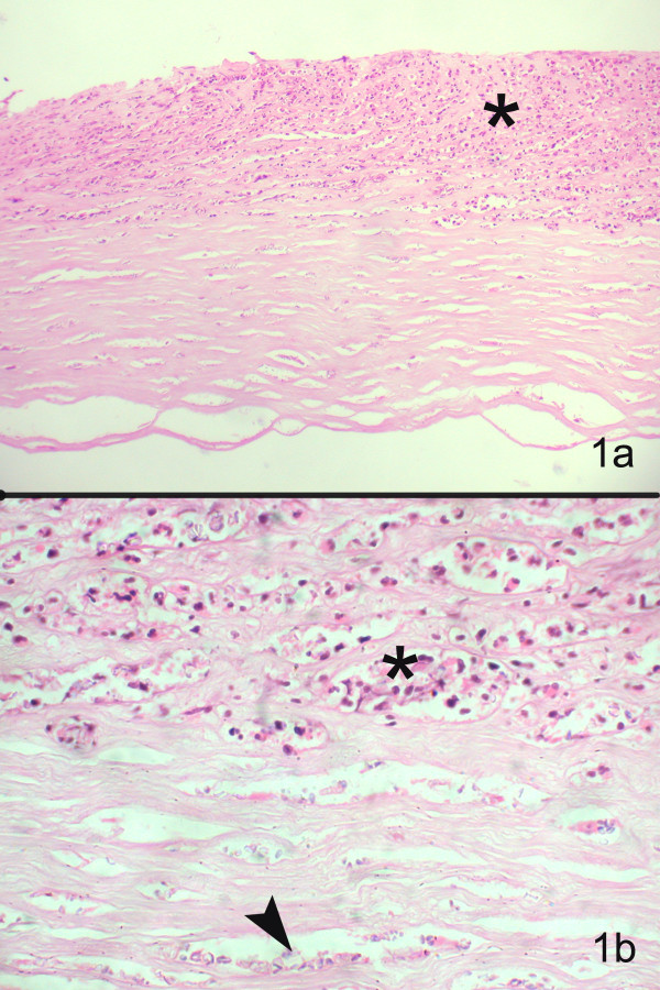
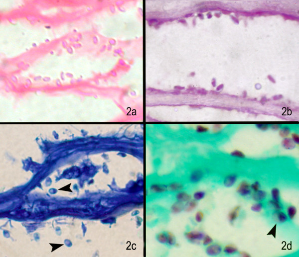
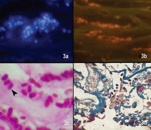
Methods: Four cases diagnosed with microsporidiosis on Hematoxylin and Eosin and Periodic Acid Schiff's stained sections of the corneal button between January 2002 and December 2004, were included. Further sections were prospectively stained with calcofluor white, Gram, Giemsa, Masson's trichrome, acridine orange, Gomori's methenamine silver, Gram's chromotrope and modified acid fast stain. The stained sections were analyzed for the spore characteristics in terms of size, shape, color contrast, cell wall morphology, waist band in cytoplasm and ease of detection.
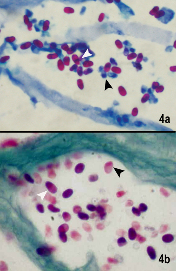
Results: All sections showed microsporidial spores as 3-5 microm, oval bodies. 1% acid fast, Gram's chromotrope and GMS stains provided a reliable diagnosis of microsporidia as diagnostic waist band could be identified and good contrast helped distinguish the spores from inflammatory debris.
Conclusion: Considering the ease of performance, cost effectiveness and rapidity of the technique, 1% acid fast stain and Gram's chromotrope stain are ideal for the detection of microsporidia.
Figures
References
-
- Weber R, Schwartz DA, Deplazes P. Laboratory Diagnosis of Microsporidiosis. In: Wittner M, Weiss LM, editor. The Microsporidia and Microsporidiosis. American Society for Microbiology, Washington D.C; 1999.
-
- Kotler DP, Orenstein JM. Prevalence of intestinal microsporidiosis in HIV-infected individuals referred for gastroenterological evaluation. Am J Gastroenterol. 1994;89:1998–2002. - PubMed
-
- Field AS, Marriott DJ, Hing MC. The Warthin-Starry stain in the diagnosis of small intestinal microsporidiosis in HIV-infected patients. Folia Parasitol (Praha) 1993;40:261–6. - PubMed
LinkOut - more resources
Full Text Sources
