Comparative Study
. 2006 Jun 21;26(25):6687-94.
doi: 10.1523/JNEUROSCI.1618-06.2006.

Kiss1 neurons in the forebrain as central processors for generating the preovulatory luteinizing hormone surge

Affiliations
Comparative Study

Kiss1 neurons in the forebrain as central processors for generating the preovulatory luteinizing hormone surge

Jeremy T Smith et al. J Neurosci. .

Abstract

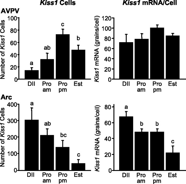
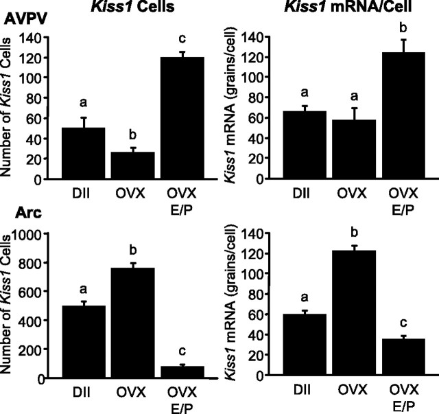
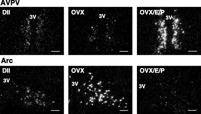
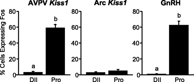
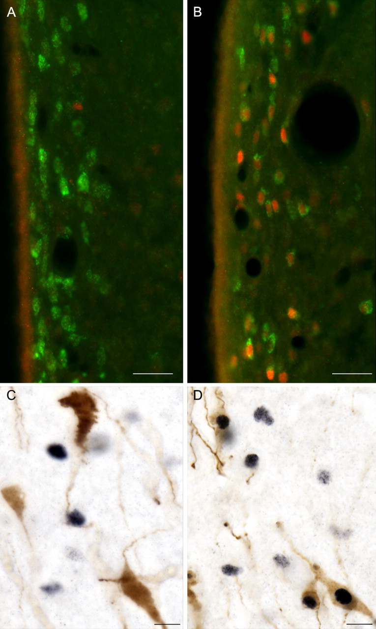
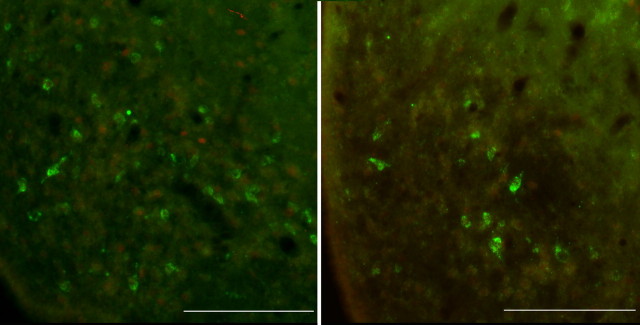
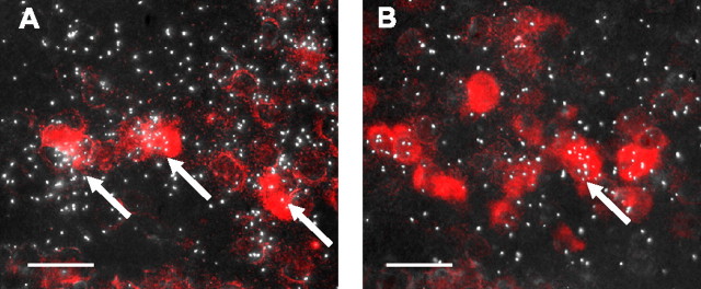
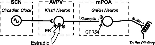
Kisspeptins are neuropeptides encoded by the Kiss1 gene, which have been implicated in the neuroendocrine regulation of gonadotropin-releasing hormone (GnRH) secretion. The goal of this study was to test the hypothesis that activation of Kiss1 neurons in the anteroventral periventricular nucleus (AVPV) is linked to the induction of the preovulatory luteinizing hormone (LH) surge in the rat. First, we determined that levels of Kiss1 mRNA in the AVPV peaked during the evening of proestrus, whereas Kiss1 mRNA in the arcuate nucleus (Arc) was at its nadir. Second, we corroborated this observation by demonstrating that Kiss1 mRNA is increased in the AVPV at the time of an estrogen (E)- and progesterone-induced LH surge in ovariectomized animals, whereas in the Arc, the expression of Kiss1 mRNA was decreased. Third, we found that most Kiss1 neurons in the AVPV coexpress the immediate early gene Fos coincidently with the LH surge, but virtually none coexpressed Fos on diestrus. In contrast, Kiss1 neurons in the Arc were Fos negative at the time of the LH surge as well as on diestrus. Finally, we found that most Kiss1 neurons in the AVPV and Arc express estrogen receptor alpha mRNA, suggesting that E acts directly on these neurons. These results suggest that Kiss1 neurons in the AVPV play an active role in mediating the effects of E on the generation of the preovulatory GnRH/LH surge on proestrus.


Figures

Figure 1.
Figure 1.
The number of identifiable Kiss1 mRNA-positive cells and grains per cell in the AVPV and Arc of the female rat during the estrous cycle. Values without common notations (a, b, c) differ significantly (p < 0.05). Values are the mean ± SEM. DII, Diestrus; Pro am, proestrus 2 h after lights on; Pro pm, proestrus 1 h before lights off; Est, estrus.
Figure 2.
Figure 2.
Kiss1 mRNA expression in the AVPV and Arc of the female rat during the E/P-induced LH surge. Values without common notations (a, b, c) differ significantly (p < 0.05). Values are the mean ± SEM. DII, Diestrus.
Figure 3.
Figure 3.
Dark-field photomicrographs showing Kiss1 mRNA-expressing cells (as reflected by the presence of white clusters of silver grains) in representative sections of the AVPV and Arc from diestrus (DII), OVX, and OVX/E/P-treated female rats. 3V, Third ventricle. Scale bars, 100 μm.
Figure 4.
Figure 4.
Quantitative analysis showing the percentage of Kiss1 neurons that coexpress Fos 30 min after lights off on diestrus II (DII) and proestrus (Pro) in the AVPV (left) and Arc (middle) and the percentage of GnRH neurons that coexpress Fos on diestrus II and proestrus (right panel). Values without common notations (a, b) differ significantly (p < 0.05). Values are the mean ± SEM.
Figure 5.
Figure 5.
A, B, Photomicrographs showing Kiss1 neurons (green) in the AVPV on diestrus II (A) and proestrus (B), with the induction of Fos (red) in Kiss1 neurons on the afternoon of proestrus (compare A, B). Scale bars, 25 μm. C, D, Photomicrographs showing GnRH neurons (brown) and Fos (black) on diestrus II (C) and their coexpression on the afternoon of proestrus (D). Scale bars, 10 μm.
Figure 6.
Figure 6.
Photomicrographs showing Kiss1 neurons (green) and nuclear Fos (red) in the arcuate nucleus on diestrus II (left) and the afternoon of proestrus (right), revealing no evidence for colocalization of Kiss1 mRNA and Fos on either day. Scale bars, 100 μm.
Figure 7.
Figure 7.
Representative photomicrographs showing coexpression of Kiss1 mRNA with ERα (A) and ERβ (B) in the anteroventral periventricular nucleus. Kiss1 mRNA-expressing cells were visualized with Vector Red substrate, and ERα (A) or ERβ (B) were marked by the presence of clusters of silver grains. Arrows indicate Kiss1 neurons that coexpress either ERα or ERβ. Scale bars, 20 μm.
Figure 8.
Figure 8.
Simplified model of the role of Kiss1 neurons in the generation of the GnRH/LH surge. According to this model, Kiss1 neurons in the AVPV receive circadian signals from the SCN and sense circulating E levels via estrogen receptors (ER), which are expressed within these neurons. When E levels and the circadian signal are both high, Kiss1 neurons become activated, causing kisspeptin to be released at synapses on GnRH neurons. Kisspeptin activates those GnRH neurons, driving the release of GnRH into the portal circulation. mPOA, Medial preoptic area.

Similar articles

Cited by

References

    1. Barbacka-Surowiak G, Surowiak J, Stoklosowa S (2003). The involvement of suprachiasmatic nuclei in the regulation of estrous cycles in rodents. Reprod Biol 3:99–129. - PubMed
    1. Barraclough CA (1961). Production of anovulatory, sterile rats by single injections of testosterone propionate. Endocrinology 68:62–67. - PubMed
    1. Berghorn KA, Bonnett JH, Hoffman GE (1994). cFos immunoreactivity is enhanced with biotin amplification. J Histochem Cytochem 42:1635–1642. - PubMed
    1. Berghorn KA, Le WW, Sherman TG, Hoffman GE (2001). Suckling stimulus suppresses messenger RNA for tyrosine hydroxylase in arcuate neurons during lactation. J Comp Neurol 438:423–432. - PubMed
    1. Brailoiu GC, Dun SL, Ohsawa M, Yin D, Yang J, Chang JK, Brailoiu E, Dun NJ (2005). KiSS-1 expression and metastin-like immunoreactivity in the rat brain. J Comp Neurol 481:314–329. - PubMed

Publication types

MeSH terms

LinkOut - more resources