Comparative Study
. 2006 Jun 21;26(25):6803-12.
doi: 10.1523/JNEUROSCI.0526-06.2006.

Vascular endothelial growth factor directly inhibits primitive neural stem cell survival but promotes definitive neural stem cell survival

Affiliations
Comparative Study

Vascular endothelial growth factor directly inhibits primitive neural stem cell survival but promotes definitive neural stem cell survival

Tamaki Wada et al. J Neurosci. .

Abstract

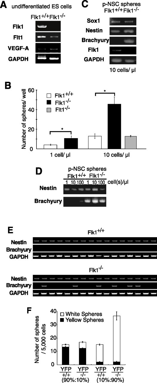
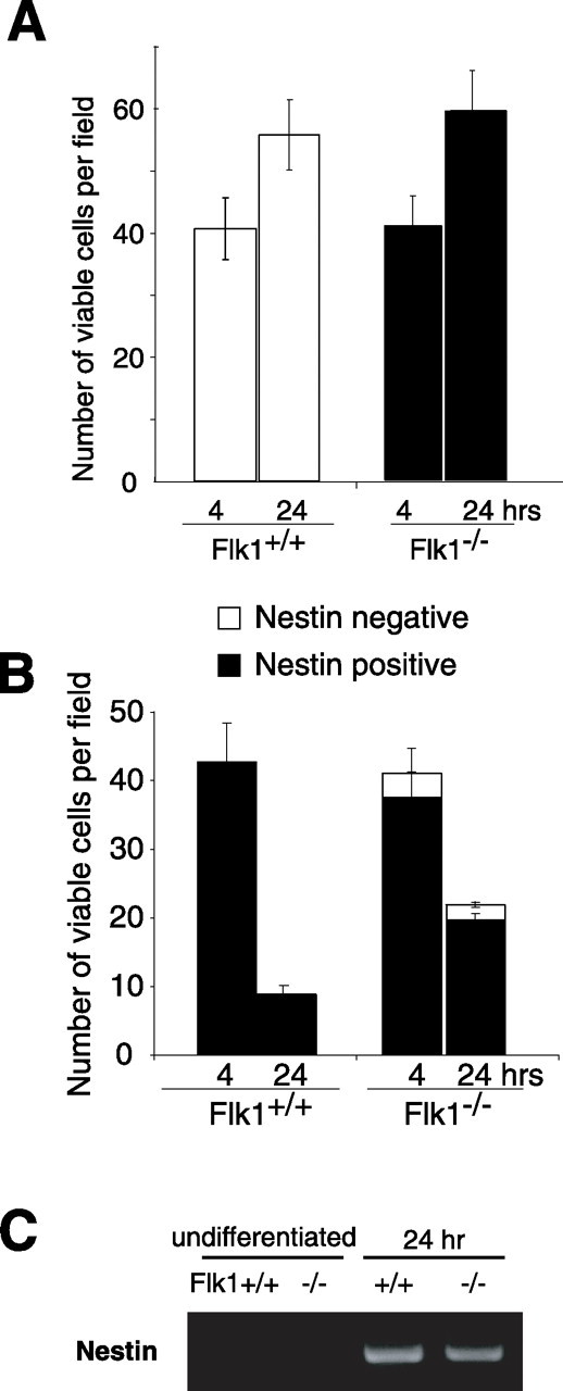
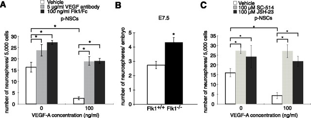
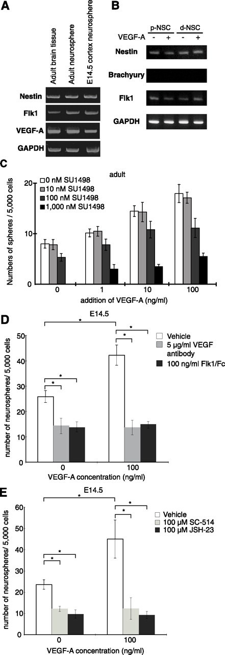
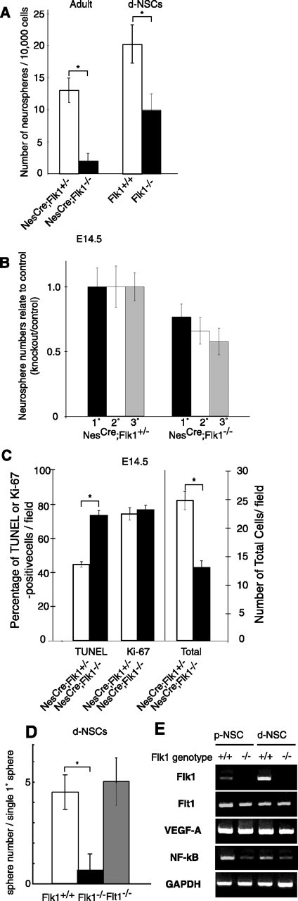
There are two types of neural stem cells (NSCs). Primitive NSCs [leukemia inhibitory factor (LIF) dependent but exogenous fibroblast growth factor (FGF) 2 independent] can be derived from mouse embryonic stem (ES) cells in vitro and from embryonic day 5.5 (E5.5) to E7.5 epiblast and E7.5-E8.5 neuroectoderm in vivo. Definitive NSCs (LIF independent but FGF2 dependent) first appear in the E8.5 neural plate and persist throughout life. Primitive NSCs give rise to definitive NSCs. Loss and gain of functions were used to study the role of vascular endothelial growth factor (VEGF)-A and its receptor, Flk1, in NSCs. The numbers of Flk1 knock-out mice embryo-derived and ES cell-derived primitive NSCs were increased because of the enhanced survival of primitive NSCs. In contrast, neural precursor-specific, Flk1 conditional knock-out mice-derived, definitive NSCs numbers were decreased because of the enhanced cell death of definitive NSCs. These effects were not observed in cells lacking Flt1, another VEGF receptor. In addition, the cell death stimulated by VEGF-A of primitive NSC and the cell survival stimulated by VEGF-A of definitive NSC were blocked by Flk1/Fc-soluble receptors and VEGF-A function-blocking antibodies. These VEGF-A phenotypes also were blocked by inhibition of the downstream effector nuclear factor kappaB (NF-kappaB). Thus, both the cell death of primitive NSC and the cell survival of definitive NSC induced by VEGF-A stimulation are mediated by bifunctional NF-kappaB effects. In conclusion, VEGF-A function through Flk1 mediates survival (and not proliferative or fate change) effects on NSCs, specifically.


Figures

Figure 1.
Figure 1.
A, Undifferentiated Flk1+/+ (but not Flk1−/−) ES cells express low levels of Flk1 mRNA. Both Flt1 and VEGF-A mRNA expression also are observed in both of the undifferentiated ES cell lines. GAPDH was used as a universal control. B, The numbers of Flk1−/− p-NSC spheres were three times greater than the numbers of Flk1+/+ p-NSC spheres at starting ES cell densities of both 1 cell/μl (t(6) = 2.2; p < 0.05) and 10 cells/μl (t(6) = 3.9; p < 0.05). The number of Flt1−/− p-NSC spheres was similar to Flt1+/+ p-NSC spheres (t(5) = 0.4; p > 0.5). C, Bulk Flk1−/− p-NSC spheres expressed the mesodermal marker gene Brachyury, which is never expressed by Flk1+/+ p-NSC spheres. Expression of the NSC markers Sox1 and Nestin was observed in both Flk1+/+ and Flk1−/− p-NSC spheres. Flk1 expression was observed in Flk1+/+ p-NSC spheres but not in Flk1−/− p-NSC spheres. GAPDH was used as a universal control. D, Nestin mRNA expression was detected in both Flk1+/+ and Flk1−/− bulk p-NSC spheres at each of the 1, 10, and 100 ES cell(s)/μl starting densities. Brachyury mRNA expression was detected in Flk1−/− p-NSC spheres at all three of the ES cell starting densities. However, only the highest ES cell starting density produced Flk1+/+ p-NSC spheres with even weak expression of Brachyury mRNA. E, Gene expression was assessed in single clonal p-NSC spheres by RT-PCR. The top three bands for each single p-NSC colony were from Flk1+/+ sphere PCRs, and the bottom three bands for each single p-NSC colony were from Flk1−/− sphere PCRs. Only Nestin (and not Brachyury) mRNA expression was observed in each of the 16 of Flk1+/+ clonal p-NSC spheres. In contrast, 6 of 16 clonal Flk1−/− p-NSC spheres expressed Brachyury mRNA, but all of them (16 of 16) expressed Nestin mRNA. F, Constitutive YFP-expressing ES cells were cocultured with either Flk1+/+ or Flk1−/− ES cells. The numbers of YFP p-NSC spheres was equivalent in both cocultures and at both YFP/Flk1−/− cell coculture ratios, but the nonfluorescent Flk1−/− p-NSC spheres showed the same threefold increase compared with nonfluorescent Flk1+/+ p-NSC spheres, as was shown in non-cocultures in B (suggesting a cell-autonomous effect of the Flk1 mutation in the p-NSC sphere-forming cells). Data are mean ± SEM. *p < 0.05.
Figure 2.
Figure 2.
A, Dissociated undifferentiated ES cells were plated on gelatin-coated chamber slides at 10 cells/μl in ES culture media containing 10% FBS. There were no significant differences in viable cells between Flk1+/+ and Flk1−/− undifferentiated ES cells at 4 h after plating. Although cell numbers were increased at 24 h after plating compared with 4 h, Flk1+/+ and Flk1−/− undifferentiated ES cells showed similar numbers of viable cells (at 4 h, t(6) = 0.2, p > 0.5; at 24 h, t(6) = 0.3, p > 0.5). B, Short-term differentiation assay from undifferentiated ES cell to p-NSC. Equal numbers of cells were present in both Flk1+/+ and Flk1−/− cultures at the 4 h after plating. However, fewer cells were found in Flk1+/+ than Flk1−/− cultures at 24 h after plating. A few Nestin-negative cells were observed only in Flk1−/− cultures at both 4 and 24 h after plating. C, RT-PCR of short-term differentiated ES cells. Nestin mRNA expression was detected in both Flk1+/+ and Flk1−/− p-NSC cells after 24 h of differentiation. Data are mean ± SEM.
Figure 3.
Figure 3.
A, The number of wild-type Flk1+/+ p-NSC spheres is decreased by the addition of 100 ng/ml VEGF-A into the ES culture minimal medium (t(4) = 2.6; p < 0.05). This effect was blocked by the addition of two VEGF-A inhibitors, VEGF-A antibodies and Flk1/Fc-soluble chimeric receptors. There was a significant interaction of VEGF-A dose and blocker (F(3,12) = 25.32; p < 0.05). Multiple comparison tests revealed that both VEGF-A antibodies (p < 0.05) and Flk1/Fc receptors (p < 0.05) increased p-NSC numbers in the presence of VEGF-A compared with VEGF-A treatment alone. Moreover, in the absence of VEGF-A, both VEGF-A antibodies (p < 0.05) and Flk1/Fc receptors (p < 0.05) produced an increase in p-NSC numbers compared with their vehicle controls. B, The numbers of neurospheres from E7.5 Flk1 conventional knock-out mice (derived clonally from single p-NSCs) increased 1.6-fold compared with the numbers from wild-type control mice (t(5) = 7.6; p < 0.05). C, The decreased number of p-NSC spheres produced by VEGF-A addition was blocked by two different NF-κB inhibitors, SC-514 and JSH-23. There was a significant interaction of VEGF-A dose and NF-κB inhibitor (F(3,80) = 15.6; p < 0.05). Multiple comparison tests revealed that both SC-514 (p < 0.05) and JSH-23 (p < 0.05) increased p-NSC numbers in the presence of VEGF-A compared with VEGF-A treatment alone. Moreover, in the absence of VEGF-A, both SC-514 (p < 0.05) and JSH-23 (p < 0.05) produced an increase in p-NSC numbers compared with their vehicle controls. Data are mean ± SEM. *p < 0.05.
Figure 4.
Figure 4.
A, Adult brain tissue as well as d-NSC-derived clonal neurospheres from the adult brain express Flk1 mRNA. Clonal E14.5 brain neurospheres also expressed Flk1 mRNA. Nestin and control GAPDH mRNAs were expressed in adult brain tissue, clonal adult brain neurospheres, and clonal E14.5 brain neurospheres. B, Nestin and Flk1 mRNA expression were not changed by VEGF-A treatment of either p-NSCs or d-NSCs. Moreover, Brachyury mRNA expression was not induced by VEGF-A treatment of either p-NSCs or d-NSCs. C, Increasing concentrations of VEGF-A enhanced the formation of neurospheres from adult mouse d-NSCs. Moreover, this VEGF-A-induced increase in neurospheres was suppressed by the addition of SU1498, a VEGF-A signaling inhibitor. SU1498 also decreased the number of neurospheres in the absence of VEGF-A in the culture media. D, The enhanced numbers of E14.5 d-NSC spheres produced by VEGF-A were blocked by the addition of two VEGF-A inhibitors, VEGF-A antibodies and Flk1/Fc-soluble chimeric receptors. There was a significant interaction of VEGF-A dose and blocker (F(3,20) = 36.86; p < 0.05). Multiple comparison tests revealed that both VEGF-A antibodies (p < 0.05) and Flk1/Fc receptors (p < 0.05) decreased d-NSC numbers in the presence of VEGF-A compared with VEGF-A treatment alone. Furthermore, in the absence of VEGF-A, both VEGF-A antibodies (p < 0.05) and Flk1/Fc chimeric receptors (p < 0.05) decrease d-NSC numbers compared with their vehicle controls. E, The increased numbers of d-NSC spheres in the presence of VEGF-A was blocked by two different NF-κB inhibitors, SC-514 and JSH-23. There was a significant interaction of VEGF-A dose and NF-κB inhibitor (F(4,56) = 18.3; p < 0.05). Multiple comparison tests revealed that both SC-514 (p < 0.05) and JSH-23 (p < 0.05) decreased d-NSC numbers in the presence of VEGF-A compared with VEGF-A treatment alone. Moreover, in the absence of VEGF-A, both SC-514 (p < 0.05) and JSH-23 (p < 0.05) produced a decrease in p-NSC numbers compared with their vehicle controls. Data are mean ± SEM. *p < 0.05.
Figure 5.
Figure 5.
A, The numbers of neurospheres from the forebrain germinal zone of adult NesCre;Flk1−/− mice were decreased dramatically compared with control NesCre;Flk1+/− mice (t(4) = 4.2; p < 0.05). Although more clonal, primary p-NSC-derived neurospheres emerge from Flk1−/− compared with Flk1+/+ ES cultures, fewer clonal, secondary d-NSC Flk1−/− than Flk1+/+ spheres arose in FGF2 and B27 from the primary p-NSC sphere dissociations (t(3) = 4.2; p < 0.05). B, There were significant decreases in the numbers of primary and passaged clonal d-NSCs derived from E14.5 NesCre;Flk1−/− conditional knock-out mice compared with control NesCre;Flk1+/− mice. C, Acute dissociation assays of adult forebrain germinal zone cells. The total numbers of NesCre;Flk1−/− cells were fewer than NesCre;Flk1+/− cells after 8 h of incubation (right; t(4) = 5.2; p < 0.05). More apoptotic, TUNEL-positive cells were seen among the NesCre;Flk1−/− cells than among the control NesCre;Flk1+/− cells (left; t(4) = 6.1; p < 0.05). However, numbers of proliferating Ki-67-positive cells were similar in NesCre;Flk1−/− and control NesCre;Flk1+/− 8 h cultures (middle; t(4) = 0.4; p > 0.5). D, Single primary p-NSC-derived d-NSC neurosphere passaging confirmed the decrease in secondary Flk1−/− compared with wild-type d-NSC neurospheres (t(3) = 3.3; p < 0.05), seen with bulk passaging (A). Similar numbers of clonal d-NSC neurospheres were produced by Flt1−/− and wild-type cells (t(4) = 0.3; p > 0.5). E, Both p-NSCs and d-NSCs derived from Flk1+/+ or Flk1−/− ES cells expressed Flt1, NF-κB, and VEGF-A mRNAs. Data are mean ± SEM. *p < 0.05.

Similar articles

Cited by

References

    1. Barkett M, Gilmore TD (1999). Control of apoptosis by Rel/NF-kappaB transcription factors. Oncogene 18:6910–6924. - PubMed
    1. Bjornson CR, Rietze RL, Reynolds BA, Magli MC, Vescovi AL (1999). Turning brain into blood: a hematopoietic fate adopted by adult neural stem cells in vivo. Science 283:534–537. - PubMed
    1. Boguslawski G, McGlynn PW, Harvey KA, Kovala AT (2004). SU1498, an inhibitor of vascular endothelial growth factor receptor 2, causes accumulation of phosphorylated ERK kinases and inhibits their activity in vivo and in vitro. J Biol Chem 279:5716–5724. - PubMed
    1. Brusselmans K, Bono F, Collen D, Herbert JM, Carmeliet P, Dewerchin M (2005). A novel role for VEGF as an autocrine survival factor for embryonic stem cells during hypoxia. J Biol Chem 280:3493–3499. - PubMed
    1. Chiasson BJ, Tropepe V, Morshead CM, van der Kooy D (1999). Adult mammalian forebrain ependymal and subependymal cells demonstrate proliferative potential, but only subependymal cells have neural stem cell characteristics. J Neurosci 19:4462–4471. - PMC - PubMed

Publication types

MeSH terms