Comparative Study
. 2006 Jun 21;26(25):6851-62.
doi: 10.1523/JNEUROSCI.5444-05.2006.

Zinc-dependent multi-conductance channel activity in mitochondria isolated from ischemic brain

Affiliations
Comparative Study

Zinc-dependent multi-conductance channel activity in mitochondria isolated from ischemic brain

Laura Bonanni et al. J Neurosci. .

Abstract

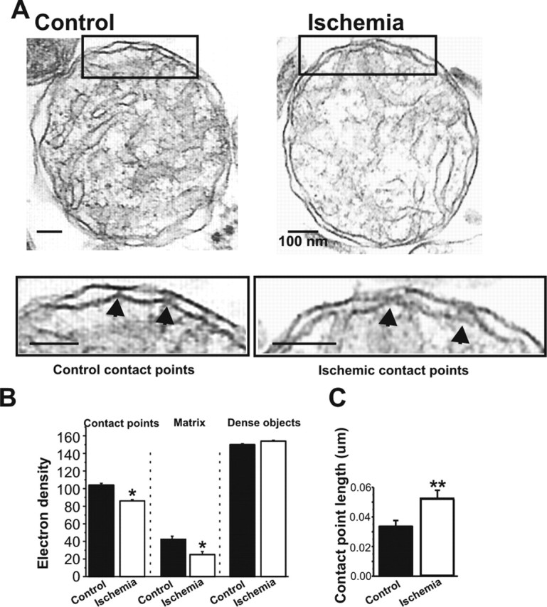
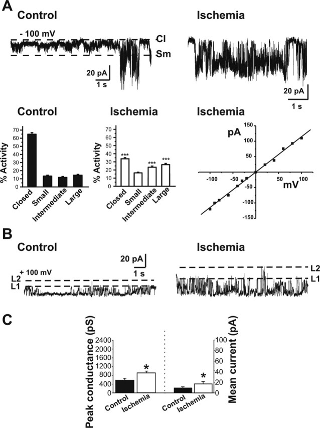
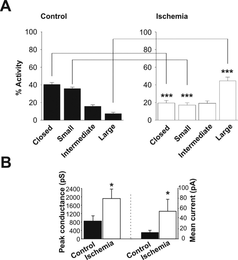
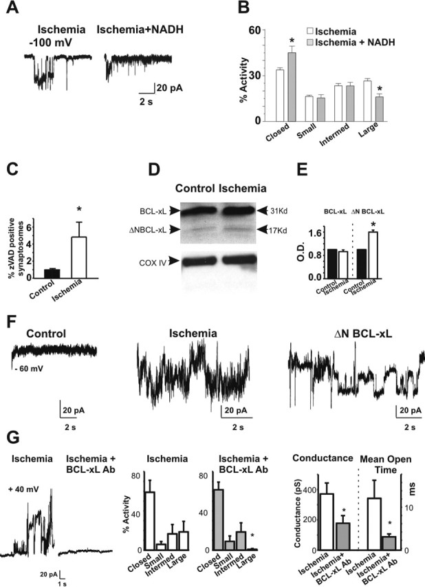
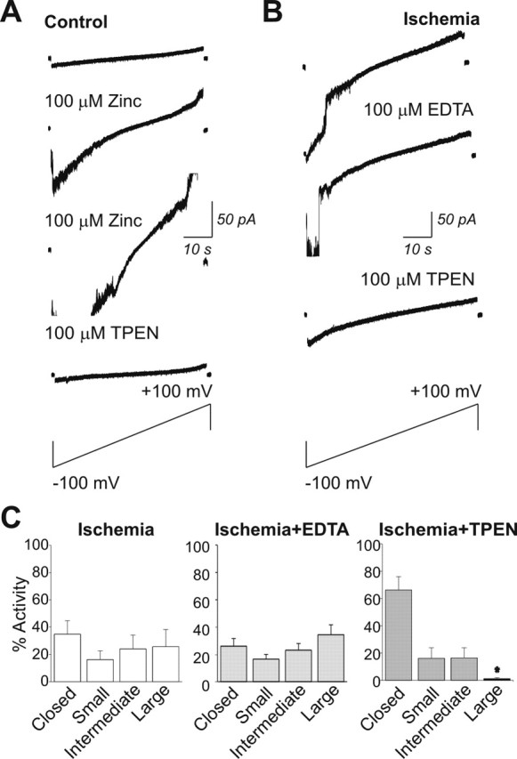
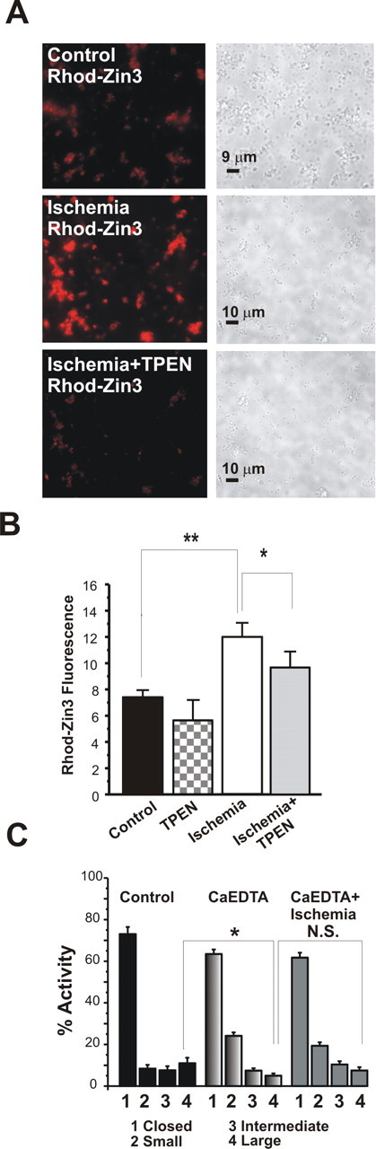
Transient global ischemia is a neuronal insult that induces delayed cell death. A hallmark event in the early post-ischemic period is enhanced permeability of mitochondrial membranes. The precise mechanisms by which mitochondrial function is disrupted are, as yet, unclear. Here we show that global ischemia promotes alterations in mitochondrial membrane contact points, a rise in intramitochondrial Zn2+, and activation of large, multi-conductance channels in mitochondrial outer membranes by 1 h after insult. Mitochondrial channel activity was associated with enhanced protease activity and proteolytic cleavage of BCL-xL to generate its pro-death counterpart, deltaN-BCL-xL. The findings implicate deltaN-BCL-xL in large, multi-conductance channel activity. Consistent with this, large channel activity was mimicked by introduction of recombinant deltaN-BCL-xL to control mitochondria and blocked by introduction of a functional BCL-xL antibody to post-ischemic mitochondria via the patch pipette. Channel activity was also inhibited by nicotinamide adenine dinucleotide, indicative of a role for the voltage-dependent anion channel (VDAC) of the outer mitochondrial membrane. In vivo administration of the membrane-impermeant Zn2+ chelator CaEDTA before ischemia or in vitro application of the membrane-permeant Zn2+ chelator tetrakis-(2-pyridylmethyl) ethylenediamine attenuated channel activity, suggesting a requirement for Zn2+. These findings reveal a novel mechanism by which ischemic insults disrupt the functional integrity of the outer mitochondrial membrane and implicate deltaN-BCL-xL and VDAC in the large, Zn2+-dependent mitochondrial channels observed in post-ischemic hippocampal mitochondria.


Figures

Figure 1.
Figure 1.
Mitochondrial morphology is altered in post-ischemic mitochondria. A, Electron micrographs of mitochondria isolated from the hippocampus of post-ischemic and control rats at 50 min after reperfusion. Insets show areas of contact between outer and inner membranes. B, The density of contact points is reduced in micrographs of post-ischemic mitochondria compared with control mitochondria (n = 6 control mitochondria, 48 contact points; n = 7 ischemic mitochondria, 65 contact points; ∗p < 0.02; mitochondria were pooled from 5 ischemic rat brains and 5 control rat brains from animals that had been treated during 1 experiment). Filled arrows in the insets of A show representative contact points used for the morphological analysis. Matrix density was lower in ischemic mitochondria compared with controls (20 measurements were made in each of three mitochondria in each of 4 micrographs of control pellets for a total of 240 control measurements; 20 measurements were made in each of 3 mitochondria in each of 6 micrographs of ischemic pellets for a total of 360 ischemic measurements; ∗p < 0.03). The density of the densest nonmitochondrial organelles in each micrograph was not different for control versus ischemic micrographs (n = 30 control measurements from 4 micrographs; n = 45 ischemic measurements from 6 micrographs; same experimental day as for contact point and matrix density measurements). C, The length of contact points in micrometers is increased in ischemic mitochondria over controls (n = 6 control mitochondria, 38 total contact points; n = 8 ischemic mitochondria, 33 total contact points; ∗∗p < 0.009; same experimental day as above). Scale bars, 100 nm.
Figure 2.
Figure 2.
Post-ischemic mitochondria exhibit an increase in large conductance channels. Recordings were made from organelle-attached patches on the membranes of isolated post-ischemic and control whole-brain mitochondria. A, Sample recordings at −100 mV in control (left) and post-ischemic (right) whole-brain mitochondria. Bar graphs show histograms of channel activity for recordings like those illustrated in A. Channel activity was defined as closed channels; small activity, <180 pS; intermediate activity, 180–760 pS; and large activity, >760 pS. Whole-brain mitochondria from post-ischemic animals contain more frequent large and intermediate conductance activity and less frequent closed times (n = 29 ischemic mitochondrial recordings; n = 26 controls; the isolations were from 2 different experiments; in 1 experiment, isolated mitochondria were pooled from 3 ischemic rat brains and 3 control rat brains; in the other experiment, mitochondria were pooled from 6 ischemic rat brains and 6 control rat brains; ∗∗∗p < 0.0001). The current–voltage relationship for a representative large-conductance channel recorded from post-ischemic mitochondria shows the absence of voltage dependence. B, Traces at +100 mV demonstrate the larger conductance and increased frequency of occurrence of discrete single channel openings in patches from post-ischemic mitochondria than in patches from controls. First and second levels (L1, L2) of amplitudes of opening of the multi-conductance single channels are indicated by dotted lines. C, Peak conductance is significantly larger in post-ischemic mitochondria than in controls (n = 36 different mitochondrial recordings; in each group, ∗p < 0.02). The mean current conducted through patches from post-ischemic mitochondria (n = 22 different recordings) is greater than that through control patches (n = 11; ∗p < 0.01). Multiplying the frequency of occurrence of a single channel event by the conductance level of that event gives the mean current conducted through a patch.
Figure 3.
Figure 3.
Post-ischemic mitochondria contain more frequent and larger conductance channel activity than do control mitochondria. A, Distribution of activity level of different conductances in recordings of isolated hippocampal mitochondria. All-points histograms show the absence of activity (Closed), activity at <180 pS (Small), activity between 180 and 760 pS (Intermediate), and activity at >760 pS (Large). Hippocampal mitochondria contain more frequent large conductance activity and less frequent small conductance activity (∗∗∗p < 0.0001). B, Peak conductance is significantly larger in post-ischemic mitochondria from hippocampus than in controls from hippocampus (n = 9 different control hippocampal recordings; n = 4 different hippocampal ischemic recordings; ∗p < 0.05). The mean current conducted through patches containing discrete single channel openings is increased in ischemic patches from hippocampal mitochondria compared with controls (n = 6 hippocampal ischemic patches; n = 10 hippocampal control patches; ∗p < 0.05). The mean current conducted through a patch was calculated by multiplying the frequency of occurrence of a single channel event by its conductance level. The hippocampal mitochondria were isolated from 10 ischemic animals and eight sham-operated controls from two independent experiments.
Figure 4.
Figure 4.
Large conductance channels are inhibited by NADH and an antibody against BCL-xL. A, Application of NADH attenuates large conductance openings of post-ischemic mitochondria from whole brain. Examples of 10 s periods of recording at the indicated voltage made from a post-ischemic mitochondrion (left) and a post-ischemic mitochondrion in the presence of 2 mm NADH in the bath and the pipette (right). B, Histograms combining all experiments show the percentage activity of different levels of conductance in the post-ischemic patches with and without NADH. Large conductances are reduced in the presence of NADH. The conductances based on peaks in amplitude histograms are as follows: closed channels, small activity at <180 pS, intermediate activity between 180 and 760 pS, and large activity at >760 pS. C, Protease activation in nerve terminals occurs early after a brief ischemic insult. A higher percentage of synaptosomes from post-ischemic whole brain or from post-ischemic hippocampus contain activated proteases compared with controls (n = 12 control samples; n = 11 post-ischemic samples from three different experiments; ∗p < 0.03; in each experiment, between 3 and 6 rat brains were used for ischemic and control conditions). The amount of fluorescent zVAD binding represents activated proteases. D, Western blots show that BCL-xL is present at high levels in mitochondria isolated from hippocampus in both post-ischemic mitochondria and controls. Protein yields are not significantly different between ischemic and control (5.4 μg/μl ischemic vs 5.46 μg/μl control), yet an N-terminal proteolytic cleavage fragment of BCL-xL is present at higher levels in post-ischemic hippocampal mitochondria than in controls. Immunoblots against COX IV are provided as an additional control for equivalence of protein samples. E, Densitometric analysis of immunoblots of BCL-xL and ΔN-BCL-xL (n = 11 controls; n = 13 post-ischemic; ∗p < 0.05). Densities of BCL-xL or ΔN-BCL-xL were normalized to values in controls. F, Recordings of isolated hippocampal mitochondria from control (left), ischemic (middle), and a control mitochondrion exposed to ΔN-BCL-xL in the patch pipette (right). Large and multiple conductances were recorded from ischemic mitochondria and from control mitochondria exposed to ΔN-BCL-xL (n = 4 recordings from control mitochondria in the presence of ΔN-BCL-xL). G, A polyclonal antibody against BCL-xL blocks large conductance activity in the outer membrane of post-ischemic mitochondria (antibody 1:1000, n = 4; or 1:500, n = 2). The left panel shows a recording without antibody, and the right panel shows a recording with antibody. Histograms combining all experiments show the frequency of channel activity in mitochondria from ischemic brain (n = 40 different mitochondria; left) and mitochondria from ischemic brain exposed to anti-BCL-xL antibody (n = 6 different mitochondria, right; ∗p < 0.0001). Closed, Absence of activity; Small, activity <180 pS; Intermed, activity between 180 and 760 pS; Large, activity >760 pS. Ischemic mitochondrial recordings in this experiment did not differ significantly from those in Figure 2; therefore, the data from the two sets of experiments were pooled. Histograms at right show the conductance level and mean channel open time of activity recorded from post-ischemic mitochondria in the presence or absence of antibody. All recordings that contained discrete single channel openings were analyzed (n = 6 ischemic mitochondrial traces in the absence of antibody; n = 7 ischemic mitochondrial traces in the presence of antibody. ∗p < 0.05).
Figure 5.
Figure 5.
Zn2+ is critical to large channel activity in the outer membrane of whole-brain mitochondria. A, Example of a patch-clamp recording from a control mitochondrion isolated from whole brain. Bath application of Zn2+ (100 μm) enhances the conductance of channels in the patch. A second application of Zn2+ further increases the conductance of channel activity in the patch. Subsequent application of the selective, membrane-permeant Zn2+ chelator TPEN reduces channel conductance in the patch. Traces shown are current responses elicited by a voltage ramp from −100 to +100 mV. B, Large conductance channel activity of post-ischemic mitochondria is attenuated by application of the membrane-permeant Zn2+ chelator TPEN. Shown are patch-clamp traces recorded during a voltage ramp from −100 to +100 mV. The membrane-permeant Zn2+ chelator TPEN, but not the membrane-impermeant Zn2+ chelator EDTA, inhibits large conductance activity in the post-ischemic mitochondrial patch. C, Histograms of activity of different size conductances recorded from post-ischemic whole-brain mitochondria before and after application of TPEN or EDTA. TPEN, but not EDTA, decreases the large conductance activity (n = 6 control and with TPEN, ∗p < 0.05; n = 3 EDTA).
Figure 6.
Figure 6.
Zn2+ fluorescence is greater in post-ischemic versus control mitochondria. A, Examples of micrographs of mitochondria isolated from the hippocampus of control (top) and post-ischemic (middle) animals and treated with the Zn2+-specific fluorescent indicator dye RhodZin-3 (100 nm). Phase images are shown to the right of fluorescent images. Middle and bottom panels show a paired experiment in which micrographs of post-ischemic hippocampal mitochondria were obtained before and 10 min after application of TPEN (20 μm). B, Data from all images were obtained by determining the intensity of fluorescence within six boxes set over clusters of mitochondria on each dish. All measurements were background subtracted (n = 61 control measurements; n = 102 post-ischemic measurements; ∗∗p < 0.003). Data were obtained from three ischemic rat brains and three control rat brains on the same day. In paired experiments, TPEN significantly decreased RhodZin-3 fluorescence in post-ischemic mitochondria (n = 14 controls after TPEN; n = 48 ischemic measurements after TPEN; fluorescence significantly decreased after TPEN in ischemic mitochondria; ∗p < 0.03). C, Appearance of large mitochondrial membrane channels is inhibited by injection of rat ventricles with the Zn2+ chelator CaEDTA 30 min before the ischemic episode. Histograms combining all recordings show the percentage activity of different levels of conductance in control patches or in post-ischemic patches with and without CaEDTA. The conductances are as follows: closed channels; small activity at <180 pS; intermediate activity between 180 and 760 pS; and large activity at >760 pS. Two rats were treated in each group (control, CaEDTA, and ischemia plus Ca EDTA). n = 10 mitochondrial recordings in the control group and n = 16 mitochondrial recordings in each of CaEDTA or ischemia plus CaEDTA (∗p < 0.05).

References

    1. Aizenman E, Stout AK, Hartnett KA, Dineley KE, McLaughlin B, Reynolds IJ (2000). Induction of neuronal apoptosis by thiol oxidation: putative role of intracellular zinc release. J Neurochem 75:1878–1888. - PubMed
    1. Alkayed NJ, Goto S, Sugo N, Joh HD, Klaus J, Crain BJ, Bernard O, Traystman RJ, Hurn PD (2001). Estrogen and Bcl-2: gene induction and effect of transgene in experimental stroke. J Neurosci 21:7543–7550. - PMC - PubMed
    1. Arslan P, Di Virgilio F, Beltrame M, Tsien RY, Pozzan T (1985). Cytosolic Ca2+ homeostasis in Ehrlich and Yoshida carcinomas. A new, membrane-permeant chelator of heavy metals reveals that these ascites tumor cell lines have normal cytosolic free Ca2+ J Biol Chem 260:2719–2727. - PubMed
    1. Assaf SY, Chung SH (1984). Release of endogenous Zn2+ from brain tissue during activity. Nature 308:734–736. - PubMed
    1. Banasiak KJ, Xia Y, Haddad GG (2000). Mechanisms underlying hypoxia-induced neuronal apoptosis. Prog Neurobiol 62:215–249. - PubMed

Publication types

MeSH terms