Comparative Study
. 2006 Jun 21;26(25):6885-92.
doi: 10.1523/JNEUROSCI.1062-06.2006.

Earlier development of the accumbens relative to orbitofrontal cortex might underlie risk-taking behavior in adolescents

Affiliations
Comparative Study

Earlier development of the accumbens relative to orbitofrontal cortex might underlie risk-taking behavior in adolescents

Adriana Galvan et al. J Neurosci. .

Abstract

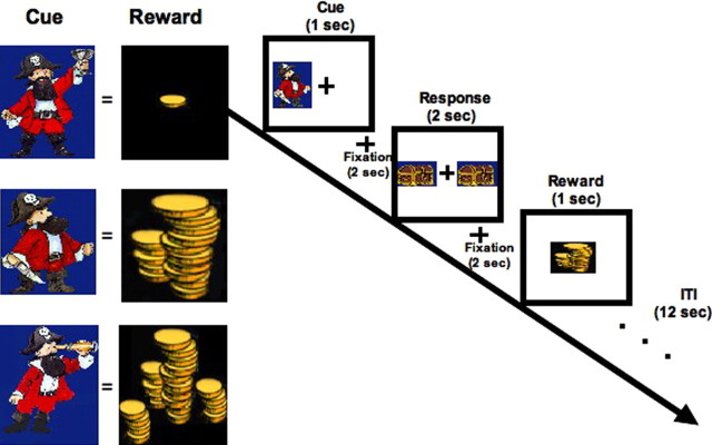
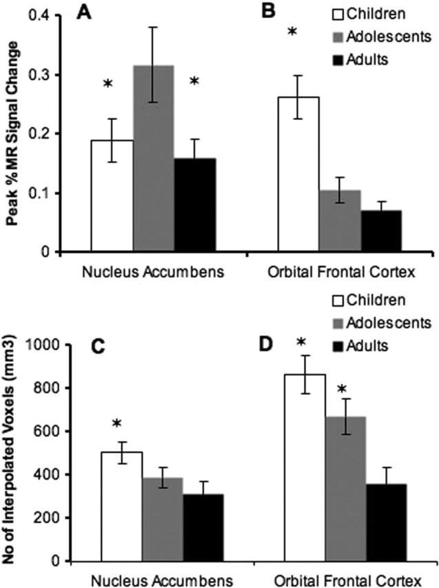
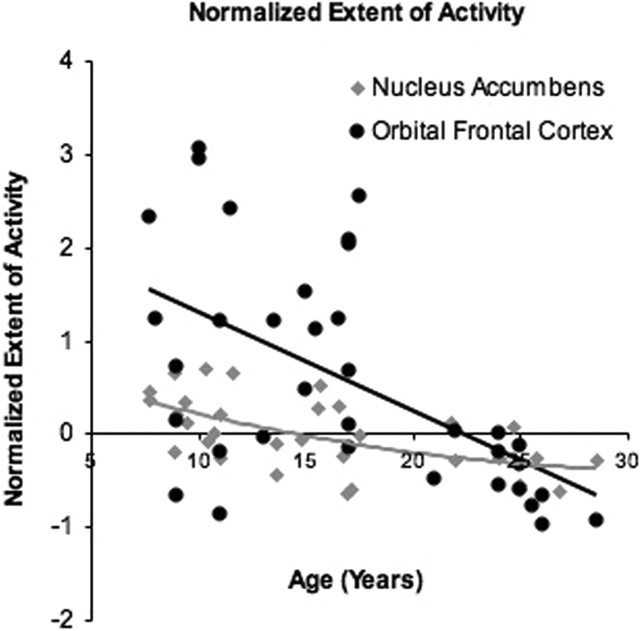
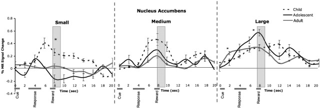
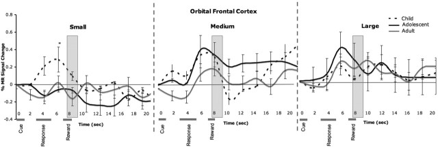
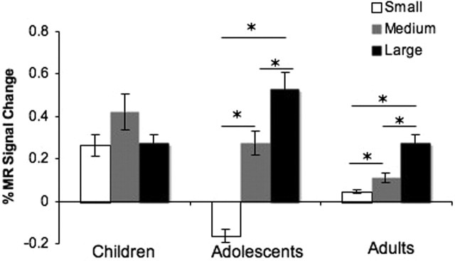
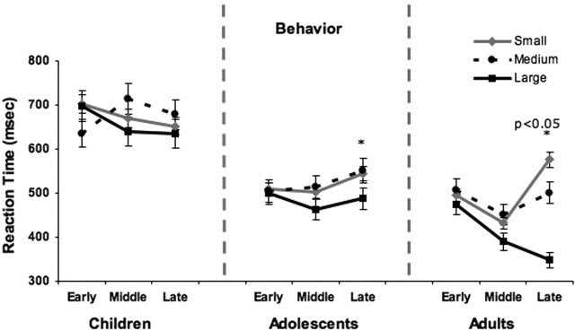
Adolescence has been characterized by risk-taking behaviors that can lead to fatal outcomes. This study examined the neurobiological development of neural systems implicated in reward-seeking behaviors. Thirty-seven participants (7-29 years of age) were scanned using event-related functional magnetic resonance imaging and a paradigm that parametrically manipulated reward values. The results show exaggerated accumbens activity, relative to prefrontal activity in adolescents, compared with children and adults, which appeared to be driven by different time courses of development for these regions. Accumbens activity in adolescents looked like that of adults in both extent of activity and sensitivity to reward values, although the magnitude of activity was exaggerated. In contrast, the extent of orbital frontal cortex activity in adolescents looked more like that of children than adults, with less focal patterns of activity. These findings suggest that maturing subcortical systems become disproportionately activated relative to later maturing top-down control systems, biasing the adolescent's action toward immediate over long-term gains.


Figures

Figure 1.
Figure 1.
Behavioral paradigm. Left panel, Three cues were each paired with a distinct reward value (counterbalanced across subjects) that remained constant throughout the experiment. Right panel, The paradigm consisted of a cue, response, and reward that were temporally separated in time with 12 s ITI. Total trial length was 20 s.
Figure 2.
Figure 2.
Localization of nucleus accumbens (A) and orbital frontal cortex (C) activation to reward. There was a main effect of reward value in the nucleus accumbens (B) [right (x = 6, y = 5, z = −2) and left (x = −8, y = 6, z = −2)] but not in the right lateral orbital frontal cortex (x = 46, y = 31, z = 1) (D). Error bars indicate SEM. Asterisks denote significant differences between small and medium, medium and large, and small and large.
Figure 3.
Figure 3.
Magnitude and extent of accumbens and OFC activity to reward. A, Adolescents showed exaggerated percent change in MR signal to large reward relative to children and adults in the accumbens. B, In the OFC, children had the greatest percent change in MR signal relative to adolescents and adults. C, Children showed the largest volume of activity in the accumbens relative to adolescents and adults. D, Children and adolescents showed greater volume of activity in the OFC relative to adults. Error bars indicate SEM. Asterisks denote significant activation differences between children and adolescents and adolescents and adults in A; greater activation in children relative to adolescents and adults in B; greater volume of activity in children relative to adolescents and adults in C; and greater volume of activity in children relative to adolescents and adolescents relative to adults in D.
Figure 4.
Figure 4.
Normalized extent of activity measure for the nucleus accumbens and OFC for all subjects, adjusted for the average extent of activity (x − mean/mean) for each region.
Figure 5.
Figure 5.
Temporal changes in the nucleus accumbens as a function of small, medium, and large reward values for late trials of the experiment for each age group. The gray bars correspond to the point(s) ∼5–6 s after the response. Error bars indicate SEM. Asterisks denote activation differences between groups.
Figure 6.
Figure 6.
Temporal changes in the orbitofrontal cortex as a function of small, medium and large reward values for late trials of the experiment for each age group. The gray bars correspond to the point(s) ∼5–6 s after the response. Error bars indicate SEM. Asterisks denote activation differences between groups.
Figure 7.
Figure 7.
Percent change in MR signal ∼5–6 s after the response relative to pretrial baseline for each age group, showing an exaggerated change in accumbens activity in adolescents relative to children or adults for the small and large reward trials. Error bars indicate SEM. Asterisks denote activation differences between groups.
Figure 8.
Figure 8.
Behavioral results. Mean reaction time as a function of small, medium, and large reward values are shown for early, middle, and late trials of the experiment for each age group. Error bars indicate SEM. Asterisks denote slower reaction time to small and medium rewards relative to small in adolescents and slower reaction time to small relative to medium and medium relative to large in adults.

References

    1. Bechara A (2005). Decision-making, impulse control and loss of willpower to resist drugs: a neurocognitive perspective. Nat Neurosci 8:1458–1463. - PubMed
    1. Bjork JM, Knutson B, Fong GW, Caggiano DM, Bennett SM, Hommer DW (2004). Incentive-elicited brain activation in adolescents: similarities and differences from young adults. J Neurosci 24:1793–1802. - PMC - PubMed
    1. Boynton GM, Engel SA, Glover GH, Heeger DJ (1996). Linear systems analysis of functional magnetic resonance imaging in human V1. J Neurosci 16:4207–4221. - PMC - PubMed
    1. Brown TT, Lugar HM, Coalson RS, Miezin FM, Petersen SE, Schlaggar BL (2005). Developmental changes in human cerebral functional organization for word generation. Cereb Cortex 15:275–290. - PubMed
    1. Bunge S, Dudukovic NM, Thomason ME, Vaidya CJ, Gabrieli JD (2002). Immature frontal lobe contributions to cognitive control in children: evidence from fMRI. Neuron 33:301–311. - PMC - PubMed

Publication types

LinkOut - more resources