

Evidence for both adenosine A1 and A2A receptors activating single vagal sensory C-fibres in guinea pig lungs
- PMID: 16793905
- PMCID: PMC1819455
- DOI: 10.1113/jphysiol.2006.109371
Evidence for both adenosine A1 and A2A receptors activating single vagal sensory C-fibres in guinea pig lungs
Abstract
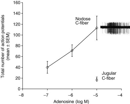
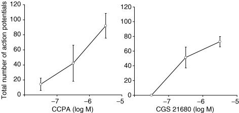
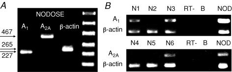
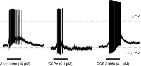
We addressed the hypothesis that single vagal afferent C-fibres can be stimulated via either the adenosine A1 or A2A receptor subtypes. The effect of adenosine on the nerve terminals of vagal sensory nerve subtypes was evaluated in an ex vivo perfused guinea pig lung preparation using extracellular recording techniques. Adenosine (10 microm) consistently evoked action potential discharge in lung C-fibre terminals arising from the nodose ganglia, but failed to evoke action potential discharge in most jugular ganglion C-fibres. Adenosine also failed to activate stretch-sensitive nodose A-fibres in the lungs. The selective A1 antagonist DPCPX (0.1 microm) or the selective A2A antagonist SCH 58261 (0.1 microm) partially inhibited the nodose C-fibre activation by adenosine, and the combination of both antagonists almost completely inhibited the response. The adenosine-induced action potential discharge in nodose C-fibres was mimicked by either the selective A1 agonist CCPA (1 microm) or the selective A2A agonist CGS 21680 (1 microm). Single cell PCR techniques revealed that adenosine A1 and A2A receptor mRNA was expressed in individual nodose neurons retrogradely labelled from the lungs. The gramicidin-perforated patch clamp technique on neurons retrogradely labelled from the lungs was employed to study the functional consequence of adenosine receptor agonists directly on neuronal membrane properties. Both the selective A1 agonist CCPA (1 microm) and the selective A2A agonist CGS 21680 (1 microm) depolarized the airway-specific, capsaicin-sensitive, nodose neurons to action potential threshold. The data support the hypothesis that adenosine selectively depolarizes vagal nodose C-fibre terminals in the lungs to action potential threshold, by stimulation of both adenosine A1 and A2A receptor subtypes located in the neuronal membrane.
Figures
References
-
- Basoglu OK, Pelleg A, Essilfie-Quaye S, Brindicci C, Barnes PJ, Kharitonov SA. Effects of aerosolized adenosine 5′-triphosphate vs adenosine 5′-monophosphate on dyspnea and airway caliber in healthy nonsmokers and patients with asthma. Chest. 2005;128:1905–1909. - PubMed
-
- Bergner A, Sanderson MJ. ATP stimulates Ca2+ oscillations and contraction in airway smooth muscle cells of mouse lung slices. Am J Physiol Lung Cell Mol Physiol. 2002;283:L1271–L1279. - PubMed
-
- Burki NK, Dale WJ, Lee LY. Intravenous adenosine and dyspnea in humans. J Appl Physiol. 2005;98:180–185. - PubMed
Publication types
MeSH terms
Substances
LinkOut - more resources
Full Text Sources
Miscellaneous