. 2006 Sep 1;575(Pt 2):583-602.
doi: 10.1113/jphysiol.2006.105106. Epub 2006 Jun 22.

Efficacy and connectivity of intracolumnar pairs of layer 2/3 pyramidal cells in the barrel cortex of juvenile rats

Affiliations

Efficacy and connectivity of intracolumnar pairs of layer 2/3 pyramidal cells in the barrel cortex of juvenile rats

Dirk Feldmeyer et al. J Physiol. .

Abstract

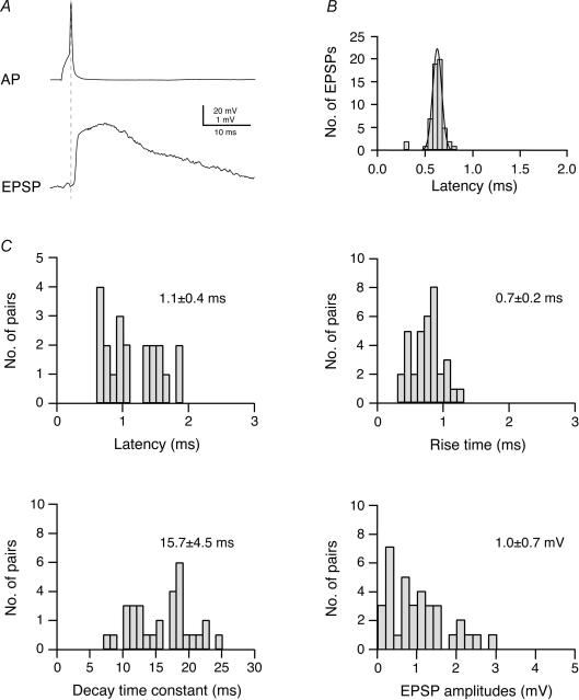
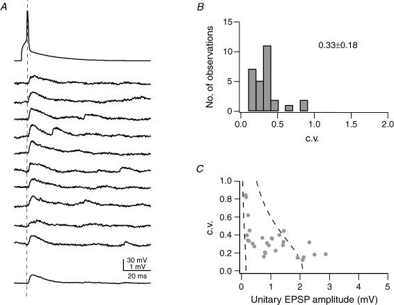
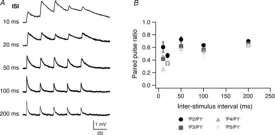
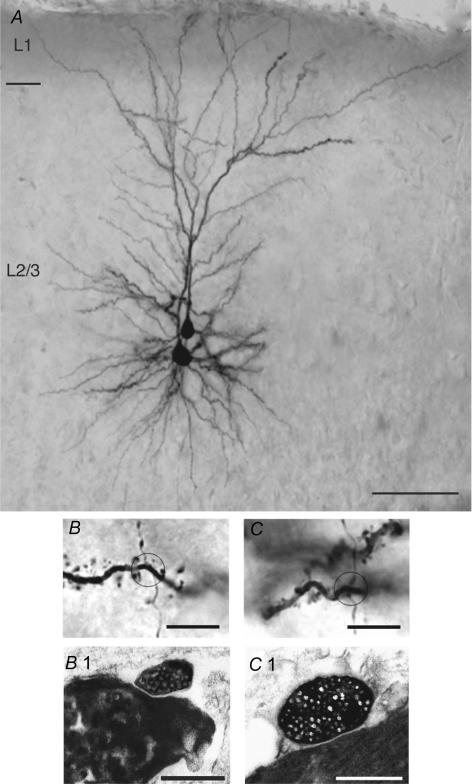
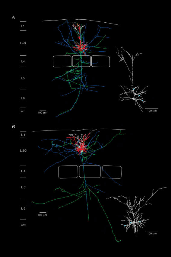
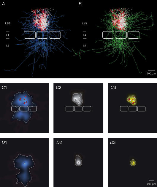
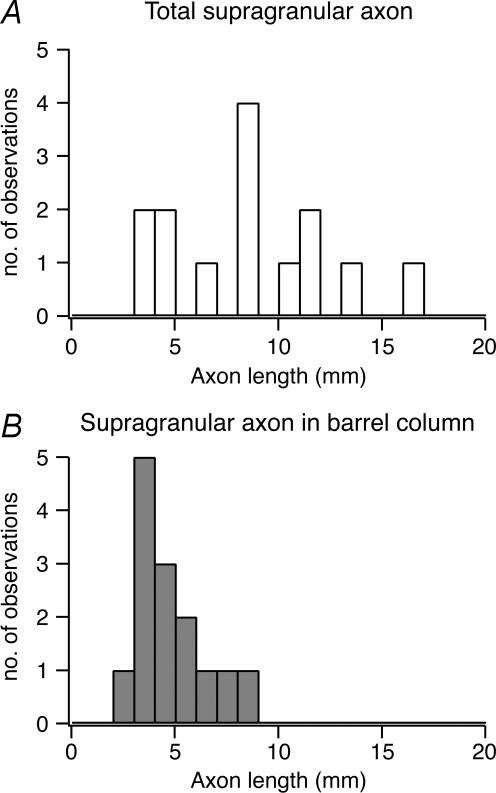
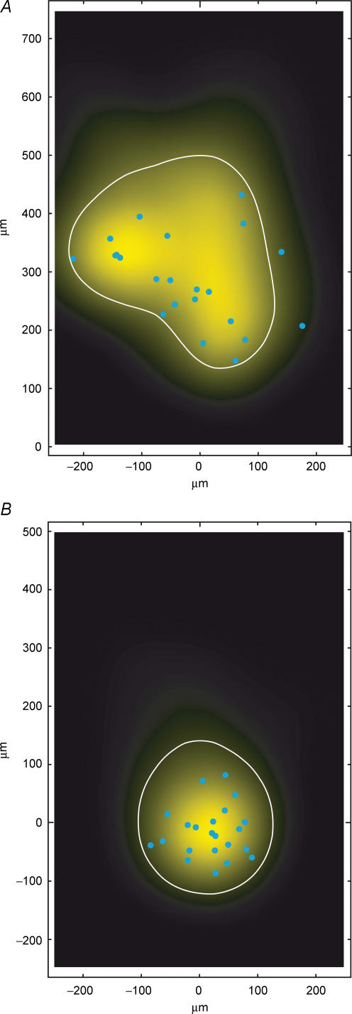
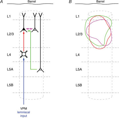
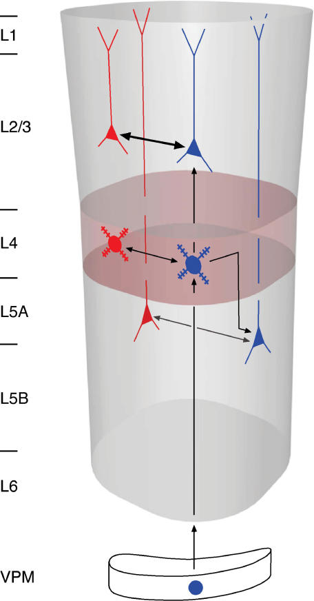
Synaptically coupled layer 2/3 (L2/3) pyramidal neurones located above the same layer 4 barrel ('barrel-related') were investigated using dual whole-cell voltage recordings in acute slices of rat somatosensory cortex. Recordings were followed by reconstructions of biocytin-filled neurones. The onset latency of unitary EPSPs was 1.1 +/- 0.4 ms, the 20-80% rise time was 0.7 +/- 0.2 ms, the average amplitude was 1.0 +/- 0.7 mV and the decay time constant was 15.7 +/- 4.5 ms. The coefficient of variation (c.v.) of unitary EPSP amplitudes decreased with increasing EPSP peak and was 0.33 +/- 0.18. Bursts of APs in the presynaptic pyramidal cell resulted in EPSPs that, over a wide range of frequencies (5-100 Hz), displayed amplitude depression. Anatomically the barrel-related pyramidal cells in the lower half of layer 2/3 have a long apical dendrite with a small terminal tuft, while pyramidal cells in the upper half of layer 2/3 have shorter and often more 'irregularly' shaped apical dendrites that branch profusely in layer 1. The number of putative excitatory synaptic contacts established by the axonal collaterals of a L2/3 pyramidal cell with a postsynaptic pyramidal cell in the same column varied between 2 and 4, with an average of 2.8 +/- 0.7 (n = 8 pairs). Synaptic contacts were established predominantly on the basal dendrites at a mean geometric distance of 91 +/- 47 mum from the pyramidal cell soma. L2/3-to-L2/3 connections formed a blob-like innervation domain containing 2.8 mm of the presynaptic axon collaterals with a bouton density of 0.3 boutons per mum axon. Within the supragranular layers of its home column a single L2/3 pyramidal cell established about 900 boutons suggesting that 270 pyramidal cells in layer 2/3 are innervated by an individual pyramidal cell. In turn, a single pyramidal cell received synaptic inputs from 270 other L2/3 pyramidal cells. The innervation domain of L2/3-to-L2/3 connections superimposes almost exactly with that of L4-to-L2/3 connections. This suggests that synchronous feed-forward excitation of L2/3 pyramidal cells arriving from layer 4 could be potentially amplified in layer 2/3 by feedback excitation within a column and then relayed to the neighbouring columns.


Figures

Figure 1
Figure 1. Time course and amplitude of EPSPs in L2/3 pyramidal cells of the barrel cortex
A, original recording of a presynaptic AP and a postsynaptic EPSP in a synaptically coupled pair of L2/3 pyramidal cells. B, distribution of EPSP latencies in an individual synaptically connected pair of L2/3 pyramidal cells; the continuous curve is a single Gaussian fit. Note that the latency distribution for this connection is narrow. C, distributions of EPSP latencies, 20–80% rise times, decay time constants and EPSP amplitudes for L2/3 pyramidal cell connections. Latencies were calculated as the time between the peak of the AP and the onset of the EPSP (indicated by the dashed line in A) and the onset of the EPSP. Decay times were obtained by fitting a single exponential to the falling phase of the EPSP.
Figure 2
Figure 2. Reliability of synaptic connections between pairs of L2/3 pyramidal cells
A, examples of 10 successive unitary EPSPs in response to a presynaptic AP (top trace); the bottom trace represents the average EPSP waveform. The dashed line indicates the peak of the AP. B, distribution of the c.v. of unitary EPSP amplitudes calculated in 28 L2/3-to-L2/3 connections from 50–200 trials (stimulation frequency 0.05–0.1 s−1); the average c.v. was 0.33 ± 0.18. C, relationship of c.v. and EPSP peak amplitude in the postsynaptic L2/3 pyramidal cell. The two dashed lines in C represent the predictions of single binomial release statistics for the c.v. as a function of EPSP amplitude assuming three synaptic contacts (close to the average number of contacts, Table 1), and qS = 0.05 mV (right curve) and qS = 0.7 mV (left curve); pr increases from 0.08 to 0.6 (right curve) and from 0.05 to 1.0 (left curve). The pr values refer to the two endpoints of each curve. Connections with large mean EPSP amplitudes are not well described by binomial release statistics.
Figure 3
Figure 3. Paired pulse ratio in pairs of L2/3 pyramidal cell
A, train of five consecutive EPSPs at different interstimulus intervals as indicated on the left. At most connections EPSP amplitude depression occurred at all frequencies tested while summation of EPSPs was apparent only at an interstimulus interval of 10–20 ms. The horizontal scale bar corresponds to the interstimulus time of each train. B, amplitude ratios (•, 2 EPSP/1 EPSP; grey square, 3 EPSP/1 EPSP; grey triangle, 4 EPSP/1 EPSP, grey inverted triangle, 5 EPSP/1 EPSP) plotted as a function of the interstimulus interval. For short interstimulus intervals (10 ms and 20 ms), the baseline to determine the EPSP amplitudes in the train was obtained by linear extrapolation of the decay phase of the preceding EPSP.
Figure 4
Figure 4. Half-tone image of a pair of synaptically coupled L2/3 pyramidal cells including the light and electron microscopic identification of synaptic contacts
A, low magnification light microscopic image of two synaptically coupled pyramidal cells filled with biocytin. Both pyramidal cells were located in the middle portion of layer 2/3. Note the elaborate symmetric basal dendritic field and the apical dendrites forming extensive tufts terminating in layer 1. Calibration bar, 100 μm. B and C, high magnification of the synaptic contacts established by en passant axonal collaterals of the presynaptic neurone on different basal dendrites of the postsynaptic L2/3 pyramidal cells. The calibration bar is 5.0 μm for both panels. B1 and C1, both light microscopically identified synaptic contacts were identified at the electron microscopic level. The synaptic boutons of the presynaptic axon collaterals are clearly identifiable by their content of transmitter vesicles. The calibration bar is 1.0 μm for both panels.
Figure 5
Figure 5. Reconstructions of two pairs of synaptically coupled L2/3 pyramidal cells
Neurolucida reconstructions of two pairs of synaptically coupled L2/3 pyramidal cells, located in the lower (A) and upper half (B) of layer 2/3, respectively. For both pairs of neurones, the dendritic configuration of the presynaptic L2/3 pyramidal cell is drawn in red with its axon in blue and the postsynaptic L2/3 pyramidal cell is drawn in white with its axon in green. Scale bar, 100 μm. Pyramidal cells in the lower half of layer 2/3 display a more prominent apical dendrite with a number of oblique collaterals, extending up to 300 μm before bifurcating close to layer 1 giving rise to a terminal tuft. In contrast, pyramidal cells in the upper half of layer 2/3 have only a short apical trunk that bifurcates after ∼30–150 μm into an extensive terminal tuft (Fig. 5B). The insets in panels A and B show the location of synaptic contacts (blue squares) on the postsynaptic neurone. Note that most synaptic contacts are found on basal dendrites.
Figure 6
Figure 6. Number and location of synaptic contacts on dendrites
A, histogram showing the geometric distances of light microscopically identified synaptic contacts from the soma in eight L2/3 pyramidal cell pairs (n = 22 contacts). Inset, distribution of the number of synaptic contacts per individual connection. B, relationship between the unitary EPSP amplitude in the postsynaptic L2/3 pyramidal cells and the number of synaptic contacts per connection. The correlation coefficient r obtained for B was −0.284. C, relationship between the unitary EPSP amplitude and the mean geometric distance from the soma of synaptic contacts in a connection. The correlation coefficient r was – 0.253. For both graphs, the correlation was statistically not significant.
Figure 7
Figure 7. Superposition of pairs of synaptically connected L2/3 pyramidal cells
A and B, 2D computer-aided reconstructions of pairs of synaptically connected L2/3 pyramidal cells superimposed and aligned with respect to the barrel centre. The dendritic domains of the presynaptic pyramidal cell are shown in red (A), those of the postsynaptic pyramidal cells in white; presynaptic axons are in blue and postsynaptic axons in green. The central barrel has the average dimensions given in Table 2. The outlines of two neighbouring barrels were added symbolically for clarity. C, 2D maps of axonal (C1) and dendritic ‘length density’ (C2) of synaptically coupled pairs of L2/3 pyramidal cells, aligned with respect to the centre of the barrel. The predicted innervation domain (C3, yellow) of postsynaptic L2/3 dendrites by presynaptic L2/3 axons is given by the product of the presynaptic L2/3 axonal density and the postsynaptic L2/3 dendritic density. Contours (thin lines) enclosing 80% of the integrated density are shown superimposed. Positions of presynaptic L2/3 pyramidal cell somata (red triangles), postsynaptic L2/3 pyramidal cell somata (white triangles) and outlines of barrels (thick white lines) are indicated symbolically. D, 2D map of axonal (D1) and dendritic ‘length density’ (D2) centred on the location of the postsynaptic L2/3 pyramidal cell soma (white triangle) of each pair of reconstructions. For these maps the same eight L2/3 pyramidal cell pairs plus two additional pairs (for which the location with respect to the barrel was not recovered) were used. Note that mainly the basal dendritic field contributes to the innervation domain (D3, yellow).
Figure 8
Figure 8. Axonal length in supragranular layers
A, histogram of the distribution of total axonal length of L2/3 pyramidal cells in the supragranular layer (n = 14 of 7 synaptic connections) and B, of the intracolumnar axonal length in the supragranular layer (i.e. the supragranular axon length within the home column). Only those synaptic connections were used for which the L2/3 pyramidal cell axon displayed no obvious truncation throughout layers 1–6. Note that the distribution of the total supragranular axon length is highly variable while that of the intracolumnar axon length is relatively narrow.
Figure 9
Figure 9. Innervation domain of presynaptic L2/3 axons on postsynaptic L2/3 pyramidal cell dendrites
A, close-up of the predicted innervation domain of postsynaptic L2/3 pyramidal cell dendrites by presynaptic L2/3 axons, aligned to the barrel centre (same data as in Fig. 7C3, i.e. n = 8, number of contacts = 22). A contour (white line) enclosing 80% of the integrated density and the locations of light microscopically identified synaptic contacts (blue circles) of eight L2/3 pyramidal cell pairs (blue cicles, n = 22) are superimposed. B, predicted innervation domain centred on the location of the postsynaptic L2/3 pyramidal cell somata (same data as for Fig. 7D3). Note that both the predicted innervation domain and the distribution of light microscopically identified synaptic contacts (blue circles) are much more compact in this representation, being confined mostly to the basal dendrites of the L2/3 pyramidal cells. All contacts were located within the contour (thin lines) enclosing 80% of the integrated density.
Figure 10
Figure 10. Schematic representation of L2/3 dendrites innervated by different cortical projections
A, schematic barrel column showing intracortical synaptic connections as well as input from the lemniscal thalamic nucleus (ventroposterior medial nucleus; VPM). The different synaptic connections are colour coded: intracortical connections are in red, violet and green; the thalamocortical inputs are drawn in dark blue (from VPM) B, innervation domains (80% contour lines) for the three types of excitatory synaptic connections in which L2/3 pyramidal cells are the postsynaptic (target) neurones (L4–L2/3 connection, red; L2/3–L2/3 connection, violet; L5A–L2/3 connection, green). Data for L4–L2/3 connections are taken from Lübke et al. (2003). Prospective L5A–L2/3 innervation domains were constructed using the dendritic domain of L2/3 pyramidal cells from Lübke et al. (2003) and this study and the axonal domain of L5A pyramidal cells from Feldmeyer et al. (2005). Note that the innervation domains show considerable overlap within the cortical column.
Figure 11
Figure 11. Schematic view of L2/3-to-L2/3 feedback connections in a cortical column
In each of the cortical layers (layer 2/3, 4 and 5A) pyramidal cells receive vertical interlaminar feed-forward input (blue cell symbols; vertical arrows) and intralaminar feedback excitation (horizontal arrows). In each layer the intralaminar connections (L2/3–L2/3, L4–L4 and L5A–L5A) could amplify vertical input. For emphasis, the arrow representing the L2/3-L2/3 connection is given in bold.

References

    1. Agmon A, Connors BW. Thalamocortical responses of mouse somatosensory (barrel) cortex in vitro. Neurosci. 1991;41:365–379. - PubMed
    1. Armstrong-James M. The nature and plasticity of sensory processing within adult rat barrel cortex. In: Jones EG, Diamond IT, editors. The Barrel Cortex of Rodents. New York: Plenum Press; 1995. pp. 333–374.
    1. Armstrong-James M, Callahan CA, Friedman MA. Thalamo-cortical processing of vibrissal information in the rat. I. Intracortical origins of surround but not centre-receptive fields of layer IV neurones in the rat S1 barrel field cortex. J Comp Neurol. 1991;303:193–210. - PubMed
    1. Armstrong-James M, Fox K. Spatiotemporal convergence and divergence in the rat S1 ‘barrel’ cortex. J Comp Neurol. 1987;263:265–281. - PubMed
    1. Armstrong-James M, Fox K, Das-Gupta A. Flow of excitation within rat barrel cortex on striking a single vibrissa. J Neurophysiol. 1992;68:1345–1358. - PubMed

Publication types

MeSH terms

LinkOut - more resources