. 2006 Dec;35(6):628-38.
doi: 10.1165/rcmb.2006-0040OC. Epub 2006 Jun 22.

Induction of the plasminogen activator system by mechanical stimulation of human bronchial epithelial cells

Affiliations

Induction of the plasminogen activator system by mechanical stimulation of human bronchial epithelial cells

Eric K Chu et al. Am J Respir Cell Mol Biol. 2006 Dec.

Abstract

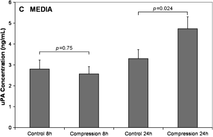
Mechanical stimulation of the airway epithelium, as would occur during bronchoconstriction, is a potent stimulus and can activate profibrotic pathways. We used DNA microarray technology to examine gene expression in compressed normal human bronchial epithelial cells (NHBE). Compressive stress applied continuously over an 8-h period to NHBE cells led to the upregulation of several families of genes, including a family of plasminogen-related genes that were previously not known to be regulated in this system. Real-time PCR demonstrated a peak increase in gene expression of 8.0-fold for urokinase plasminogen activator (uPA), 16.2-fold for urokinase plasminogen activator receptor (uPAR), 4.2-fold for plasminogen activator inhibitor-1 (PAI-1), and 3.9-fold for tissue plasminogen activator (tPA). Compressive stress also increased uPA protein levels in the cell lysates (112.0 versus 82.0 ng/ml, P = 0.0004), and increased uPA (4.7 versus 3.3 ng/ml, P = 0.02), uPAR (1.3 versus 0.86 ng/ml, P = 0.007), and PAI-1 (50 versus 36 ng/ml, P = 0.006) protein levels in cell culture media. Functional studies demonstrated increased urokinase-dependent plasmin generation in compression-stimulated cells (0.0090 versus 0.0033 OD/min, P = 0.03). In addition, compression led to increased activation of matrix metalloproteinase (MMP)-9 and MMP-2 in a urokinase-dependent manner. In postmortem human lung tissue, we observed an increase in epithelial uPA and uPAR immunostaining in the airways of two patients who died in status asthmaticus compared with minimal immunoreactivity noted in airways from seven lung donors without asthma. Together these observations suggest an integrated response of airway epithelial cells to mechanical stimulation, acting through the plasminogen-activating system to modify the airway microenvironment.


Figures

<b>Figure 1.</b>
Figure 1.
Protocols for compressive stress. Black triangles indicate harvest of samples for analysis.
<b>Figure 2.</b>
Figure 2.
Hierarchical clustering of 1,024 differentially regulated genes in response to compressive stress. Gene expression patterns are listed from top to bottom, and the time course of compression is presented as time zero (before compression) on the left with increasing durations of compression up to 8 h to the right. Upregulated genes are indicated in red and downregulated genes are indicated in green.
<b>Figure 3.</b>
Figure 3.
PCA performed on the 1st and 2nd principal components of the 1,024 most differentially regulated genes. Plasminogen activator genes are indicated by red circles (uPA, UPAR [2 probes], PAI-1, tPA).
<b>Figure 4.</b>
Figure 4.
Real-time PCR results of gene expression of plasminogen activator genes in response to 8 h of continuous compressive stress. uPA, uPAR, PAI-1, and tPA were all significantly increased by compression. Significant increases compared with time zero are indicated by solid black symbols with the magnitude of the significance indicated respectively by the signs above: *P < 0.05, +P < 0.01, #P < 0.001.
<b>Figure 5.</b>
Figure 5.
uPA, uPAR, PAI-1 protein expression in response to compressive stress. (A) Compressive stress increases urokinase protein concentrations in cell lysates at 8 h and 24 h after the onset of compression. uPAR and PAI-1 concentrations are not altered by compression in the cell lysates (data not shown). Compressive stress increases cell culture media protein concentrations of: (B) soluble uPAR at 8 h and 24 h after the onset of compression, (C) uPA at 24 h after the onset of compression, and (D) PAI-1 at 24 h after the onset of compression. (Continued).
<b>Figure 6.</b>
Figure 6.
Plasminogen activator activity is increased in compressed cells. Colorimetric assay (405 nm) indicates increased plasmin generation over time (A) in NHBE cell lysates. Plasmin generation is greater in compression stimulated cells compared to unstimulated controls. (B) This effect is seen after both 8 and 24 h after the onset of compression, but only reaches statistical significance at 24 h. An antibody that blocks the activity of uPA confirms that the plasmin generation is due to uPA activity.
<b>Figure 7.</b>
Figure 7.
MMP-9/MMP-2 activity by Zymography. (A) pro–MMP-9/MMP-2 and active MMP-9/MMP-2 expression at various time points after the onset of compressive stress. Compressive stress was applied for up to 8 h, and cell lysates were examined for MMP-9/MMP-2 activity at baseline, 4, 8, 12, and 24 h after the onset of compressive stress. Pro–MMP-9 (92 kD) and pro–MMP-2 (72 kD) are present at all time points. Activated MMP-9 (82 kD) and MMP-2 (62 kD) are present from 8–24 h after the onset of compression, but were most apparent at 24 h. (B) MMP-9 and MMP-2 activities were examined in the presence of a urokinase inhibitor, a protease inhibitor that blocks plasmin (aprotinin), and a nonspecific control antibody (IgG1) as shown in this representative zymogram. Both pro–MMP-9 (92 kD) and the activated MMP-9 (82kD) are increased by compressive stress. Similarly pro–MMP-2 (72 kD) and the activated MMP-2 (62 kD) are also increased by compressive stress. Addition of an inhibitory antibody to urokinase as well as aprotinin reduced the activated MMP-9 and activated MMP-2 down to control levels.
<b>Figure 8.</b>
Figure 8.
Immunostaining for uPA and uPAR in human lung tissue. Representative images are shown from one of six lung donors, and one of two patients who died in status asthmaticus. Low-power fields (original magnification: ×10) for uPA (A, B) and uPAR (C, D) reveal increased staining in status asthmaticus (B, D) relative to lung donor (A, C) tissue. Higher-power fields (original magnification: ×40) highlight the increased epithelial staining for uPA (E, F) and uPAR (G, H) in status asthmaticus (F, H) relative to lung donor (E, G) airways. A relative absence of epithelial staining was found with a nonspecific isotype control antibody in both lung donor (I) and status asthmaticus (J) airway epithelium, although some nonspecific reactivity was seen in airway smooth muscle.

References

    1. Elias JA, Lee CG, Zheng T, Ma B, Homer RJ, Zhu Z. New insights into the pathogenesis of asthma. J Clin Invest 2003;111:291–297. - PMC - PubMed
    1. Davies DE, Wicks J, Powell RM, Puddicombe SM, Holgate ST. Airway remodeling in asthma: new insights. J Allergy Clin Immunol 2003; 111:215–225; quiz 226. - PubMed
    1. Swartz MA, Tschumperlin DJ, Kamm RD, Drazen JM. Mechanical stress is communicated between different cell types to elicit matrix remodeling. Proc Natl Acad Sci USA 2001;98:6180–6185. - PMC - PubMed
    1. Tschumperlin DJ, Shively JD, Kikuchi T, Drazen JM. Mechanical stress triggers selective release of fibrotic mediators from bronchial epithelium. Am J Respir Cell Mol Biol 2003;28:142–149. - PubMed
    1. Wiggs BR, Hrousis CA, Drazen JM, Kamm RD. On the mechanism of mucosal folding in normal and asthmatic airways. J Appl Physiol 1997;83:1814–1821. - PubMed

Publication types

MeSH terms