. 2006 Jul 12;25(13):3234-44.
doi: 10.1038/sj.emboj.7601191. Epub 2006 Jun 22.

Biosynthesis of tumorigenic HER2 C-terminal fragments by alternative initiation of translation

Affiliations

Biosynthesis of tumorigenic HER2 C-terminal fragments by alternative initiation of translation

Judit Anido et al. EMBO J. .

Abstract

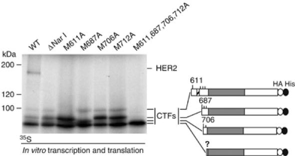
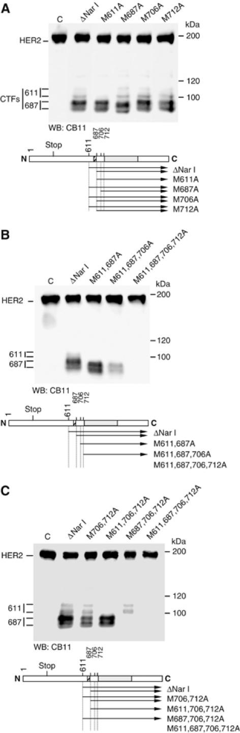
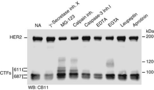
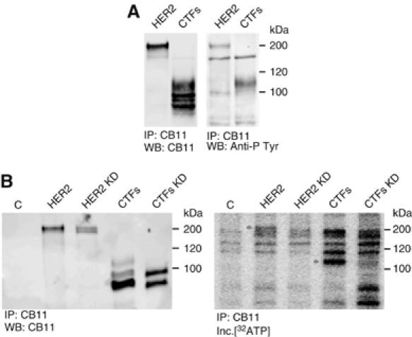
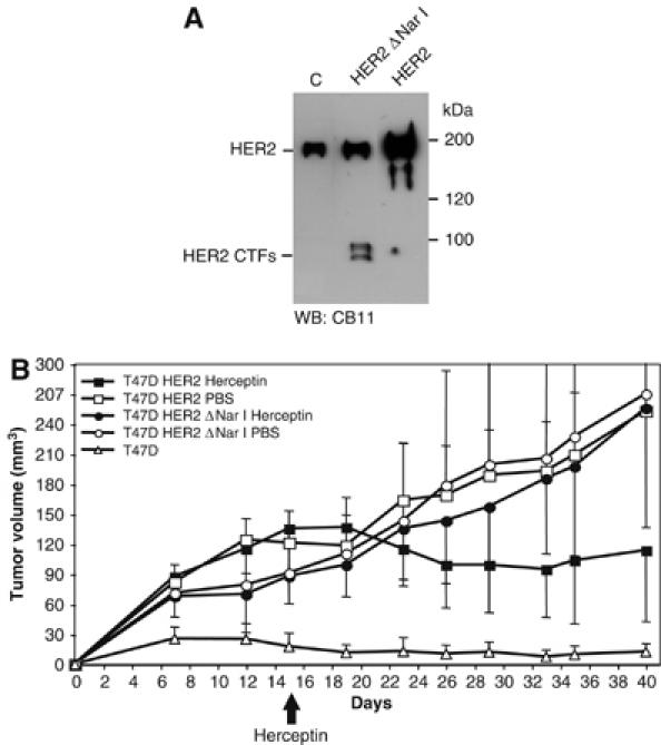
The overactivation of the HERs, a family of tyrosine kinase receptors, leads to the development of cancer. Although the canonical view contemplates HER receptors restricted to the secretory and endocytic pathways, full-length HER1, HER2 and HER3 have been detected in the nucleoplasm. Furthermore, limited proteolysis of HER4 generates nuclear C-terminal fragments (CTFs). Using cells expressing a panel of deletion and point mutants, here we show that HER2 CTFs are generated by alternative initiation of translation from methionines located near the transmembrane domain of the full-length molecule. In vitro and in vivo, HER2 CTFs are found in the cytoplasm and nucleus. Expression of HER2 CTFs to levels similar to those found in human tumors induces the growth of breast cancer xenografts in nude mice. Tumors dependent on CTFs are sensitive to inhibitors of the kinase activity but do not respond to therapeutic antibodies against HER2. Thus, the kinase domain seems necessary for the activity of HER2 CTFs and the presence of these HER2 fragments could account for the resistance to treatment with antibodies.


Figures

Figure 1
Figure 1
Spontaneous generation of CTFs in cells stably transfected with HER2. (A) Schematic of HER2 showing the N-terminus (N), the transmembrane domain (hatched box), the intracellular kinase domain (shaded box), the hemaglutinin antigen (HA) and hexahistidine (His) tags and the C-terminus (C). The localization of epitopes recognized by the monoclonal antibodies Herceptin® and Ab3 are shown. The monoclonal antibodies L87 and CB11 recognize undefined epitopes located in the extracellular and intracellular domain, respectively. (B) Analysis by Western blot with the indicated antibodies of parental CHO cells (C) and different clones of the same cells stably transfected with HER2 tagged with HA and His. (C) Schematic of peptides detected by mass spectrometry analysis in CTFs purified by Ni-chromatography. The sequence from lysine 676 to lysine 753 is shown. Methionines 687, 706 and 712 are shown in bold. The hatched and shaded boxes represent the transmembrane and tyrosine kinase domains, respectively. Peptides detected by mass spectrometry are indicated with thick lines. (D) Lysates from CHO cells stably transfected with untagged HER2 and from a human mammary tumor expressing CTFs were analyzed by Western blot with the antibody CB11.
Figure 2
Figure 2
CTFs are not generated by ectodomain shedding. (A) Stably transfected CHO cells expressing different HER2 species were incubated overnight with the indicated concentrations of the metalloprotease inhibitor BB-94 and analyzed by Western blot with the monoclonal anti-HER2 antibody CB11. (B) Stably transfected CHO cells expressing CTFs (clone #1) were treated for 24 h without (NA) or with 125 nM of γ-secretase inhibitor X, 10 μM of Calpain inhibitor or 10 μM of Caspase-3 inhibitor I and analyzed by Western blot with CB11 antibodies. (C) Parental CHO cells (C) or the same cells as in (A) were metabolically labeled with 35S-Translabel for 15 min, washed, lysed and immunoprecipitated with anti-HA antibodies. Immunoprecipitates were analyzed by SDS–PAGE and fluorography. (D) Top, CHO cells were transiently transfected with a control plasmid (C) or with plasmids encoding HER2 ΔNarI or HER2 as indicated. Cell lysates from transiently transfected cells were analyzed by Western blot with CB11 antiobodies. Bottom: Schematics showing the HER2 constructs transfected. The position of the Nar I sites used to make the HER2 ΔNar I construct are shown in wild-type HER2. The HER2 ΔNar I construct contains a stop codon after glycin 312 and generates a predicted protein of 32 kDa (solid line) the rest of the sequence in this construct is out of frame and is shown in dotted lines. The N-terminus (N), the transmembrane domain (hatched box), the intracellular kinase domain (shaded box) and the C-terminus (C) are shown. The position of the first in-frame methionine is indicated with the number 1. (E) T47D cells permanently transfected with wild-type HER2 mRNA were analyzed by Western blotting with antibodies against the intracellular domain of HER2 or by Northern blotting with probes specific for the extracellular (ECD) or intracellular (ICD) domains of HER2, respectively.
Figure 3
Figure 3
Biosynthesis of HER2 species from different mutant cDNAs in vitro. cDNAs encoding wild-type HER2, a Nar I deletion construct, or the indicated point mutants, also containing the Nar I deletion, were transcribed and translated in vitro in the presence of 35S-translabel and analyzed by SDS–PAGE and fluorography. Right: schematic showing the hypothetical HER2 CTFs generated, the numbers indicate the positions of different methionines (see also, Figure 1).
Figure 4
Figure 4
Biosynthesis of HER2 species from different mutant cDNAs in vivo. (A–C) MCF7 cells transiently transfected with a control plasmid (C) with the indicated single, double, triple or quadruple mutant constructs were lysed and cell lysates were analyzed by Western blot with CB11 antibodies. All the mutant constructs contain the ΔNar I deletion. Each panel contains the corresponding diagram showing the CTFs generated by the different constructs.
Figure 5
Figure 5
Effect of different protease inhibitors on the expression of CTFs. T47D cells permanently transfected with the ΔNar I construct were treated for 24 h without (NA) or with 125 nM of γ-secretase inhibitor X, 5 μM of the proteasome inhibitor MG132, 10 μM of Calpain inhibitor, 10 μM of Caspase-3 inhibitor I, 10 mM of EDTA, 10 mM of EGTA, 100 μM of leupeptin or 10 μg/ml of aprotinin. Treated cells were lysed and cell lysated analyzed with CB11 antibodies.
Figure 6
Figure 6
Subcellular localization of different HER2 species. (A) Stably transfected CHO cells expressing predominantly HA-tagged full-length HER2 (#3) and HER2 CTFs (#1), respectively, were fixed, permeabilized and stained with anti-HA and FITC-labeled anti-mouse antibodies. (B) The same cells were homogenized and fractionated by ultracentrifugation. Aliquots from soluble and membrane fractions were analyzed by Western blotting and probed with CB11 antibody or, as a control, with anti-β-tubulin antibodies. (C) MDA-MB-468 cells stably expressing CTFs WT or KD versions were fixed, permeabilized and stained with anti-HER2 (CB11) and FITC-labeled anti-mouse antibodies. (D) Homogenates from tumor samples were analyzed by Western blot with the anti-HER2 monoclonal antibody CB11. (E) Immunohistochemical staining with CB11 antibodies of the same tumor samples. In the magnification, nuclear staining is marked with arrows. (F) Tumor 48 was homogenized as in B and the different fractions analyzed by Western with the monoclonal antibody CB11.
Figure 7
Figure 7
Phosphorylation of CTFs. (A) MDA-MB-468 cells transfected with wild-type HER2 or a deletion construct that encodes CTFs were lysed. Cell lysates were immunoprecipitated with CB11 antibodies and analyzed by Western blot with the same antibodies or anti-phosphotyrosine antibodies as indicated. (B) Cell lysates from parental cells MDA-MB-468 (C) or the same cells transfected with constructs expressing HER2 or CTFs (WT or KD versions) were lysed and immunoprecipitated as in (A). Immunoprecipitates were incubated with [γ32ATP] and analyzed by SDS–PAGE and autoradiography.
Figure 8
Figure 8
Analysis of the tumors induced by parental T47D cells and the same cells expressing full-length HER2 or HER2 CTFs. (A) Parental T47D cells (C) or the same cells stably transfected with cDNAs encoding HER2 ΔNar I or HER2 were lysed and the cell lysates analyzed by Western blot with antibodies against the cytoplasmic domain of HER2 (CB11). (B) The same cells were subcutaneously injected into nude mice. Tumor volumes were measured at the indicated days after injection. Herceptin was given (i.p.) from day 15 (arrow) twice a week at a dose of 30 mg/kg. The control group was treated with PBS. Each point represents the mean of six individual determination±s.d.
Figure 9
Figure 9
Effect of Lapatinib on the phosphorylation of CTFs and on tumour growth. (A) Cells stably transfected with cDNAs encoding HER2 ΔNar I were treated with lapatinib (10 μM) during 8 h. Cell lysates were immunoprecipitated with anti-HA antibodies and analyzed by Western blot with the same antibody or anti-phosphotyrosine antibodies as indicated. Note that the T47D clone used in (A) express higher levels of CTFs than that used in Figure 8B. (B) The same cells T47D cells expressing CTFs used in Figure 8A and B were subcutaneously injected into nude mice. Tumor volumes were measured at the indicated days after injection. Lapatinib was given orally from day 22th (arrow) twice a day at a dose of 75 mg/kg. The vehicle was administered to the control group.

References

    1. Arribas J, Borroto A (2002) Protein ectodomain shedding. Chem Rev 102: 4627–4638 - PubMed
    1. Arribas J, Coodly L, Vollmer P, Kishimoto TK, Rosejohn S, Massagué J (1996) Diverse cell surface protein ectodomains are shed by a system sensitive to metalloprotease inhibitors. J Biol Chem 271: 11376–11382 - PubMed
    1. Arribas J, Massagué J (1995) Transforming growth factor alpha and beta amyloid precursor protein share a secretory mechanism. J Cell Biol 128: 433–441 - PMC - PubMed
    1. Bagatell R, Khan O, Paine-Murrieta G, Taylor CW, Akinaga S, Whitesell L (2001) Destabilization of steroid receptors by heat shock protein 90-binding drugs: a ligand-independent approach to hormonal therapy of breast cancer. Clin Cancer Res 7: 2076–2084 - PubMed
    1. Baselga J, Norton L (2002) Focus on breast cancer. Cancer Cell 1: 319–322 - PubMed

Publication types