. 2006 Dec 1;99(5):1285-97.
doi: 10.1002/jcb.20904.

Biologic properties of mesenchymal stem cells derived from bone marrow and adipose tissue

Affiliations

Biologic properties of mesenchymal stem cells derived from bone marrow and adipose tissue

Reza Izadpanah et al. J Cell Biochem. .

Abstract

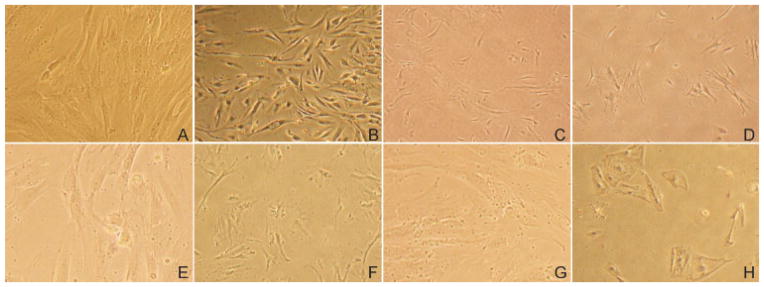
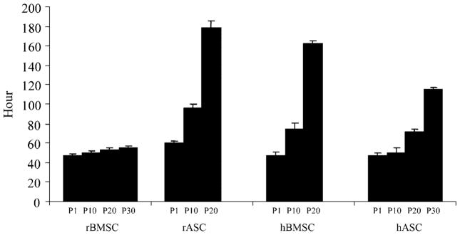
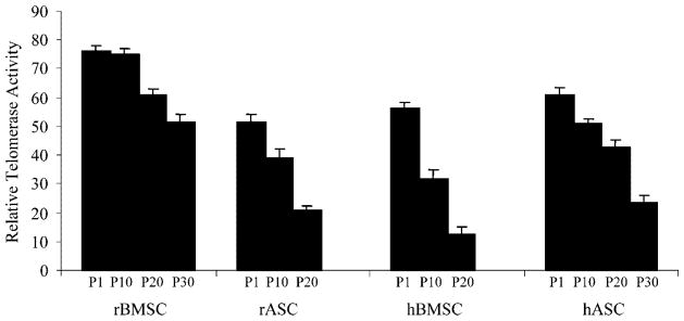
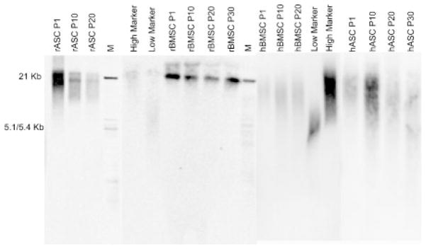
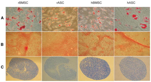
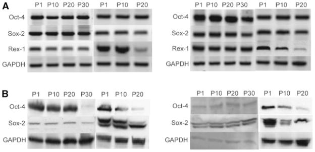
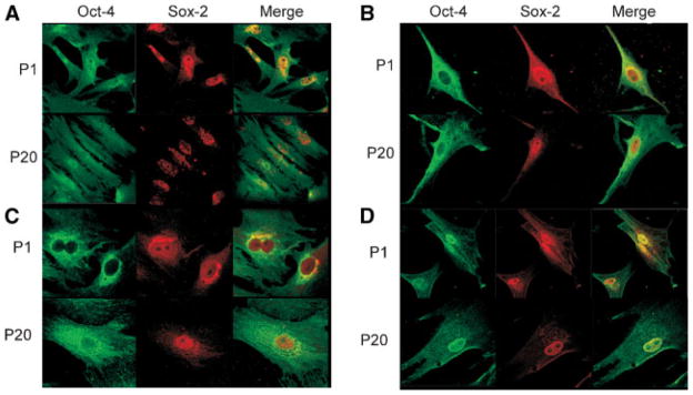
The biologic characteristics of mesenchymal stem cells (MSCs) isolated from two distinct tissues, bone marrow and adipose tissue were evaluated in these studies. MSCs derived from human and non-human primate (rhesus monkey) tissue sources were compared. The data indicate that MSCs isolated from rhesus bone marrow (rBMSCs) and human adipose tissue (hASCs) had more similar biologic properties than MSCs of rhesus adipose tissue (rASCs) and human bone marrow MSCs (hBMSCs). Analyses of in vitro growth kinetics revealed shorter doubling time for rBMSCs and hASCs. rBMSCs and hASCs underwent significantly more population doublings than the other MSCs. MSCs from all sources showed a marked decrease in telomerase activity over extended culture; however, they maintained their mean telomere length. All of the MSCs expressed embryonic stem cell markers, Oct-4, Rex-1, and Sox-2 for at least 10 passages. Early populations of MSCs types showed similar multilineage differentiation capability. However, only the rBMSCs and hASCs retain greater differentiation efficiency at higher passages. Overall in vitro characterization of MSCs from these two species and tissue sources revealed a high level of common biologic properties. However, the results demonstrate clear biologic distinctions, as well.


Figures

Fig. 1
Fig. 1
MSC morphology in low and high passages. Microscopic photographs of four types of MSCs cultures at low and high passages. rBMSCs and hASCs passage 1 (A and C) and passage 30 (B and D). Cultures of hBMSCs and rASCs at Passage 1 (E and G) and passage 20 (F and H), respectively.
Fig. 2
Fig. 2
Population doubling time of MSCs. A known number of MSCs types from different passages were cultured. The total number of cells was determined at different time points to obtain the doubling time.
Fig. 3
Fig. 3
Telomerase activity in MSCs. Telomerase enzyme activity measured using a photometric enzyme immunoassay on samples isolated at different passages for each line of MSCs. The bars indicate the mean enzyme activity out of three replicates for every passage of each cell type.
Fig. 4
Fig. 4
Telomere length at multiple passages of MSCs. Overall telomere length in passages of rASCs, rBMSCs, hASCs, and hBMSCs. Genomic DNA isolated from each indicated passage was digested with HinfI and RsaI and electrophoresed in a 0.8% agarose gel. Terminal restriction fragments were visualized, using a labeled (TTAGGG) probe.
Fig. 5
Fig. 5
Differentiation of MSCs along mesodermal lineages. rBMSCs, rASCs, hBMSCs, and hASCs were differentiated along adipogenesis and formed lipid vesicles stained with Oil red-O (A). Mineralization revealed in osteogenic-differentiated MSCs using Alizarin red staining (B). MSCs under chondrogenesis condition promoted the formation of chondrocytes, sectioned paraffin-embedded micromass pellets were stained with Toluidine Blue (C).
Fig. 6
Fig. 6
Transcription factor mRNA and expression in MSCs. Total cellular RNA analyzed by RT-PCR for Oct-4, Sox-2, and Rex-1 mRNA expression (A). The cell lysate from MSC types at different passages were subjected to Western blot analysis using Oct-4 and Sox-2 monoclonal antibodies to determine the level of expression of these two proteins (B).
Fig. 7
Fig. 7
Oct-4 and Sox-2 localization in MSCs. Immunohistochemical staining of rBMSCs (A), hBMSCs (B), rASCs (C), and hASCs (D). Cells were fixed, permeabilized, and stained to visualize Oct-4 and Sox-2. Distribution and localization of these transcription factors exhibited at passage 1 and 20 each MSC types. Merge panel showed co-localization of these two proteins.

References

    1. Allsopp RC, Vaziri H, Patterson C, Goldstein S, Younglai EV, Futcher AB, Greider CW, Harley CB. Telomere length predicts replicative capacity of human fibroblasts. Proc Natl Acad Sci USA. 1992;89:10114–10118. - PMC - PubMed
    1. Arnhold S, Klein H, Semkova I, Addicks K, Schraermeyer U. Neurally selected embryonic stem cells induce tumor formation after long-term survival following engraftment into the subretinal space. Invest Ophthalmol Vis Sci. 2004;45:4251–4255. - PubMed
    1. Aust L, Devlin B, Foster SJ, Halvorsen YD, Hicok K, du Laney T, Sen A, Willingmyre GD, Gimble JM. Yield of human adipose-derived adult stem cells from liposuction aspirates. Cytotherapy. 2004;6:7–14. - PubMed
    1. Bianco P, Gehron Robey P. Marrow stromal stem cells. J Clin Invest. 2000;105:1663–1668. - PMC - PubMed
    1. Bodnar AG, Ouellette M, Frolkis M, Holt SE, Chiu CP, Morin GB, Harley CB, Shay JW, Lichtsteiner S, Wright WE. Extension of life-span by introduction of telomerase into normal human cells. Science. 1998;279:349–352. - PubMed

Publication types

LinkOut - more resources